. 2023:2545:401-412.
doi: 10.1007/978-1-0716-2561-3_21.

Measuring Cellular Ploidy In Situ by Light Microscopy

Affiliations

Measuring Cellular Ploidy In Situ by Light Microscopy

Delisa E Clay et al. Methods Mol Biol. 2023.

Abstract

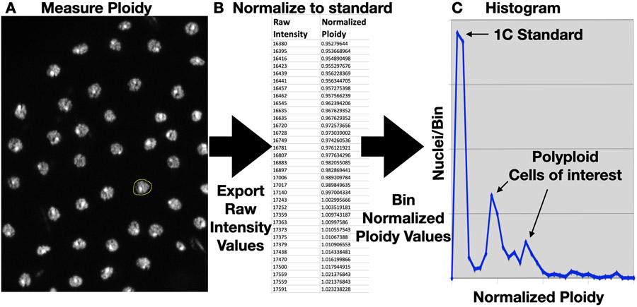
Determining cellular DNA content is valuable in the study of numerous biological processes, including organ development and injury repair. While FACS analysis of dissociated cells is a widely used method for assaying ploidy in a tissue cell population, for many tissue samples, it is possible and convenient to measure ploidy in situ using light microscopy. Here, we present two protocols for measuring cellular ploidy in tissues. These protocols are based on our studies in Drosophila melanogaster, but these are applicable to other settings as well. We present example results from Drosophila hindgut, midgut, and wing imaginal disc as examples. The first protocol focuses on measuring DNA content from decondensed interphase nuclei, while the second protocol details the visualization of condensed chromosomes for ploidy determination, either from mitotic cells or from interphase cells with drug-induced chromosome condensation. These techniques can be completed in 1 day and require standard lab supplies as well as a fluorescence light microscope.

Keywords: Cellular ploidy; Microscopy; Polyploidy.


Figures

Figure 1-
Figure 1-. DAPI stained haploid sperm nuclei.
Scale bar= 20 microns.
Figure 2-
Figure 2-. The Squash procedure.
A. Aspirating a bead of liquid on a siliconized coverslip with a gel loading tip. B. Picking up the coverslip with a positively charged slide. C. Tracing a grid over the coverslip with a metal probe. D. Tracing a grid on the bottom of the slide (covered with a paper towel) with a probe handle. E. Placing the slide in the vise. F. Removing the coverslip with a razor blade.
Figure 3-
Figure 3-. Producing ploidy histogram data from samples prepared using Protocol 3.1.
A. ROI selection (yellow) of a DAPI-stained nucleus of interest in ImageJ. B. Example range of raw integrated density and converted ploidy data (C), normalized to a haploid standard. C. An example histogram plot that includes both a 1C standard and polyploid cells of interest.
Figure 4-
Figure 4-. Condensed chromosome imaging of Drosophila melanogaster cells, prepared using protocol 3.2.
A. A metaphase spread from a 2N/4C XY mitotic rectal epithelial cell. B. A metaphase spread from a 2N/4C XX mitotic midgut intestinal stem cell. C. A metaphase spread from an 8N/16C XY hindgut rectal papillar cell. D. An interphase wing imaginal disc cell without Calyculin A. E. An interphase XX wing imaginal disc cell treated with Calyculin A to reveal 2N/2C ploidy (note- the small 4th chromosomes are not present in this image). F. An interphase hindgut rectal papillar cell of similar ploidy to the cell in C, treated with Calyculin A. The centromeric regions on each chromosome become un-paired in this assay[8]. For all images where distinct chromosomes are seen, the chromosome type is labeled. Chromosome types are pseudo-colored as follows: X/Y: purple, 2nd or 3rd autosomes: green or red, autosome 4: blue. Scale bar= 25 microns.

References

    1. Peterson NG & Fox DT (2021) Communal living: the role of polyploidy and syncytia in tissue biology. Chromosome Research, in press - PMC - PubMed
    1. Øvrebø JI, Edgar BA (2018) Polyploidy in tissue homeostasis and regeneration. Development 145: dev.156034. - PMC - PubMed
    1. Fox DT, Soltis DE, Soltis PS, et al. (2020) Polyploidy: A Biological Force From Cells to Ecosystems. Trends Cell Biology 30: 688–694. - PMC - PubMed
    1. Gatti M, Bonaccorsi S, Pimpinelli S (1994) Looking at Drosophila Mitotic Chromosomes. Methods Cell Biol 44:371–391. - PubMed
    1. Kennison JA (2008) Dissection of larval salivary glands and polytene chromosomes preparation. Cold Spring Harb Protoc 3:pdb.prot4708. - PubMed

Publication types

LinkOut - more resources