. 2023 Jan 31;13(1):985.
doi: 10.1038/s41598-023-28147-5.

Food protein-derived amyloids do not accelerate amyloid β aggregation

Affiliations

Food protein-derived amyloids do not accelerate amyloid β aggregation

M Mahafuzur Rahman et al. Sci Rep. .

Abstract

The deposition of proteins in the form of amyloid fibrils is closely associated with several serious diseases. The events that trigger the conversion from soluble functional proteins into insoluble amyloid are not fully understood. Many proteins that are not associated with disease can form amyloid with similar structural characteristics as the disease-associated fibrils, which highlights the potential risk of cross-seeding of disease amyloid by amyloid-like structures encountered in our surrounding. Of particular interest are common food proteins that can be transformed into amyloid under conditions similar to cooking. We here investigate cross-seeding of amyloid-β (Aβ), a peptide known to form amyloid during the development of Alzheimer's disease, by 16 types of amyloid fibrils derived from food proteins or peptides. Kinetic studies using thioflavin T fluorescence as output show that none of the investigated protein fibrils accelerates the aggregation of Aβ. In at least two cases (hen egg lysozyme and oat protein isolate) we observe retardation of the aggregation, which appears to originate from interactions between the food protein seeds and Aβ in aggregated form. The results support the view that food-derived amyloid is not a risk factor for development of Aβ pathology and Alzheimer's disease.


Conflict of interest statement

The authors declare no competing interests.

Figures
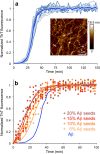
Figure 1
Figure 1
Aggregation of Aβ1–42 with and without Aβ1–42 seeds. (a) ThT kinetics of Aβ1–42 without any added seeds. The figure shows 15 traces from 5 experiments and the average in clear blue. Inset: representative AFM image of Aβ1–42 amyloid fibrils. (b) ThT kinetics of Aβ1–42 with 5%, 10%, 15% or 20% Aβ1–42 seeds added. The average data for Aβ1–42 without seeds is shown in blue. Experimental data for three replicates at each condition (filled circles) and sigmoidal curve fits of the average data (solid lines) are shown.
Figure 2
Figure 2
AFM images of amyloid fibrils from the different protein sources used to prepare seeds for the study. (a) Lysozyme, (b) pure β-lactoglobulin, (c) WPI (straight morphology), (d) WPI (worm-like morphology), (e) soybean, (f) mung bean, (g) fava bean, (h) lupine, (i) potato, and (j) oat.
Figure 3
Figure 3
ThT kinetic traces for Aβ1–42 without seeds (blue) and seeded with 5% (orange) or 10% (red) seeds from (a) lysozyme, (b) pure β-lactoglobulin, (c) WPI (straight morphology), (d) WPI (worm-like morphology), (e) soybean, (f) mung bean, (g) fava bean, (h) lupine, (i) potato, and (j) oat. Experimental data for three replicates at each condition (filled circles) and sigmoidal curve fits of the average data (solid lines) are shown.
Figure 4
Figure 4
AmyloFit analysis of Aβ1–42 seeded with 5%, 10%, 15% and 20% seeds of (a) lysozyme and (b) oat protein. Experimental data for three replicates at each condition (circles) and the model fit (solid lines) are shown.
Figure 5
Figure 5
Seeding with amyloid fibrils from β-lactoglobulin-derived peptides. (a) Sequences and locations of the peptides in the native structure of β-lactoglobulin. (b,c) AFM images of amyloid fibrils from the (b) βLG11-20 and (c) βLG8-33 peptides, respectively. (d,e) ThT kinetic traces for Aβ1–42 without seeds (blue) and seeded with 5% (orange) or 10% (red) seeds from (d) βLG11-20 and (e) βLG8-33, respectively. Experimental data for three replicates at each condition (filled circles) and a sigmoidal curve fit of the average data (solid lines) are shown.
Figure 6
Figure 6
Seeding with amyloid fibrils from soy protein-derived peptides. (a) Sequences and location in the native structure of soy storage proteins. GG1, and GG2 originates from glycinin, BA1 from β-conglycinin subunit α, and BB2 from β-conglycinin subunit β. (be) AFM images of amyloid fibrils from the (b) GG1, (c) BA1, (d) GG2 and (e) BB2 peptides, respectively. (fi) ThT kinetic traces for Aβ1–42 without seeds (blue) and seeded with 5% (orange) or 10% (red) seeds from (f) GG1, (g) BA1, (h) GG2 and (i) BB2 peptides, respectively. Experimental data for three replicates at each condition (filled circles) and a sigmoidal curve fit of the average data (solid lines) are shown.
Figure 7
Figure 7
Summary of t1/2 for all seeding conditions presented in this study. For each type of seed the values are shown from the highest concentration (left) to the lowest concentration (right). The blue horizontal region corresponds to t1/2 for unseeded Aβ1–42 ± standard deviation.

Similar articles

Cited by

References

    1. Ke PC, et al. Half a century of amyloids: Past, present and future. Chem. Soc. Rev. 2020;49:5473–5509. doi: 10.1039/C9CS00199A. - DOI - PMC - PubMed
    1. Knowles TPJ, Vendruscolo M, Dobson CM. The amyloid state and its association with protein misfolding diseases. Nat. Rev. Mol. Cell Biol. 2014;15:384–396. doi: 10.1038/nrm3810. - DOI - PubMed
    1. Chiti F, Dobson CM. Protein misfolding, functional amyloid, and human disease. Annu. Rev. Biochem. 2006;75:333–366. doi: 10.1146/annurev.biochem.75.101304.123901. - DOI - PubMed
    1. Chiti F, Dobson CM. Protein misfolding, amyloid formation, and human disease: A summary of progress over the last decade. Annu. Rev. Biochem. 2017;86:27–68. doi: 10.1146/annurev-biochem-061516-045115. - DOI - PubMed
    1. Dobson CM. Protein misfolding, evolution and disease. Trends Biochem. Sci. 1999;24:329–332. doi: 10.1016/S0968-0004(99)01445-0. - DOI - PubMed

Publication types