. 2023 Jan 31;13(1):1807.
doi: 10.1038/s41598-023-28822-7.

Induction of psoriasis- and atopic dermatitis-like phenotypes in 3D skin equivalents with a fibroblast-derived matrix

Affiliations

Induction of psoriasis- and atopic dermatitis-like phenotypes in 3D skin equivalents with a fibroblast-derived matrix

Bianka Morgner et al. Sci Rep. .

Abstract

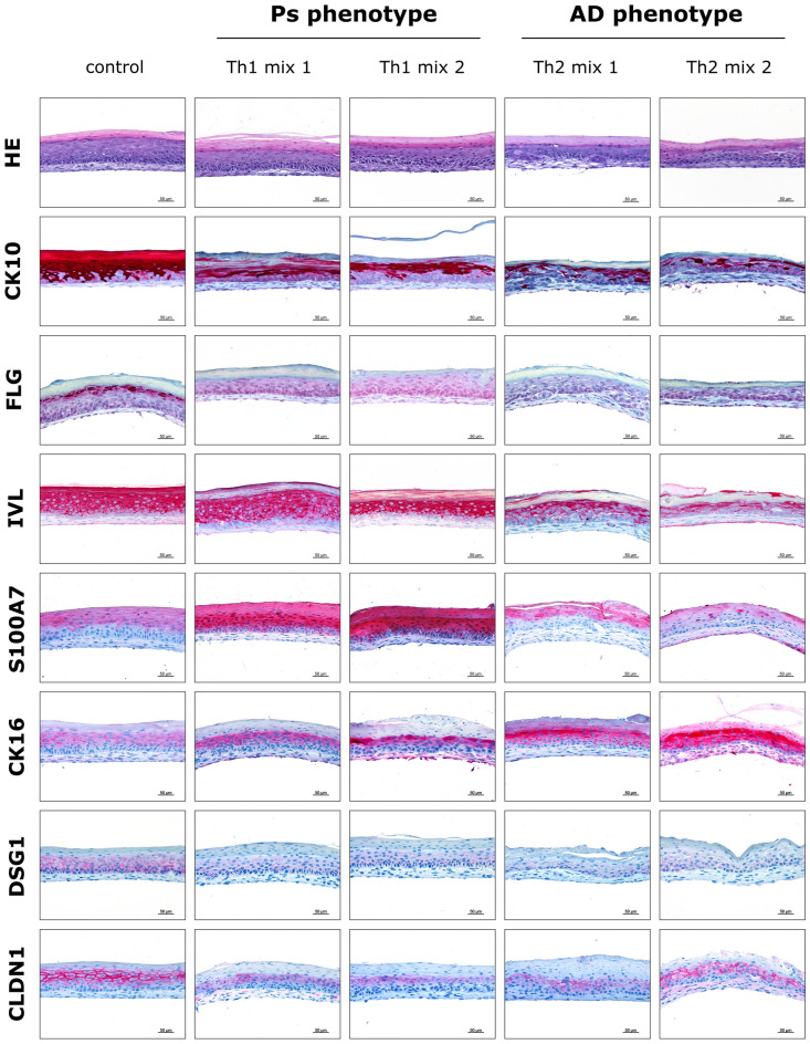
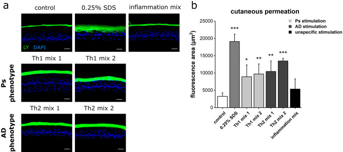
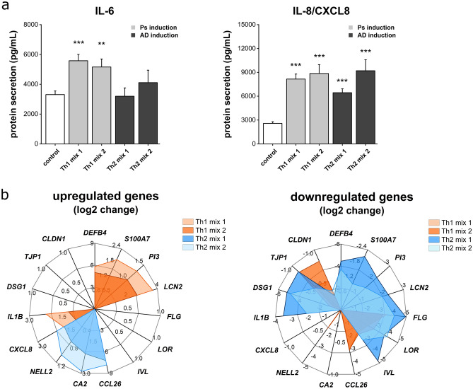
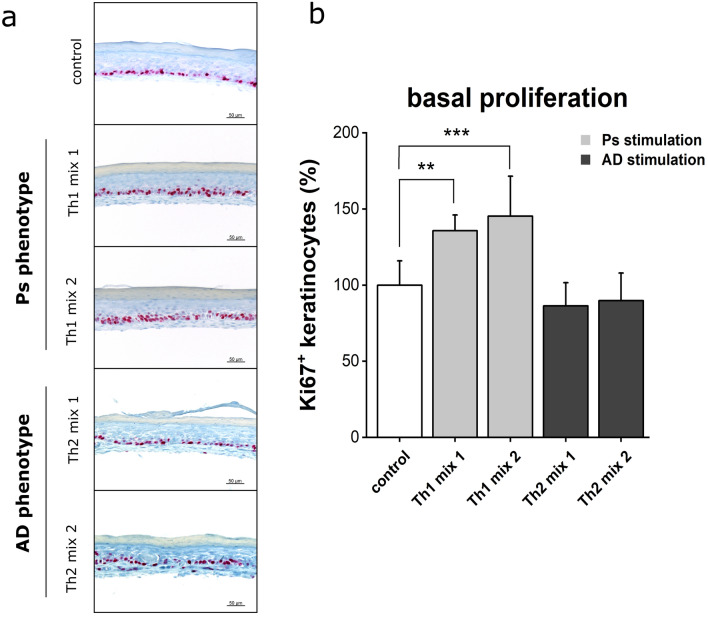
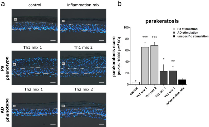
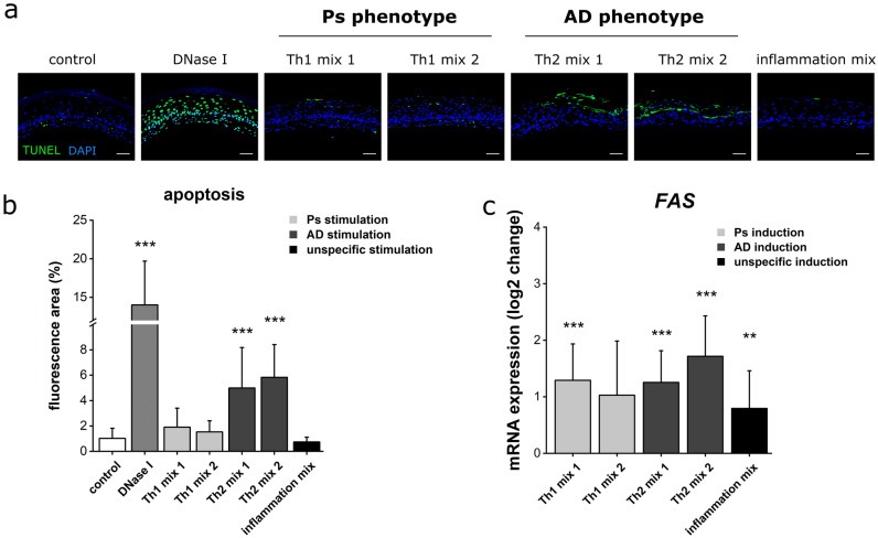
Skin homeostasis is a complex regulated process relying on the crosstalk of keratinocytes, fibroblasts and immune cells. Imbalances of T-cell subsets and the cytokine environment can lead to inflammatory skin diseases such as psoriasis (Ps) and atopic dermatitis (AD). Modern tissue engineering provides several in vitro models mimicking Ps and AD phenotypes. However, these models are either limited in their pathological features, life span, sample availability, reproducibility, controlled handling or simplicity. Some models further lack intensive characterization as they solely focus on differentiation and proliferation aspects. This study introduces a self-assembly model in which the pathological T-cell-signalling of Ps and AD was simulated by subcutaneous Th1 and Th2 cytokine stimulation. The self-established dermal fibroblast-derived matrices of these models were hypothesized to be beneficial for proximal cytokine signalling on epidermal keratinocytes. Comprehensive histological and mRNA analyses of the diseased skin models showed a weakened barrier, distinct differentiation defects, reduced cellular adhesion, inflammation and parakeratosis formation. A keratin shift of declining physiological cytokeratin-10 (CK10) towards increasing inflammatory CK16 was observed upon Th1 or Th2 stimulation. Antimicrobial peptides (AMPs) were upregulated in Ps and downregulated in AD models. The AD biomarker genes CA2, NELL2 and CCL26 were further induced in AD. While Ps samples featured basal hyperproliferation, cells in AD models displayed apoptotic signs. In accordance, these well-controllable three-dimensional in vitro models exhibited Ps and AD-like phenotypes with a high potential for disease research and therapeutic drug testing.


Conflict of interest statement

The authors declare no competing interests.

Figures

Figure 1
Figure 1
Histological alterations in disease-associated skin models. Microanatomy of the skin models was revealed by HE staining. IHC staining was used for visualization of differentiation markers/structural proteins (CK10, FLG, IVL, CK16), antimicrobial peptides (S100A7) and cellular contact proteins (DSG1, CLDN1) of skin equivalents stimulated with Th1 or Th2 cytokine mixes compared to an unstimulated physiological control. Scale bar: 50 µm; pictures are representatives of n = 2 experiments with a total of 4 skin models (keratinocytes derived from one donor).
Figure 2
Figure 2
Impaired cutaneous barrier function of disease-associated skin models. (a) Penetration of the Lucifer Yellow (LY) fluorescent dye (green) was used to visualize the permeability of skin equivalents stimulated with Th1, Th2 or unspecific inflammatory cytokine mixes compared to an unstimulated physiological negative control. Pretreatment with 0.25% SDS served as permeability control. DAPI (blue) was used for nuclear staining. Scale bar: 50 µm, (b) Quantification of LY fluorescence area (mean + SD) of n = 2 experiments with a total of 4 skin models (keratinocytes derived from two different donors). Statistics: one-way ANOVA with Dunett‘s T3 post hoc test; p ≤ 0.05 *, p ≤ 0.01 **, p ≤ 0.001 *** compared to control.
Figure 3
Figure 3
Release of pro-inflammatory mediators and disease-associated gene expression patterns. (a) Secretion of the cytokines IL-6 and IL-8/CXCL8 in undernatants of skin equivalents stimulated with Th1 or Th2 cytokine mixes compared to an unstimulated physiological control was measured by ELISA. Values are mean + SEM of n = 2 experiments with a total of 8 skin models (keratinocytes derived from one donor). Statistics: Mann–Whitney U test; p ≤ 0.05 *, p ≤ 0.01 **, p ≤ 0.001 *** compared to control. (b) Log2 change of mRNA expression of genes encoding antimicrobial peptides (DEFB4, S100A7, PI3, LCN2), structural proteins (FLG, IVL, LOR), chemokines & cytokines (CXCL8, IL1B, CCL26), AD biomarkers (CA2, NELL2) and protein components of cellular adhesions (DSG1, TJP1, CLDN1) was determined by real-time qPCR. Values are mean of n = 2 experiments with a total of 4 skin models.
Figure 4
Figure 4
Basal hyperproliferation in psoriasis-like skin models. (a) Mitotic cells of skin equivalents (unstimulated physiological control or stimulated with Th1, Th2 cytokine mixes) were visualized using Ki67 immunohistochemistry staining. Scale bar: 50 µm, (b) Quantification of Ki67 positive keratinocytes in % to unstimulated control (mean + SD) of n = 2 experiments with a total of 4 skin models (keratinocytes derived from one donor). Statistics: one-way ANOVA with Bonferroni post hoc test; p ≤ 0.05 *, p ≤ 0.01 **, p ≤ 0.001 *** compared to control.
Figure 5
Figure 5
Pathological signs of parakeratosis in disease-associated skin models. (a) Nuclei of sections obtained from skin equivalents (unstimulated physiological control or stimulated with Th1 or Th2 cytokine mixes) were visualized using DAPI fluorescence staining. The solid line marks the basal membrane as dermal-epidermal interface and the dashed line indicates the stratum corneum (SC) layer above. Scale bar: 50 µm, (b) Quantification of nuclei per 10,000 µm2 SC area given as parakeratosis score (mean + SD) of n = 2 experiments with a total of 4 skin models (keratinocytes derived from two different donors). Statistics: one-way ANOVA with Dunett‘s T3 post hoc test; p ≤ 0.05 *, p ≤ 0.01 **, p ≤ 0.001 *** compared to control.
Figure 6
Figure 6
Increased apoptotic signs in AD-like skin models. (a) Apoptotic areas within skin equivalents (unstimulated physiological control or stimulated with Th1, Th2 cytokine mixes) were visualized using TUNEL staining (green fluorescence). DAPI was used for nuclear staining (blue fluorescence). (b) Quantification of apoptotic TUNEL fluorescence area (mean + SD) of n = 2 experiments with a total of 4 skin models (keratinocytes derived from one donor) presented as % per skin model. Statistics: Mann–Whitney U test; p ≤ 0.05 *, p ≤ 0.01 **, p ≤ 0.001 *** compared to control. (c) Log2 change of mRNA expression of the FAS cell death receptor gene was determined by real-time qPCR. Values are mean + SD of n = 2 experiments with a total of 4 skin models. Statistics: Mann–Whitney U test; p ≤ 0.05 *, p ≤ 0.01 **, p ≤ 0.001 *** compared to control.
Figure 7
Figure 7
Characterization of Ps- and AD-like skin models (a) Graphical summary showing disease-associated attributes of the cutaneous in vitro models mimicing Ps or AD phenotypes. Ps skin models are characterized by differentiation defects, inflammation, impaired cellular attachment, hyperproliferation, parakeratosis and increased AMP production. AD skin models feature differentiation defects, inflammation, diminished cellular attachment, apoptotic events, parakeratosis and decreased AMP formation. (b) Color code-based semiquantitative assessment of investigated parameters. Abbrevaitions: SC—stratum corneum, LY—Lucifer Yellow, TUNEL—dT-mediated dUTP nick-end labeling.

Similar articles

Cited by

References

    1. Parisi R, et al. National, regional, and worldwide epidemiology of psoriasis: systematic analysis and modelling study. BMJ. 2020;369:m1590. doi: 10.1136/bmj.m1590. - DOI - PMC - PubMed
    1. Nutten S. Atopic dermatitis: global epidemiology and risk factors. Ann. Nutr. Metab. 2015;66(Suppl 1):8–16. doi: 10.1159/000370220. - DOI - PubMed
    1. Oliveira Mde F, Rocha Bde O, Duarte GV. Psoriasis: classical and emerging comorbidities. An. Bras. Dermatol. 2015;90:9–20. doi: 10.1590/abd1806-4841.20153038. - DOI - PMC - PubMed
    1. Silverberg JI, et al. Association of atopic dermatitis with allergic, autoimmune, and cardiovascular comorbidities in US adults. Ann. Allergy Asthma Immunol. 2018;121:604–612. doi: 10.1016/j.anai.2018.07.042. - DOI - PubMed
    1. Leung S, et al. The cytokine milieu in the interplay of pathogenic Th1/Th17 cells and regulatory T cells in autoimmune disease. Cell Mol. Immunol. 2010;7:182–189. doi: 10.1038/cmi.2010.22. - DOI - PMC - PubMed

Publication types