. 2023 Jul;44(7):1416-1428.
doi: 10.1038/s41401-023-01056-z. Epub 2023 Jan 31.

Bavachin protects against diet-induced hepatic steatosis and obesity in mice

Affiliations

Bavachin protects against diet-induced hepatic steatosis and obesity in mice

Xiang Wei et al. Acta Pharmacol Sin. 2023 Jul.

Abstract

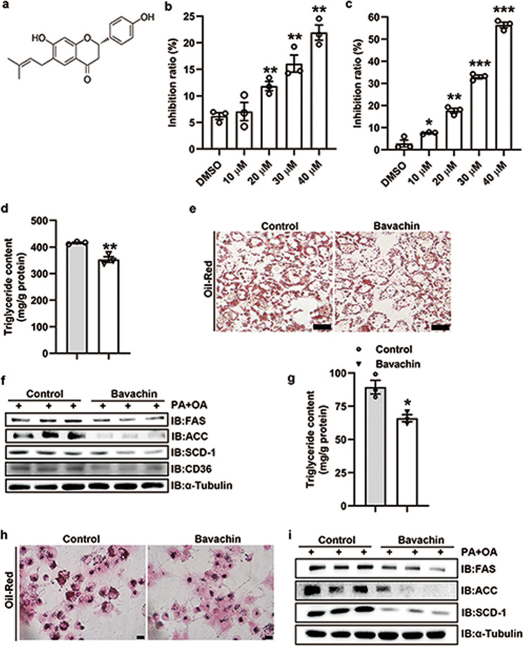
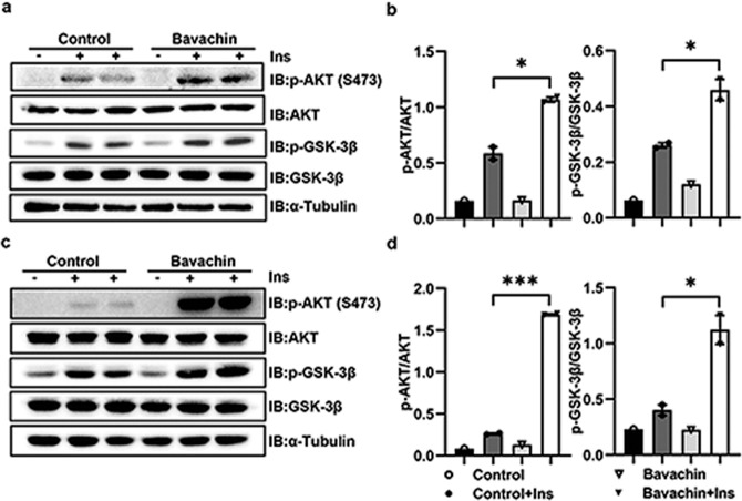
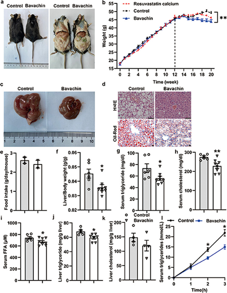
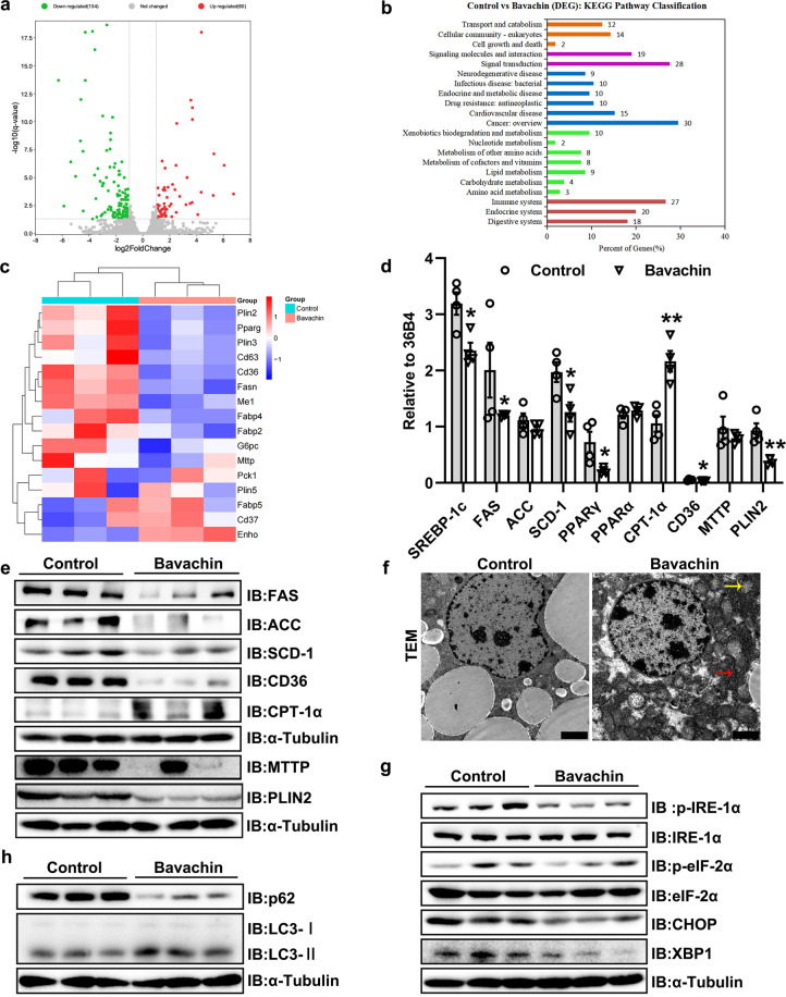
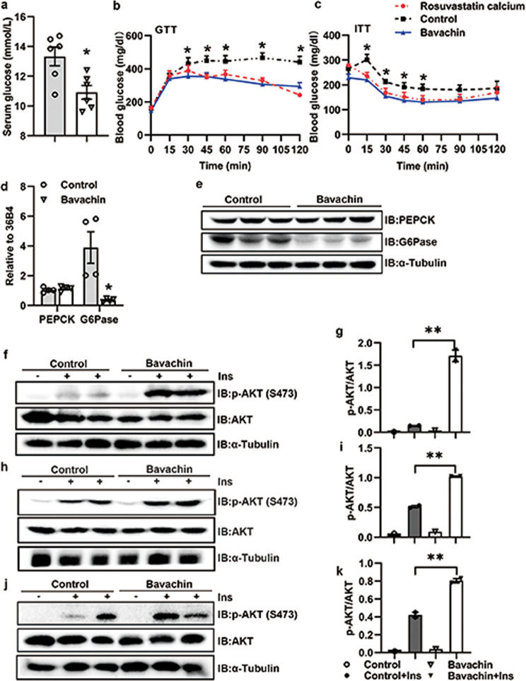
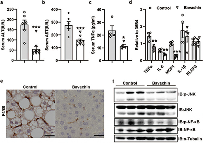
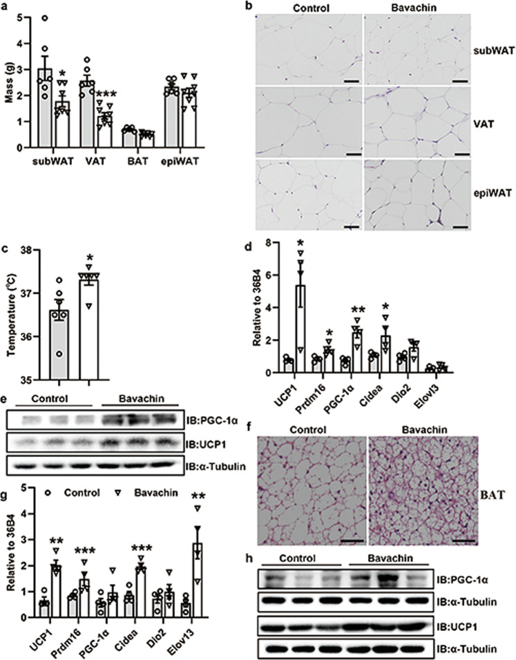
Non-alcoholic fatty liver disease (NAFLD) is a major health concern worldwide, and the incidence of metabolic disorders associated with NAFLD is rapidly increasing because of the obesity epidemic. There are currently no approved drugs that prevent or treat NAFLD. Recent evidence shows that bavachin, a flavonoid isolated from the seeds and fruits of Psoralea corylifolia L., increases the transcriptional activity of PPARγ and insulin sensitivity during preadipocyte differentiation, but the effect of bavachin on glucose and lipid metabolism remains unclear. In the current study we investigated the effects of bavachin on obesity-associated NAFLD in vivo and in vitro. In mouse primary hepatocytes and Huh7 cells, treatment with bavachin (20 μM) significantly suppressed PA/OA or high glucose/high insulin-induced increases in the expression of fatty acid synthesis-related genes and the number and size of lipid droplets. Furthermore, bavachin treatment markedly elevated the phosphorylation levels of AKT and GSK-3β, improving the insulin signaling activity in the cells. In HFD-induced obese mice, administration of bavachin (30 mg/kg, i.p. every other day for 8 weeks) efficiently attenuated the increases in body weight, liver weight, blood glucose, and liver and serum triglyceride contents. Moreover, bavachin administration significantly alleviated hepatic inflammation and ameliorated HFD-induced glucose intolerance and insulin resistance. We demonstrated that bavachin protected against HFD-induced obesity by inducing fat thermogenesis and browning subcutaneous white adipose tissue (subWAT). We revealed that bavachin repressed the expression of lipid synthesis genes in the liver of obese mice, while promoting the expression of thermogenesis, browning, and mitochondrial respiration-related genes in subWAT and brown adipose tissue (BAT) in the mice. In conclusion, bavachin attenuates hepatic steatosis and obesity by repressing de novo lipogenesis, inducing fat thermogenesis and browning subWAT, suggesting that bavachin is a potential drug for NAFLD therapy.

Keywords: AKT; GSK-3β; bavachin; insulin sensitivity; lipogenesis; mouse primary hepatocytes; non-alcoholic fatty liver disease; obesity; subcutaneous white adipose tissue; thermogenesis.


Conflict of interest statement

The authors declare no competing interests.

Figures

Fig. 1
Fig. 1. Bavachin attenuates lipid accumulation in primary mouse hepatocytes and Huh7 cells.
a Chemical structure of bavachin. MTT was used to detect the effect of different bavachin concentrations on the cytotoxicity of primary mouse hepatocytes b and Huh7 cells c. d TG content of primary mouse hepatocytes after PA/OA stimulation with or without bavachin treatment. e Oil Red O staining of primary mouse hepatocytes after PA/OA stimulation with or without bavachin treatment. f Western blot analysis of proteins involved in lipogenesis and fatty acid transport in primary mouse hepatocytes after PA/OA stimulation with or without bavachin treatment. g TG content of Huh7 cells after PA/OA stimulation with or without bavachin treatment. h Oil Red O staining of Huh7 cells after PA/OA stimulation with or without bavachin administration. i Western blot analysis of proteins involved in lipogenesis and fatty acid transport in Huh7 cells after PA/OA stimulation with or without bavachin administration. Data are presented as mean ± SEM; *P < 0.05, **P < 0.01, ***P < 0.001 by Student’s t-test.
Fig. 2
Fig. 2. Bavachin improves insulin sensitivity in vitro.
a AKT and GSK-3β phosphorylation were analysed by Western blot in primary mouse hepatocytes exposed to solvent or bavachin (20 μM) for 24 h and subsequently treated with 100 nM Insulin (Ins) for 5 min. b AKT and GSK-3β phosphorylation densitometry in a. c AKT and GSK-3β phosphorylation were analysed by Western blot in Huh7 cells exposed to solvent or bavachin (20 μM) for 24 h then treated with 100 nM Insulin (Ins) for 5 min. d AKT and GSK-3β phosphorylation densitometry in c. Data are presented as mean ± SEM; *P < 0.05, ***P < 0.001 by Student’s t-test.
Fig. 3
Fig. 3. Bavachin ameliorates hepatic steatosis caused by HFD feeding.
a The representative gross morphology of mice fed a HFD at 0 for 20 weeks and treated with intraperitoneal bavachin at week 12 for 8 weeks. b The body weights of control and bavachin-treated mice were measured weekly from weeks 1–20. Representative results of c morphology, d H&E staining of sections (top panel), and Oil Red O staining (bottom panel) of livers from mice in the indicated groups; scale bars, 20 μm. e Daily food intake of in the indicated mice. f The ratio of the liver weight to body weight in the indicated mice. gi Changes in the fasting serum TG levels, TC levels and FFA levels in the indicated mice. j, k Changes in the fasting hepatic TG levels and TC levels in the indicated mice. l Hepatic VLDL-TG secretion in the indicated mice. Data are presented as mean ± SEM; *P < 0.05, **P < 0.01 by Student’s t-test. One-way analysis of variance (ANOVA) was used to compare the three groups of data.
Fig. 4
Fig. 4. Bavachin represses the hepatic lipogenesis in HFD-induced mice.
a Scatterplot of differentially expressed genes identified by RNA-seq in the livers of 20 weeks HFD-fed mice with or without bavachin treatment. b KEGG pathway analysis showing the involved pathways of DEGs identified by RNA-seq. c Heatmap showing the expression of genes involved in hepatic lipid metabolism on RNA-seq. qPCR (d) and Western blot (e) analysis of genes involved in lipogenesis, fatty acid oxidation, and transport in the livers of control and bavachin-treated mice after 20 weeks of HFD. f Transmission EM observation of the hepatic microstructure, yellow arrows indicate the mitochondria, red arrows indicate the endoplasmic reticulum; scale bars, 2 μm. g Western blot analysis of proteins involved in ER stress in the livers of control and bavachin-treated mice after 20 weeks of HFD. h Western blot analysis of proteins involved in autophagy in the livers of control and bavachin-treated mice after 20 weeks of HFD. Data are presented as the mean ± SEM; *P < 0.05, **P < 0.01, ***P < 0.001 by Student’s t-test.
Fig. 5
Fig. 5. Bavachin improves glucose homeostasis and insulin resistance in HFD-fed mice.
a Blood glucose levels of control and bavachin-treated mice after 20 weeks of HFD followed by a 6 h fast. GTT analyses (b) and ITT analyses (c) were performed in control and bavachin-treated mice after 20 weeks of HFD. qPCR (d) and Western blot (e) were used to analyze the expression of gluconeogenetic genes in the liver of control and bavachin-treated mice after 20 weeks of HFD. f Western blot analysis of insulin-stimulated AKT phosphorylation in the liver of control and bavachin-treated mice after 20 weeks of HFD. g Quantification of the Western blot data shown in f. h Western blot analysis of insulin-stimulated AKT phosphorylation in the subcutaneous fat tissue of control and bavachin-treated mice after 20 weeks of HFD. i Quantification of the Western blot data shown in h. j Western blot analysis of insulin-stimulated AKT phosphorylation in the muscle of control and bavachin-treated mice after 20 weeks of HFD. k Quantification of the Western blot data shown in j. Data are presented as mean ± SEM; *P < 0.05, **P < 0.01 by Student’s t-test. One-way analysis of variance (ANOVA) was used to compare the three groups of data.
Fig. 6
Fig. 6. Bavachin alleviates HFD-induced hepatic inflammation.
Serum ALT (a), AST (b) and TNFα (c) levels of control and bavachin-treated mice after 20 weeks of HFD followed by a 6 h fast. d mRNA expression of pro-inflammatory cytokines in the liver of control and bavachin-treated mice after 20 weeks of the HFD. e Histological F4/80 staining in liver sections of control and bavachin-treated mice fed a HFD for 20 weeks; scale bars, 25 μm. f Western blot analysis of JNK and NF-κB protein and phosphorylation levels in the liver of control and bavachin-treated mice after 20 weeks of the HFD. Data are presented as mean ± SEM; *P < 0.05, **P < 0.01, ***P < 0.001 by Student’s t-test.
Fig. 7
Fig. 7. Bavachin increases thermogenesis and browning of the white adipose tissue in mice fed HFD.
a The weight of different fat pads in control and bavachin-treated mice fed a HFD for 20 weeks. b H&E staining of subWAT, VAT, and epiWAT of control and bavachin-treated mice; scale bars, 50 μm. c Rectal body temperature in control and bavachin-treated mice. d qPCR analysis of genes involved in thermogenesis, beiging, and mitochondrial respiration in the subWAT of control and bavachin-treated mice after 20 weeks of HFD. e Western blot analysis of UCP1 and PGC-1α protein levels in the subWAT of control and bavachin-treated mice after 20 weeks of HFD. f H&E staining of the BAT of control and bavachin-treated mice; scale bars, 50 μm. g qPCR analysis of genes involved in thermogenesis, beiging, and mitochondrial respiration in the BAT of control and bavachin-treated mice after 20 weeks of HFD. h Western blot analysis of UCP1 and PGC-1α protein levels in the BAT of control and bavachin-treated mice after 20 weeks of HFD. Data are presented as mean ± SEM; *P < 0.05, **P < 0.01, ***P < 0.001 by Student’s t-test.

References

    1. Cotter TG, Rinella M. Nonalcoholic fatty liver disease 2020: the state of the disease. Gastroenterology. 2020;158:1851–64. doi: 10.1053/j.gastro.2020.01.052. - DOI - PubMed
    1. Overi D, Carpino G, Franchitto A, Onori P, Gaudio E. Hepatocyte injury and hepatic stem cell niche in the progression of non-alcoholic steatohepatitis. Cells. 2020;9:590. doi: 10.3390/cells9030590. - DOI - PMC - PubMed
    1. Younossi ZM, Koenig AB, Abdelatif D, Fazel Y, Henry L, Wymer M. Global epidemiology of nonalcoholic fatty liver disease-Meta-analytic assessment of prevalence, incidence, and outcomes. Hepatology. 2016;64:73–84. doi: 10.1002/hep.28431. - DOI - PubMed
    1. Friedman SL, Neuschwander-Tetri BA, Rinella M, Sanyal AJ. Mechanisms of NAFLD development and therapeutic strategies. Nat Med. 2018;24:908–22. doi: 10.1038/s41591-018-0104-9. - DOI - PMC - PubMed
    1. Liu ZL, Xie LZ, Zhu J, Li GQ, Grant SJ, Liu JP. Herbal medicines for fatty liver diseases. Cochrane Database Syst Rev. 2013;8:D9059. - PubMed