. 2023 Jul;44(7):1366-1379.
doi: 10.1038/s41401-023-01057-y. Epub 2023 Jan 31.

Notoginsenoside R1 protects against myocardial ischemia/reperfusion injury in mice via suppressing TAK1-JNK/p38 signaling

Affiliations

Notoginsenoside R1 protects against myocardial ischemia/reperfusion injury in mice via suppressing TAK1-JNK/p38 signaling

Jing-Jing Zeng et al. Acta Pharmacol Sin. 2023 Jul.

Abstract

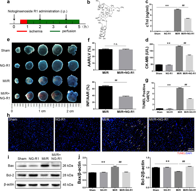
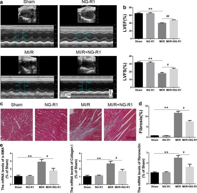
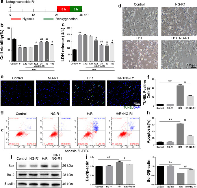
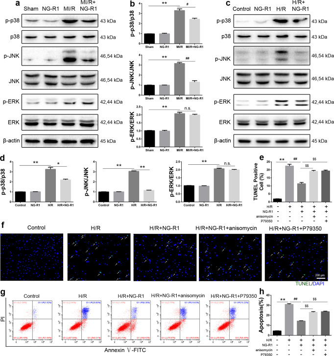
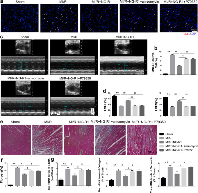
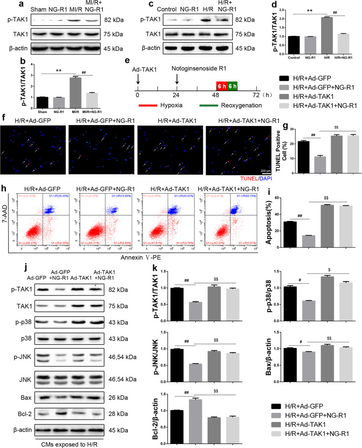
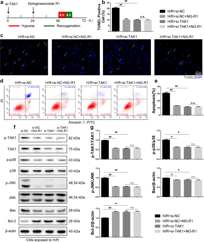
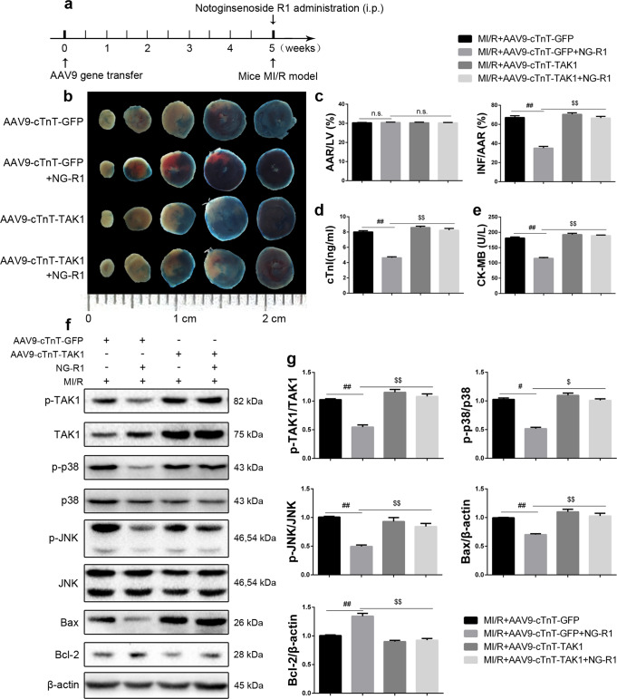
Previous studies show that notoginsenoside R1 (NG-R1), a novel saponin isolated from Panax notoginseng, protects kidney, intestine, lung, brain and heart from ischemia-reperfusion injury. In this study we investigated the cardioprotective mechanisms of NG-R1 in myocardial ischemia/reperfusion (MI/R) injury in vivo and in vitro. MI/R injury was induced in mice by occluding the left anterior descending coronary artery for 30 min followed by 4 h reperfusion. The mice were treated with NG-R1 (25 mg/kg, i.p.) every 2 h for 3 times starting 30 min prior to ischemic surgery. We showed that NG-R1 administration significantly decreased the myocardial infarction area, alleviated myocardial cell damage and improved cardiac function in MI/R mice. In murine neonatal cardiomyocytes (CMs) subjected to hypoxia/reoxygenation (H/R) in vitro, pretreatment with NG-R1 (25 μM) significantly inhibited apoptosis. We revealed that NG-R1 suppressed the phosphorylation of transforming growth factor β-activated protein kinase 1 (TAK1), JNK and p38 in vivo and in vitro. Pretreatment with JNK agonist anisomycin or p38 agonist P79350 partially abolished the protective effects of NG-R1 in vivo and in vitro. Knockdown of TAK1 greatly ameliorated H/R-induced apoptosis of CMs, and NG-R1 pretreatment did not provide further protection in TAK1-silenced CMs under H/R injury. Overexpression of TAK1 abolished the anti-apoptotic effect of NG-R1 and diminished the inhibition of NG-R1 on JNK/p38 signaling in MI/R mice as well as in H/R-treated CMs. Collectively, NG-R1 alleviates MI/R injury by suppressing the activity of TAK1, subsequently inhibiting JNK/p38 signaling and attenuating cardiomyocyte apoptosis.

Keywords: JNK; TAK1; apoptosis; myocardial ischemia/reperfusion injury; notoginsenoside R1; p38.


Conflict of interest statement

The authors declare no competing interests.

Figures

Fig. 1
Fig. 1. NG-R1 ameliorates MI/R injury in vivo.
a Schematic illustration of the NG-R1 treatment protocol. NG-R1 (25 mg·kg−1 dissolved in normal saline) was intraperitoneally injected into C57/BL6 mice a total of three times. The mice were divided into the sham group (Sham), NG-R1 group (NG-R1), MI/R group (MI/R), and MI/R + NG-R1 group (MI/R + NG-R1). Normal saline was administered in the sham group. b Molecular structure of NG-R1. c Cardiac troponin-I (cTnI) levels in plasma 4 h after perfusion. d Cardiac creatine kinase-MB (CK-MB) levels in plasma 4 h after perfusion. e Representative infarct images obtained by Evans blue/TTC staining in each group. The infarct size (INF: white area), the area at risk (AAR: red and white area), and the nonischemic area (blue). Scale bar = 1 cm. f The ratio of the area at risk to the left ventricle and the ratio of the infarct size to the area at risk based on. g, h Representative immunofluorescence images showing TUNEL (red) staining in the infarct border zone. Scale bar = 200 µm. i, j Protein expression levels of Bax and Bcl-2 were analyzed by Western blotting. β-actin served as a loading control. The data are shown as the mean ± SEM (n = 6 for each group). **P < 0.01 vs. the Sham group. ##P < 0.01 vs. the MI/R group.
Fig. 2
Fig. 2. NG-R1 prevents MI/R injury-induced cardiac remodeling and dysfunction.
a Representative images of M-mode echocardiography in each group. b Quantitative echocardiographic analysis of left ventricular ejection fraction (LVEF) and left ventricular fractional shortening (LVFS). c, d Representative images of Masson trichrome-stained MI/R hearts and quantification of the fibrosis area (%). Scale bar = 100 µm. e Quantitative reverse transcription polymerase chain reaction analysis of α-SMA, collagen I and fibronectin. The data are represented as the mean ± SEM (n = 6 for each group). **P < 0.01 vs. the Sham group. #P < 0.05 and ##P < 0.01 vs. the MI/R group.
Fig. 3
Fig. 3. NG-R1 treatment attenuates neonatal cardiomyocytes (CMs) death in mice subjected to H/R.
a Schematic illustration of H/R injury in CMs. b The viability of CMs after NG-R1 pretreatment (0, 3.12, 6.25, 12.5, 25, 50, 100 μM) for 24 h was analyzed by MTS assays. c Cell damage was determined by an LDH release assay. d Representative morphology of CMs in the control group (control), NG-R1 group (NG-R1), H/R group (H/R) and H/R + NG-R1 group (H/R + NG-R1). Scale bar = 250 μm. e Immunofluorescence staining of TUNEL-positive cells. Scale bar = 200 µm. f Quantitative analysis of (e). g, h Representative images showing apoptosis were determined by FITC-PI/Annexin V staining. The apoptosis rate of each group was determined. i, j The expression levels of Bcl-2 and Bax were measured by Western blotting. β-actin served as a loading control. The data are shown as the mean ± SEM (n = 6 for each group). **P < 0.01 vs. the Control group. #P < 0.05 and ##P < 0.01 vs. the H/R group.
Fig. 4
Fig. 4. NG-R1 inhibits activation of the JNK/p38 signaling pathway after MI/R.
a Western blot analysis of phosphorylated and total p38, JNK and ERK protein levels in ischemic myocardial tissue 4 h after perfusion. β-actin served as a loading control. b Quantitative analysis of a. c Western blot analysis of phosphorylated and total p38, JNK and ERK protein levels in vitro. d Quantitative analysis of c. e, f Representative images and quantitative analysis of TUNEL-positive cells. Scale bar = 200 µm. g Representative images of apoptosis were determined by FITC-PI/Annexin V staining. h Statistical analysis of apoptosis in the five groups. The data are shown as the mean ± SEM (n = 6 for each group). *P < 0.05 and **P < 0.01 vs. the Sham group or the Control group. #P < 0.05 and ##P < 0.01 vs. the MI/R group or the H/R group. $$P < 0.01 vs. the H/R + NG-R1 group.
Fig. 5
Fig. 5. The protective effects of NG-R1 involve JNK/p38 signaling.
a Representative immunofluorescence images showing TUNEL (red) staining in the infarct border zone in the various groups. Scale bar = 200 µm. b Quantitative analysis of a. c Representative images of M-mode echocardiography in each group. d Quantitative echocardiographic analysis of LVEF and LVFS. e, f Representative images of Masson trichrome-stained MI/R hearts and quantification of the fibrosis area (%). Scale bar = 100 µm. g Quantitative reverse transcription polymerase chain reaction analysis of α-SMA, collagen I and fibronectin. The data are represented as the mean ± SEM (n = 6 for each group). **P < 0.01 vs. the Sham group. #P < 0.05 and ##P < 0.01 vs. the MI/R group. $P < 0.05 and $$P < 0.01 vs. the MI/R + NG-R1 group.
Fig. 6
Fig. 6. TAK1 overexpression reversed the antiapoptotic effect of NG-R1 on CMs.
a Western blot analysis of phosphorylated and total TAK1 protein levels in ischemic myocardial tissue 4 h after perfusion. β-actin served as a loading control. b Quantitative analysis of a. c Western blot analysis of phosphorylated and total TAK1 protein levels in vitro. d Quantitative analysis of c. e Schematic illustration of TAK1 adenoviral infection. The TAK1 overexpression adenoviral vector or control GFP adenovirus (20 MOI) was used to infect CMs 48 h before H/R injury. f Immunofluorescence staining of TUNEL-positive cells. Scale bar = 200 µm. g Quantitative analysis of f. h Representative images of apoptosis, as determined by PE-7-AAD/Annexin V staining. I Statistical analysis of apoptosis in the four groups. j The expression levels of p-TAK1, TAK1, p-p38, p38, p-JNK, JNK, Bcl-2 and Bax were determined by Western blotting. k Quantitative analysis of j. The data are shown as the mean ± SEM (n = 6 for each group). **P < 0.01 vs. the Sham group or the Control group. #P < 0.05 and ##P < 0.01 vs. the MI/R group, the H/R group or the H/R + Ad-GFP group. $P < 0.05 and $$P < 0.01 vs. The H/R + Ad-GFP + NG-R1 group.
Fig. 7
Fig. 7. NG-R1 inhibits H/R-induced apoptosis in CMs by suppressing TAK1.
a Schematic illustration of si-TAK1 transfection. CMs were transfected with si-TAK1 48 h before H/R injury. b, c Representative images and quantitative analysis of TUNEL-positive cells. Scale bar = 200 µm. d Representative images of apoptosis, as determined by FITC-PI/Annexin V staining. e Statistical analysis of apoptosis in the four groups. f The protein levels of p-TAK1, TAK1, p-p38, p38, p-JNK, JNK, Bcl-2 and Bax were determined by Western blotting. g Quantitative analysis of f. The data are shown as the mean ± SEM (n = 6 for each group). #P < 0.05 and ##P < 0.01 vs. the H/R + si-NC group.
Fig. 8
Fig. 8. TAK1 overexpression reversed the protective effect of NG-R1 on MI/R injury.
a The time course of AAV9-mediated TAK1 overexpression. Recombinant AAV9 vectors harboring TAK1 or GFP with a c-TnT promoter [3 × 1011 vector genomes/mouse] were injected into the tail veins of the mice 5 weeks before MI/R. b, c Representative infarct images obtained by Evans blue/TTC staining in each group. d Cardiac troponin-I (cTnI) levels in plasma 4 h after perfusion. e Cardiac creatine kinase-MB (CK-MB) levels in plasma 4 h after perfusion. f, g Western blot images and quantitative analysis of p-TAK1, TAK1, p-p38, p38, p-JNK, JNK, Bcl-2 and Bax protein expression in ischemic myocardial tissue. The data are shown as the mean ± SEM (n = 6 for each group). #P < 0.05 and ##P < 0.01 vs. the MI/R + AAV9-cTnT-GFP group. $P < 0.05 and $$P < 0.01 vs. the MI/R + AAV9-cTnT-GFP + NG-R1 group.

References

    1. Benjamin EJ, Blaha MJ, Chiuve SE, Cushman M, Das SR, Deo R, et al. Heart Disease and Stroke Statistics-2017 Update: A Report From the American Heart Association. Circulation. 2017;135:e146–146e603. doi: 10.1161/CIR.0000000000000485. - DOI - PMC - PubMed
    1. Hausenloy DJ, Yellon DM. Targeting myocardial reperfusion injury–the search continues. N Engl J Med. 2015;373:1073–5. doi: 10.1056/NEJMe1509718. - DOI - PubMed
    1. Hausenloy DJ, Yellon DM. Ischaemic conditioning and reperfusion injury. Nat Rev Cardiol. 2016;13:193–209. doi: 10.1038/nrcardio.2016.5. - DOI - PubMed
    1. Heusch G. The coronary circulation as a target of cardioprotection. Circ Res. 2016;118:1643–58. doi: 10.1161/CIRCRESAHA.116.308640. - DOI - PubMed
    1. Ruan Y, Zeng J, Jin Q, Chu M, Ji K, Wang Z, et al. Endoplasmic reticulum stress serves an important role in cardiac ischemia/reperfusion injury (Review) Exp Ther Med. 2020;20:268. doi: 10.3892/etm.2020.9398. - DOI - PMC - PubMed