. 2023 Mar;32(3):e4586.
doi: 10.1002/pro.4586.

Molecular structures reveal the origin of spectral variation in cryptophyte light harvesting antenna proteins

Affiliations

Molecular structures reveal the origin of spectral variation in cryptophyte light harvesting antenna proteins

Katharine A Michie et al. Protein Sci. 2023 Mar.

Abstract

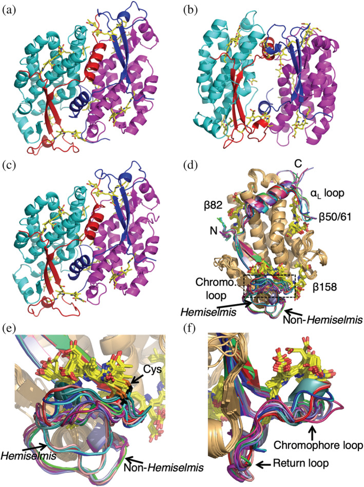
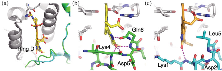
In addition to their membrane-bound chlorophyll a/c light-harvesting antenna, the cryptophyte algae have evolved a unique phycobiliprotein antenna system located in the thylakoid lumen. The basic unit of this antenna consists of two copies of an αβ protomer where the α and β subunits scaffold different combinations of a limited number of linear tetrapyrrole chromophores. While the β subunit is highly conserved, encoded by a single plastid gene, the nuclear-encoded α subunits have evolved diversified multigene families. It is still unclear how this sequence diversity results in the spectral diversity of the mature proteins. By careful examination of three newly determined crystal structures in comparison with three previously obtained, we show how the α subunit amino acid sequences control chromophore conformations and hence spectral properties even when the chromophores are identical. Previously we have shown that α subunits control the quaternary structure of the mature αβ.αβ complex (either open or closed), however, each species appeared to only harbor a single quaternary form. Here we show that species of the Hemiselmis genus contain expressed α subunit genes that encode both distinct quaternary structures. Finally, we have discovered a common single-copy gene (expressed into protein) consisting of tandem copies of a small α subunit that could potentially scaffold pairs of light harvesting units. Together, our results show how the diversity of the multigene α subunit family produces a range of mature cryptophyte antenna proteins with differing spectral properties, and the potential for minor forms that could contribute to acclimation to varying light regimes.

Keywords: cryptophyte; evolution; light harvesting protein; phycobiliprotein; x-ray crystallography.


Conflict of interest statement

The authors declare no competing interests.

Figures

FIGURE 1
FIGURE 1
Structure of α subunit precursor proteins and alignment of mature α subunit sequences from x‐ray structures. (a–c) Precursors are synthesized on cytoplasmic ribosomes and directed across the endoplasmic reticulum by a typical ER signal peptide (SP) and then across three membranes into the plastid stroma where the transit peptide (TP) is removed. The lumenal targeting domain (LTD) with twin Arg motif (RR) directs the assembled and chromophorylated holoprotein into the thylakoid lumen where it is cleaved by the thylakoidal processing protease, leaving the mature protein (blue). (a) Typical αL and (b) αS sequences from Rhodomonas sp. CS24 PE545; (c) Hemiselmis α sequence with Asp insertion (blue arrowhead). Red arrow: bilin binding site. The variable chromophore loop region is between the bilin‐binding site and the second sheet region. (d) Alignment of mature α subunit sequences from x‐ray structures. Boxes indicate conserved blocks corresponding to secondary structure elements or specific sequence motifs. Amino acids are colored by side chain properties. Red arrow: Cys19 that attaches the bilin chromophore. PE545, Rhodomonas sp. CS24; PE566, Cryptomonas pyrenoidifera CCAP 979/61; PC645, “Chroomonas” sp. CCMP270; PC630, Chroomonas gentoftensis CCAC1627; PC577, H. pacifica CCMP706; PC612, H. virescens CCAC1635; PE555, H. andersenii CCMP644.
FIGURE 2
FIGURE 2
Crystal structures of cryptophyte light harvesting proteins. (a) Structure of the closed form PC630. (b) Structure of the open form PC577. (c) Structure of the closed form PE566. β subunits are colored magenta and cyan, while α subunits are colored blue and red. In closed form structures, αL is colored blue while αS is red. (d) An overlay of all unique αβ protomer structures determined to date. All β subunits are wheat. Chromophores are shown as stick models. (e) Expanded view of the overlay focusing on the α chromophore loop. The outward chromophore loop (middle, corresponding to the boxed area in panel (d)) from the cysteine (shown as sticks with Cβ black and Sγ orange) to which the chromophore is attached follows many different paths. The return loop (bottom) shows three distinct paths: two for Hemiselmis and one non‐Hemiselmis structures. (f) The same overlay as per (e) but rotated by 90°. The outbound chromophore loop cradles the α chromophore while the return loop hugs the underlying β subunit.
FIGURE 3
FIGURE 3
Chromophore geometry. An overlay of chromophores using the central pyrrole B and C rings for superposition. Chromophores have been stripped of any external chemical groups for visual simplicity. Chromophores are grouped by location in the structure, with (a) α chromophore, (b) bilin β50/61, (c) bilin β82, and (d) bilin β158. Only gross patterns are shown; some chromophores have finer hidden patterns that are explored in Figures S2–S6. The left‐hand side shows the overview overlay while the right‐hand side panels show details of pyrrole ring A and D rotations with reference to the planar central rings B and C. Conjugated and non‐conjugated chromophores are grouped for ring D as the analysis is different. Angle measures are either given as dihedral angle pairs (θ inner, θ outer) for conjugated ring pairs (see Section 4, with standard deviation taken over the set of all angles for each group) or as a single angle, φ, between two ring planes for non‐conjugated ring pairs (see Section 4, with standard deviation taken over the set of all angles for each group). In panel (c), right hand side, clusters are labeled based on the amino acid at residue α5/6 in the α subunit that interacts with the β82 chromophore. PC630 in dark green, PC577 in cyan, PC645 in light blue, PC612 in dark blue, PE545 in red and PE566 in orange. Standard deviation is over all members of each cluster.
FIGURE 4
FIGURE 4
Sequence differences between the N‐termini of PC630 and PC645 α subunits rotate pyrrole ring D in the β82 PCB chromophore. Panel (a) shows an overlay of the β82 chromophore site where the PC630 chromophore is shown in yellow CPK while the PC645 chromophore is shown in orange CPK colors. PC630 α subunit is shown in green while the PC645 α subunit is cyan. (b) Multiple hydrogen bonds between PC630 α subunit (green CPK) and β82 pyrrole ring D stabilize the chromophore conformation. (c) PC645 α subunit forms one backbone hydrogen bond with pyrrole ring D, however, the side chain of Leu5 from the α subunit makes van der Waals contact, maintaining the chromophore conformation. The figure shows models for αS, however, the same interactions are seen in αL.
FIGURE 5
FIGURE 5
Sequence differences between PC577 and PC612 in the α subunit chromophore loop rotate pyrrole ring D in the α20 PCB chromophore. Panels (a, b) show cartoon representations of the α20 PCB chromophore site in PC577 and PC612, respectively. The chromophore loop of PC577 contains a one‐turn helix (a), which is not seen in PC612 (b). (c, d) Atomic models of the α20 chromophore site in PC577 and PC612, respectively. The view is identical to panels (a) and (b). In (c), the chromophore loop interacts with the α20 PCB where the backbone carbonyl of Gly24 (α subunit) makes a hydrogen bond with the nitrogen atom of pyrrole ring D, resulting in a rotation of this ring when compared to PC612 (panel (d)). Panel (d) shows the same view for PC612. Note, there is a gap between the α subunit chromophore loop and the chromophore surface, which is filled by ordered water molecules (red spheres). (e) Structure‐based sequence alignment of the α subunits of PC577 and PC612 (red—identical; and green—high similarity).
FIGURE 6
FIGURE 6
Comparison between PE566 and PE545. (a,b) Overlay the α subunit chromophore sites for αL and αS, respectively. Although the chromophore loop chains differ, a planar salt bridge lies across the face of the central pyrrole rings B and C, maintaining the α chromophore in the same conformation. Panel (a) compares αL structures. Here in PE545, Arg21 and Glu25 form a salt bridge, while in PE566, His22 replaces Arg21, while maintaining the same planar salt bridge structure. Panel (b) compares αS where the Arg‐Glu salt bridge is formed in both proteins (PE545: Arg21‐Glu25; PE566: Arg21‐Glu27). (c) The αS helix of PE566, showing an unremarkable distribution of side chains. (d) In contrast, the αS helix of PE545 shows one face with essentially no side chains (either Gly or Ala, only). (e) Structure‐based alignment of αL and αS sequences for PE566 and PE545. Sequences are broken to indicate secondary structure boundaries. In (a) and (b), ribbons are colored by chain as per Figure 2a–c, with PE566 in full colors and PE545 in pastel shades. Stick figures are colored with carbon gray for PE545 and carbon yellow for PE566.
FIGURE 7
FIGURE 7
Tryptic peptides from H. virescens CCMP706 show that at least one closed form gene is expressed. Each peptide sequence found by LC–MS/MS is shown as a boxed section in its respective transcript sequence. Sequences are identified by their MMETSP1356 transcript number except for Hvir‐PCR‐1, the PCR‐derived sequence which is identical to the crystal structure sequence and to transcripts Hvir‐0052 and Hvir‐10022 (not shown). Note that the closed form sequence Hvir‐10352 is identified by two unique peptides. This sequence clusters with ChroomonasHemiselmis αS while the other two closed form sequences (Hvir‐8822 and Hvir‐7568) cluster with ChroomonasHemiselmis αL (see Figure 8). Dashes in the sequences are introduced for alignment.
FIGURE 8
FIGURE 8
Phylogenetic tree of Chroomonas and Hemiselmis α‐subunit sequences shows open and closed forms are clearly distinguished. Open form Hemiselmis sequences form a single clade with a bootstrap support of 90. Closed form sequences break into αL and αS clades, where Hemiselmis sequences are grouped with those from Chroomonas. Four outlier sequences are classified as “indeterminate”. Cmes, Chroomonas mesostigmatica; C270, Chroomonas sp. CCMP270; C1312, Chroomonas sp. CCAC1312; C1627, C. gentoftensis; H. vir, Hemiselmis virescens; H.and, H. andersenii; H. tep, H. tepida; H. ruf, H. rufescens; H. cry, H. cryptochromatica. Scale bar: changes per 100 residues.
FIGURE 9
FIGURE 9
Internal duplication creating tandem α sequence conserved in four PE species from three clades. (a) The four sequences have been split into N‐terminal half (N) and C‐terminal half (C) to show the duplication (boxed). G. theta CCMP2712 CpeA10 peptide sequences identified via LC–MS/MS (Kieselbach et al., 2018) are underlined and italicized. (b) AlphaFold2 model of G. theta CpeA10 (red) bound to two β subunits (green and cyan). (c) Addition of two αLβ protomers (αL subunit purple, β subunit wheat) to create two complete complexes that are linked by CpeA10. Two views in (b) and (c) are related by a 90° rotation about the vertical axis in the page. The four tandem sequences are: Gt‐CpeA10, G. theta CCMP2712; Hphi, Hanusia phi CCMP325 MMETSP1048 transcript; Rsal, Rhodomonas salina CCMP1319 MMETSP1047 transcript; and Cry‐cpeAc PCR sequence from Cryptomonas pyrenoidifera CCAP 979/61. Rhodomonas sp. CS24 PE545 single α domain sequences are included for comparison. Red arrows, chromophore attachment cysteine site.

References

    1. Adams PD, Grosse‐Kunstleve RW, Hung LW, Ioerger TR, McCoy AJ, Moriarty NW, et al. PHENIX: building new software for automated crystallographic structure determination. Acta Crystallogr D Biol Crystallogr. 2002;58:1948–54. - PubMed
    1. Apt KE, Collier JL, Grossman AR. Evolution of the phycobiliproteins. J Mol Biol. 1995;248:79–96. - PubMed
    1. Archibald JM. Cryptomonads. Curr Biol. 2020;30:R1114–6. - PubMed
    1. Arpin PC, Turner DB, McClure SD, Jumper CC, Mirkovic T, Challa JR, et al. Spectroscopic studies of cryptophyte light harvesting proteins: vibrations and coherent oscillations. J Phys Chem B. 2015;119:10025–34. - PubMed
    1. Broughton MJ, Howe CJ, Hiller RG. Distinctive organization of genes for light‐harvesting proteins in the cryptophyte alga Rhodomonas. Gene. 2006;369:72–9. - PubMed

Publication types