

Clinical anatomy of hepatic vessels by computed tomography angiography: A minireview
- PMID: 36721671
- PMCID: PMC9884335
- DOI: 10.4329/wjr.v15.i1.1
Clinical anatomy of hepatic vessels by computed tomography angiography: A minireview
Abstract
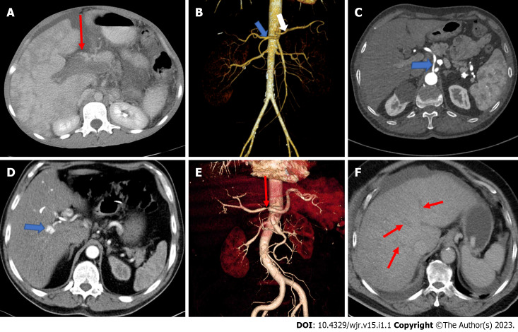
The liver has a complex vascular anatomy with a unique dual blood supply. Clinical conditions of the liver vary widely and include disorders originating in the vascular and biliary systems as well as the parenchyma. In most vascular disorders, the effects on the liver are generally subclinical because of its abundant blood supply. However, early diagnosis of such vascular diseases can significantly reduce patient morbidity and mortality. Because imaging findings of vascular disease are not always readily apparent, diagnosis can be difficult. Computed tomography angiography is an excellent imaging modality for visualizing the vascular anatomy of patients for treatment planning. In this review article, we focus on the vascular anatomy of the liver and the imaging findings in some acute hepatic vascular diseases.
Keywords: Computed tomography angiography; Hepatic artery; Periportal region; Portal triad; Portal vein; Sinusoid.
©The Author(s) 2023. Published by Baishideng Publishing Group Inc. All rights reserved.
Conflict of interest statement
Conflict-of-interest statement: The authors declare that they have no conflict of interest to disclose.
Figures
References
-
- Colagrande S, Centi N, La Villa G, Villari N. Transient hepatic attenuation differences. AJR Am J Roentgenol. 2004;183:459–464. - PubMed
-
- Price M, Patino M, Sahani D. Computed Tomography Angiography of the Hepatic, Pancreatic, and Splenic Circulation. Radiol Clin North Am. 2016;54:55–70. - PubMed
-
- Standring S. Gray’s Anatomy The Anatomical Basis of Clinical Practice. 42nd ed. London: Churchill Livingstone Elsevier, 2021: 327-329, 1205-1217.
-
- Sun P, Zhang G, Su X, Jin C, Yu B, Yu X, Lv Z, Ma H, Zhang M, Wei W, Li W. Maintenance of Primary Hepatocyte Functions In Vitro by Inhibiting Mechanical Tension-Induced YAP Activation. Cell Rep. 2019;29:3212–3222.e4. - PubMed
Publication types
LinkOut - more resources
Full Text Sources
