

Advances in imaging techniques to assess kidney fibrosis
- PMID: 36723057
- PMCID: PMC9897785
- DOI: 10.1080/0886022X.2023.2171887
Advances in imaging techniques to assess kidney fibrosis
Abstract
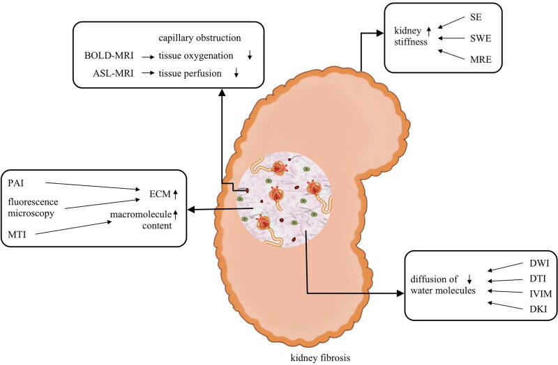
As a sign of chronic kidney disease (CKD) progression, renal fibrosis is an irreversible and alarming pathological change. The accurate diagnosis of renal fibrosis depends on the widely used renal biopsy, but this diagnostic modality is invasive and can easily lead to sampling error. With the development of imaging techniques, an increasing number of noninvasive imaging techniques, such as multipara meter magnetic resonance imaging (MRI) and ultrasound elastography, have gained attention in assessing kidney fibrosis. Depending on their ability to detect changes in tissue stiffness and diffusion of water molecules, ultrasound elastography and some MRI techniques can indirectly assess the degree of fibrosis. The worsening of renal tissue oxygenation and perfusion measured by blood oxygenation level-dependent MRI and arterial spin labeling MRI separately is also an indirect reflection of renal fibrosis. Objective and quantitative indices of fibrosis may be available in the future by using novel techniques, such as photoacoustic imaging and fluorescence microscopy. However, these imaging techniques are susceptible to interference or may not be convenient. Due to the lack of sufficient specificity and sensitivity, these imaging techniques are neither widely accepted nor proposed by clinicians. These obstructions must be overcome by conducting technology research and more prospective studies. In this review, we emphasize the recent advancement of these noninvasive imaging techniques and provide clinicians a continuously updated perspective on the assessment of kidney fibrosis.
Keywords: Imaging techniques; kidney fibrosis; magnetic resonance imaging; ultrasound elastography.
Conflict of interest statement
No potential conflict of interest was reported by the author(s).
Figures
References
-
- Falke LL, Gholizadeh S, Goldschmeding R, et al. . Diverse origins of the myofibroblast-implications for kidney fibrosis. Nat Rev Nephrol. 2015;11(4):233–244. - PubMed
-
- Orlacchio A, Chegai F, Del Giudice C, et al. . Kidney transplant: usefulness of real-time elastography (RTE) in the diagnosis of graft interstitial fibrosis. Ultrasound Med Biol. 2014;40(11):2564–2572. - PubMed
-
- Yoon H, Lee YS, Lim BJ, et al. . Renal elasticity and perfusion changes associated with fibrosis on ultrasonography in a rabbit model of obstructive uropathy. Eur Radiol. 2020;30(4):1986–1996. - PubMed
-
- Bob F, Grosu I, Sporea I, et al. . Ultrasound-based shear wave elastography in the assessment of patients with diabetic kidney disease. Ultrasound Med Biol. 2017;43(10):2159–2166. - PubMed
Publication types
MeSH terms
LinkOut - more resources
Full Text Sources
Other Literature Sources
Medical
Research Materials