Observational Study
. 2023 May 1;18(5):613-625.
doi: 10.2215/CJN.0000000000000106. Epub 2023 Feb 1.

Kidney Biopsy Findings in Patients with SARS-CoV-2 Infection or After COVID-19 Vaccination

Collaborators, Affiliations
Observational Study

Kidney Biopsy Findings in Patients with SARS-CoV-2 Infection or After COVID-19 Vaccination

Maria de Las Mercedes Noriega et al. Clin J Am Soc Nephrol. .

Abstract

Background: Emerging case series described a temporal association between severe acute respiratory syndrome coronavirus 2 (SARS-CoV-2) vaccination and de novo or relapsing kidney diseases. We aimed to further understand vaccination- and coronavirus disease 2019 (COVID-19)-associated kidney diseases.

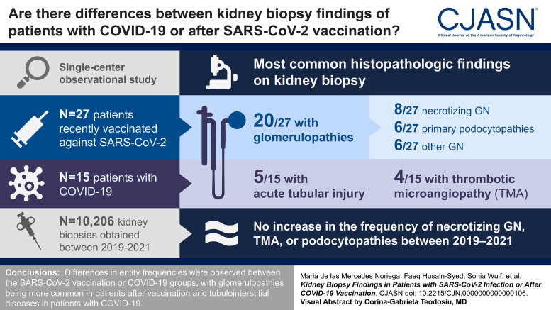
Methods: We present findings from native kidney biopsies of patients recently vaccinated against SARS-CoV-2 ( n =27) and those with COVID-19 ( n =15), reviewed at a single German center. Diagnoses were compared among all native kidney biopsies ( n =10,206) obtained between the prepandemic (2019), pandemic (2020), and vaccination periods (2021) to determine whether there was an increase in kidney diseases in the observed periods.

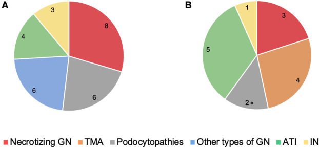
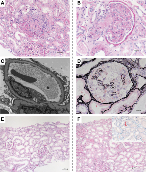
Results: Biopsy indication was increased serum creatinine and/or new-onset proteinuria. Glomerulopathies (20/27, 74%) were more common than tubulointerstitial diseases in postvaccination patients, with necrotizing GN (8/27, 30%) and primary podocytopathies and other GN types (6/27, 22% each) the most common forms. Acute tubular injury was the most common kidney disease in patients with COVID-19, followed by thrombotic microangiopathy (TMA) and necrotizing GN. The postvaccination and COVID-19 infection groups had similar kidney function recovery rates (69% and 73%, respectively). Furthermore, the frequencies of necrotizing GN, pauci-immune GN, TMA, or primary podocytopathies at our center did not increase between 2019 and 2021.

Conclusions: We observed differences in entity frequencies between the SARS-CoV-2 vaccination or COVID-19 groups, with glomerulopathies being more common in patients after vaccination and tubulointerstitial diseases in patients with COVID-19. Cases of TMA were observed only in the COVID-19 group. We detected no increase in the frequency of necrotizing GN, TMA, or podocytopathies between 2019 and 2021.

Clinical trial registry name and registration number: Kidney Histopathology After COVID-19 and SARS-CoV-2 Vaccination, NCT05043168.

Podcast: This article contains a podcast at https://dts.podtrac.com/redirect.mp3/www.asn-online.org/media/podcast/CJASN/2023_05_08_CJN0000000000000106.mp3.


Figures

None
Graphical abstract
Figure 1
Figure 1
Frequencies of diagnosis in postvaccination and COVID-19 kidney biopsies. The figure illustrates biopsy-based kidney disease frequencies in patients with a temporal association to SARS-CoV-2 vaccination (A) or COVID-19 infection (B). *Including one case of collapsing glomerulopathy. ATI, acute tubular injury; COVID-19, coronavirus disease 2019; IN, interstitial nephritis; SARS-CoV-2, severe acute respiratory syndrome coronavirus 2; TMA, thrombotic microangiopathy.
Figure 2
Figure 2
Representative kidney biopsy findings of postvaccination case and those with COVID-19. The figure illustrates representative kidney histopathology findings in patients after SARS-CoV-2 vaccination (panels A, C, E) or with COVID-19 (panels B, D, F). (A) Light microscopy image showing ANCA-associated necrotizing GN and ATI in case V1. PAS staining. Magnification, ×200. (B) Light microscopy image showing TMA in case C7. PAS staining. Magnification, ×200. (C) Diffuse foot process effacement (minimal changes) in case V11. Transmission electron microscopy. Magnification, ×8000. (D) Light microscopy image showing a collapsing glomerulopathy lesion characterized by glomerular epithelial cell hyperplasia and underlying glomerular capillary collapse in case C8 of African ancestry. Jones methenamine silver staining. Magnification, ×400. (E) Light microscopy image showing ATI with dilatation, flattened epithelium, and brush border defects in case V21 that had ATI. PAS staining. Magnification, ×100. (F) Light microscopy image showing ATI and myoglobin casts (inset shows myoglobin immunohistochemistry staining) in case C12. PAS. Magnification, ×100. PAS, periodic acid–Schiff.

References

    1. Hirsch JS Ng JH Ross DW, et al. . Acute kidney injury in patients hospitalized with COVID-19. Kidney Int. 2020;98(1):209–218. doi: 10.1016/j.kint.2020.05.006 - DOI - PMC - PubMed
    1. Sharma P Uppal NN Wanchoo R, et al. . COVID-19-Associated kidney injury: a case series of kidney biopsy findings. J Am Soc Nephrol. 2020;31(9):1948–1958. doi: 10.1681/ASN.2020050699 - DOI - PMC - PubMed
    1. Kudose S Batal I Santoriello D, et al. . Kidney biopsy findings in patients with COVID-19. J Am Soc Nephrol. 2020;31(9):1959–1968. doi: 10.1681/ASN.2020060802 - DOI - PMC - PubMed
    1. Akilesh S Nast CC Yamashita M, et al. . Multicenter clinicopathologic correlation of kidney biopsies performed in COVID-19 patients presenting with acute kidney injury or proteinuria. Am J Kidney Dis. 2021;77(1):82–93.e1. doi: 10.1053/j.ajkd.2020.10.001 - DOI - PMC - PubMed
    1. May RM Cassol C Hannoudi A, et al. . A multi-center retrospective cohort study defines the spectrum of kidney pathology in Coronavirus 2019 Disease (COVID-19). Kidney Int. 2021 10;100(6):1303–1315. doi: 10.1016/j.kint.2021.07.015 - DOI - PMC - PubMed

Publication types

Substances

Associated data