. 2023 Mar 3;12(2):97-111.
doi: 10.1093/stcltm/szac088.

Prophylactic Administration of Mesenchymal Stromal Cells Does Not Prevent Arrested Lung Development in Extremely Premature-Born Non-Human Primates

Affiliations

Prophylactic Administration of Mesenchymal Stromal Cells Does Not Prevent Arrested Lung Development in Extremely Premature-Born Non-Human Primates

Marius A Möbius et al. Stem Cells Transl Med. .

Abstract

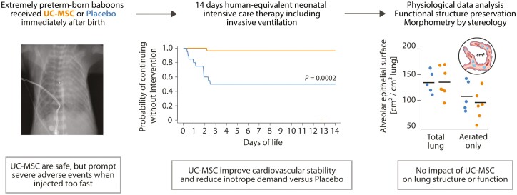
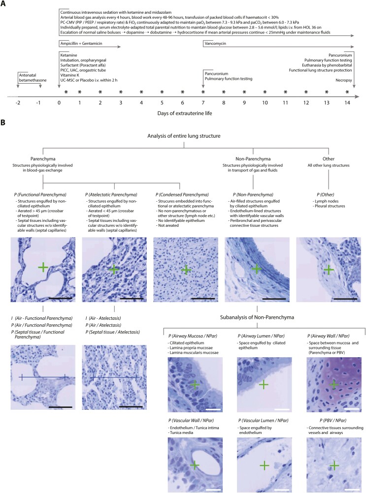
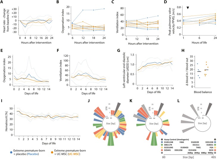
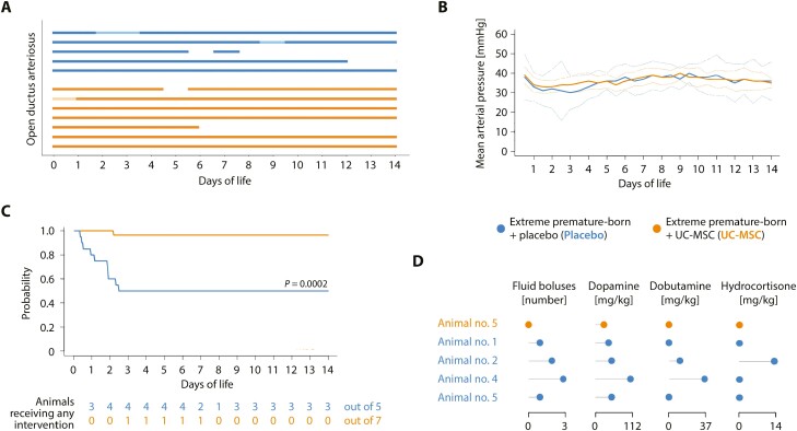
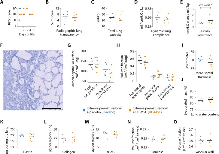
Premature birth is a leading cause of childhood morbidity and mortality and often followed by an arrest of postnatal lung development called bronchopulmonary dysplasia. Therapies using exogenous mesenchymal stromal cells (MSC) have proven highly efficacious in term-born rodent models of this disease, but effects of MSC in actual premature-born lungs are largely unknown. Here, we investigated thirteen non-human primates (baboons; Papio spp.) that were born at the limit of viability and given a single, intravenous dose of ten million human umbilical cord tissue-derived MSC per kilogram or placebo immediately after birth. Following two weeks of human-equivalent neonatal intensive care including mechanical ventilation, lung function testing and echocardiographic studies, lung tissues were analyzed using unbiased stereology. We noted that therapy with MSC was feasible, safe and without signs of engraftment when administered as controlled infusion over 15 minutes, but linked to adverse events when given faster. Administration of cells was associated with improved cardiovascular stability, but neither benefited lung structure, nor lung function after two weeks of extrauterine life. We concluded that a single, intravenous administration of MSC had no short- to mid-term lung-protective effects in extremely premature-born baboons, sharply contrasting data from term-born rodent models of arrested postnatal lung development and urging for investigations on the mechanisms of cell-based therapies for diseases of prematurity in actual premature organisms.

Keywords: adverse events; bronchopulmonary dysplasia; cell therapy; extreme premature birth; lung development; unbiased stereology.


Conflict of interest statement

M.A.M. and D.F. are founders and co-owners of MDTB Cells GmbH, a spin-off company from the Technische Universität Dresden producing mesenchymal cells for clinical and laboratory research purposes. M.A.M., D.F., and M.R. are inventors of granted patent DE102016114043B3 and pending patents US20190185809A1, CA3032048A1, EP3491113A1, and WO2018020008A1, which are all property of MDTB Cells GmbH. S.S. declared research funding from Ottawa Health Research Institute (not commercial) and stock ownership in Pfizer. C.B. declared non-related research speaker reimbursement from Prolacta Bioscience. All other authors declared no potential conflicts of interest.

Figures

Graphical Abstract
Graphical Abstract
Figure 1.
Figure 1.
Design of the study. (A) Clinical management of extremely premature-born, mechanically ventilated baboons. Abbreviations: FiO2: fraction of inspired oxygen; HOL: hour of life; paO2/paCO2: partial pressures of oxygen/carbon dioxide in the arterial blood; PC-CMV: pressure-controlled continuous mandatory ventilation; PEEP: positive end-expiratory pressure; PICC: peripheral inserted central venous catheter; PIP: positive inspiratory pressure; UAC: umbilical arterial catheter. UC-MSC: umbilical cord tissue-derived mesenchymal stromal cells. Asterisks (*) indicate timepoints of echocardiographic studies. (B) Sequential deconstruction of lung tissue into unambiguously identifiable structures. Counting of lung-testsystem interactions (points: P and intersections: I) enabled quantification of absolute and relative volumes, surfaces and thicknesses unbiased by design. PBV: peribronchial/perivascular connective tissue. Analyses and acquisition of reference volumes was performed at magnifications of 10 × 10 (entire lung structure) or 40 × 10 (subanalysis of non-parenchymatous tissues). Black scale bars: 100µm, white scale bars: 25µm. Staining toluidine blue on Technovit 7100.
Figure 2.
Figure 2.
Controlled administration of MSC is safe in critically ill, extremely premature-born baboons. Progression of the (A) heart rate, (B) oxygenation index, (C) ventilation index and the (D) peak pulmonary valve velocity (PPVEL) after administration of UC-MSC or placebo, respectively. PPVEL integrates pulmonary perfusion and pulmonary arterial pressure and is expected to drop in cases of substantial changes in pulmonary vascular resistance or right ventricular function (eg, in cases of significant pulmonary arterial obstructions due to the intravenously administered cells). Every graph/ line in (B-D) represents data from one animal; data is presented as averaged over 120 minutes (A) or six hours (B, C). Echocardiographic studies depicted in (D) were either performed prior to (three animals receiving placebo, three UC-MSC) or four hours after the intervention (two animals receiving placebo, three UC-MSC) and again after 24 hours of life in all animals. The timepoint of intervention is indicated with a triangle (▼). See Supplementary Fig. S1 for information on the clinical course and progression of adverse events in a single extremely premature-born baboon receiving UC-MSC over two minutes and Supplementary Fig. S2 for information on the renal function of the investigated animals. Progression of (E) the oxygenation and (F) the ventilation index over 2 weeks of neonatal critical care, depicted as group means (solid curves) and 95% confidence interval of the means (dotted curves), averaged over 24-hour intervals. (G) Progression of the left ventricular end-diastolic diameter (LVEDD), an estimator of ventricular volume in situations with active left-to-right shunting through an open ductus arteriosus. Data is also depicted as group mean with CI95%. The first echocardiographic study was performed between the first and sixth hour of life (timepoint zero), thereafter every day. (H) Blood balance (ie, the difference between transfused and withdrawn blood volumes) on day of life 14, summed up over the entire course of neonatal intensive care. Horizontal bars indicate group means. (I) Temporal progression of the hematocrit during neonatal critical care, averaged over 12-hour intervals. No significant (P < .05) differences between groups by Mann-Whitney-U (H) or Welch’s two-sided, unequal variance t-test with test level adjustment using Šidák’s correction, comparing data for every timepoint (A-G, I). Circular barplots (J-L) depicting size (in base pairs—bp—on the circular x-axis) and number of short tandem repeats (y-axis) on each allele of investigated short tandem repeat (STR)-loci. Equally coloured bars of comparable transcript size indicate the two alleles of an STR-locus, the single dark-grey bar the assay control (amelogenin) and the light-grey bars the four baboon-specific, constant amplificates. STR signature of (J) the employed cell product and (K) an arterial blood sample drawn after approximately one third of the cell dose was given intravenously (positive control). (L) Representative result of an STR analysis of blood drawn 24 hours (n = 7) or 72 hours (n = 7) after cell administration; or of systematic—uniform sampled pieces of spleen (n = 4), liver (n = 4), or lung (n = 7) obtained on day of life 14 after cell administration. All samples were analysed individually, and 2-4 of the baboon-specific, constant amplificates (light grey) were found per sample and animal besides the assay control (dark grey). Moreover, several human STR-patterns not matching the pattern of the injected cell product were detected in samples and tracked down to the researchers performing the necropsies (not shown).
Figure 3.
Figure 3.
Umbilical cord tissue-derived mesenchymal stromal cells benefit cardiovascular stability in critically ill, extremely premature-born baboons. (A) Presence of an open ductus arteriosus, assessed by daily echocardiography. Every bar represents an animal; solid lines an open, gaps a closed ductus arteriosus. Opaque parts indicate days where the ductus was not assessable by echocardiography. All observed shunting occurred from the systemic into the pulmonal circulation (left-to-right). No significant differences between groups, comparing data for every 24-hour interval. (B) Temporal progression of the mean arterial pressure, depicted as group mean (solid curves) with 95% confidence interval of the mean (CI95%; dotted curves). No significant differences between groups, comparing data for every twelve-hour interval. (C) Event probability for continuing the experiment without requiring escalating interventions (normal saline bolus, dopamine, dobutamine, hydrocortisone) to maintain mean arterial blood pressures ≥ 25 mmHg. Only the start of a new intervention was defined as event; event frequencies were compared on day of life 14. (D) Cumulative doses of medications in animals requiring interventions for cardiovascular insufficiency. Every row in the lollipop chart represents one animal. See Supplementary Fig. S3 for further information on the volume status of the animals. All data derives from five placebo and seven MSC-treated animals. Statistics: Event frequencies in (A) and (C) were compared using Fisher’s exact method. Welch’s two-sided, unequal variance t-test followed by multiple testing adjustment using Šidák’s correction was used to compare data presented in (B).
Figure 4.
Figure 4.
No pulmonary effects of exogenous MSC in extremely premature-born, ventilated baboons. (A) Maximum stage of neonatal respiratory distress syndrome (RDS) in the first five days of life and (B) lung transparency sum scores on day of life 14. All animals had radiographic signs of RDS from birth on; see Supplementary Fig. S4 for representative baboon chest X-ray films. Datapoints represent results from two individual, blinded image analyses per animal. (C) Total lung capacity, (D) dynamic lung compliance and (E) airway resistance after 14 days of mechanical ventilation. (F) Low-magnification photomicrograph showcasing the heterogenous pulmonary architecture an extremely premature-born, 14-day ventilated baboon lung with a side-by-side of atelectatic and emphysematous lung areas. Scale bar = 500 µm. Staining with toluidine blue on 1.5 µm semi-thin Technovit 7100 sections. Also refer to Supplementary Fig. S5. Results of unbiased stereological analyses of the pulmonary structure: (G) Total alveolar epithelial surfaces in the functional and atelectatic parenchyma (total lung surface) as well as in the functional parenchyma only; (H) volume fractions of functional, atelectatic and condensed parenchyma, and (I) mean septal thickness in the functional parenchyma in the lungs of extremely premature-born, ventilated non-human primates 14 days after injection of UC-MSC or placebo. Every datapoint represents mean results of 9-16 (range) isotropic uniformly-randomized, systematic uniformly randomized-sampled pieces of lung tissue (IUR-SURS) per animal; 270 ± 41 (mean ± standard deviation) systematic, uniformly-randomized (SUR) sampled fields of view were analyzed per lung. Refer to Supplementary Fig. S6 and Table S1 for results of further stereological analyses. (J) Analysis of lung water content. Every datapoint represents the mean evaporated mass from of three individual SUR-sampled lung pieces per animal. Quantification of the pulmonary extracellular matrix components (K) elastin, (L) collagen and (M) sulphated glycosaminoglycans (sGAG), expressed as µg per mg dried lung tissue. The mean of three individually analysed SUR-sampled lung pieces make one datapoint. Quantification of the (O) airway mucosa volume and (P) the pulmonary vascular wall volume in relation to the overall vascular or airway volume, respectively. Datapoints represent results from analyses of 622 ± 78 (mean ± standard deviation) SUR-sampled fields of view on 9-16 (range) IUR-SURS per animal. Also see Supplementary Fig. S7. Horizontal bars indicate group means in all graphs and every dot represents data from one animal. No significant (P < .05) differences between UC-MSC and placebo-receiving animals for any of the presented data by Welch’s 2-sided, unequal variance t-test following Shapiro-Wilk testing.

References

    1. Lawn JE, Kinney M.. Preterm birth: now the leading cause of child death worldwide. Sci Transl Med 2014;6(263):263ed21; 10.1126/scitranslmed.aaa2563. - DOI - PubMed
    1. Thébaud B, Goss KN, Laughon M, et al. . Bronchopulmonary dysplasia. Nat Rev Dis Primers 2019;5(1):78; 10.1038/s41572-019-0127-7. - DOI - PMC - PubMed
    1. Lignelli E, Palumbo F, Myti D, et al. . Recent advances in our understanding of the mechanisms of lung alveolarization and bronchopulmonary dysplasia. Am J Physiol Lung Cell Mol Physiol 2019;317(6):L832–L887; 10.1152/ajplung.00369.2019. - DOI - PubMed
    1. Abiramalatha T, Ramaswamy VV, Bandyopadhyay T, et al. . Interventions to prevent bronchopulmonary dysplasia in preterm neonates: an umbrella review of systematic reviews and meta-analyses. JAMA Pediatr 2022;176(5):502–516; 10.1001/jamapediatrics.2021.6619. - DOI - PubMed
    1. Horbar JD, Edwards EM, Greenberg LT, et al. . Variation in performance of neonatal intensive care units in the United States. JAMA Pediatr 2017;171(3):e164396; 10.1001/jamapediatrics.2016.4396. - DOI - PubMed

Publication types