. 2023 Dec 18;62(50):20567-20581.
doi: 10.1021/acs.inorgchem.2c03931. Epub 2023 Feb 1.

Sc-HOPO: A Potential Construct for Use in Radioscandium-Based Radiopharmaceuticals

Affiliations

Sc-HOPO: A Potential Construct for Use in Radioscandium-Based Radiopharmaceuticals

Michael D Phipps et al. Inorg Chem. .

Abstract

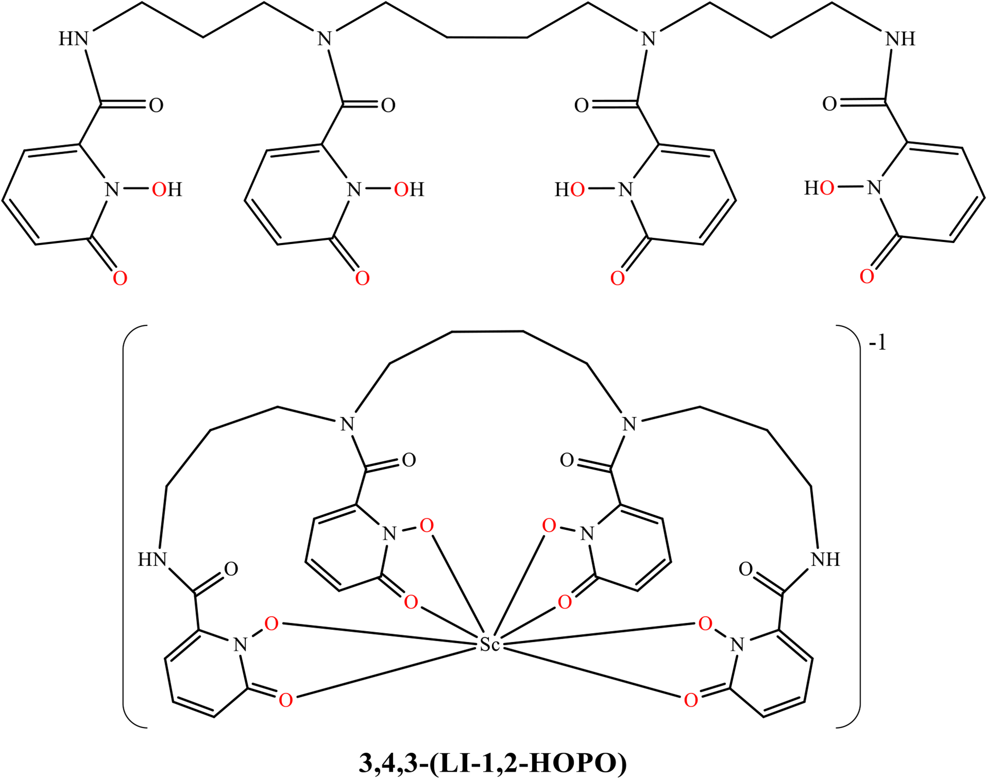
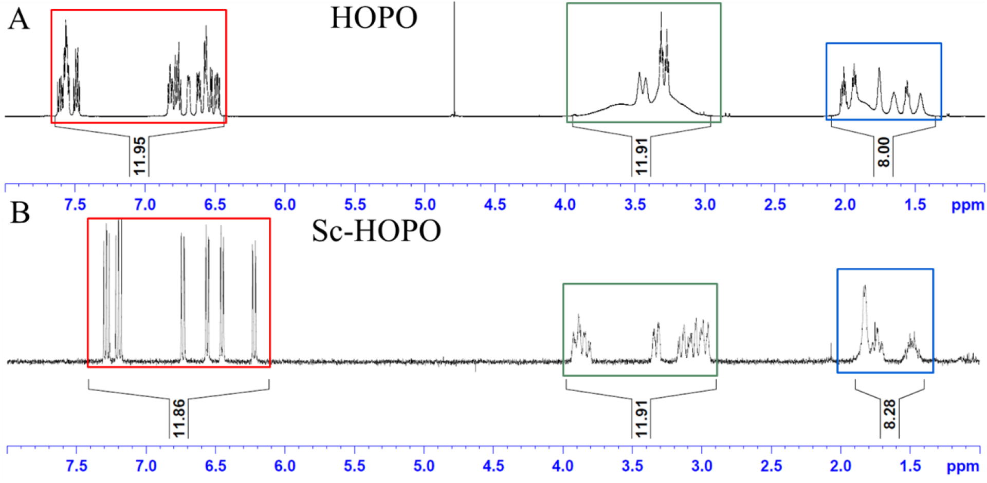
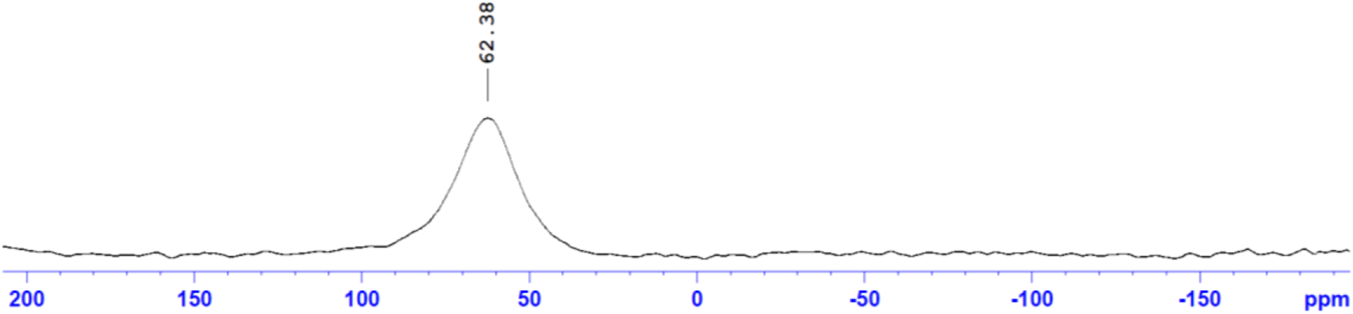
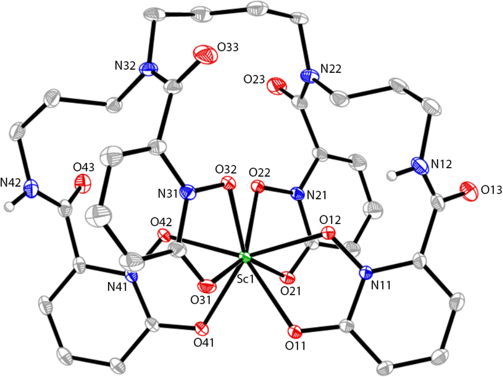
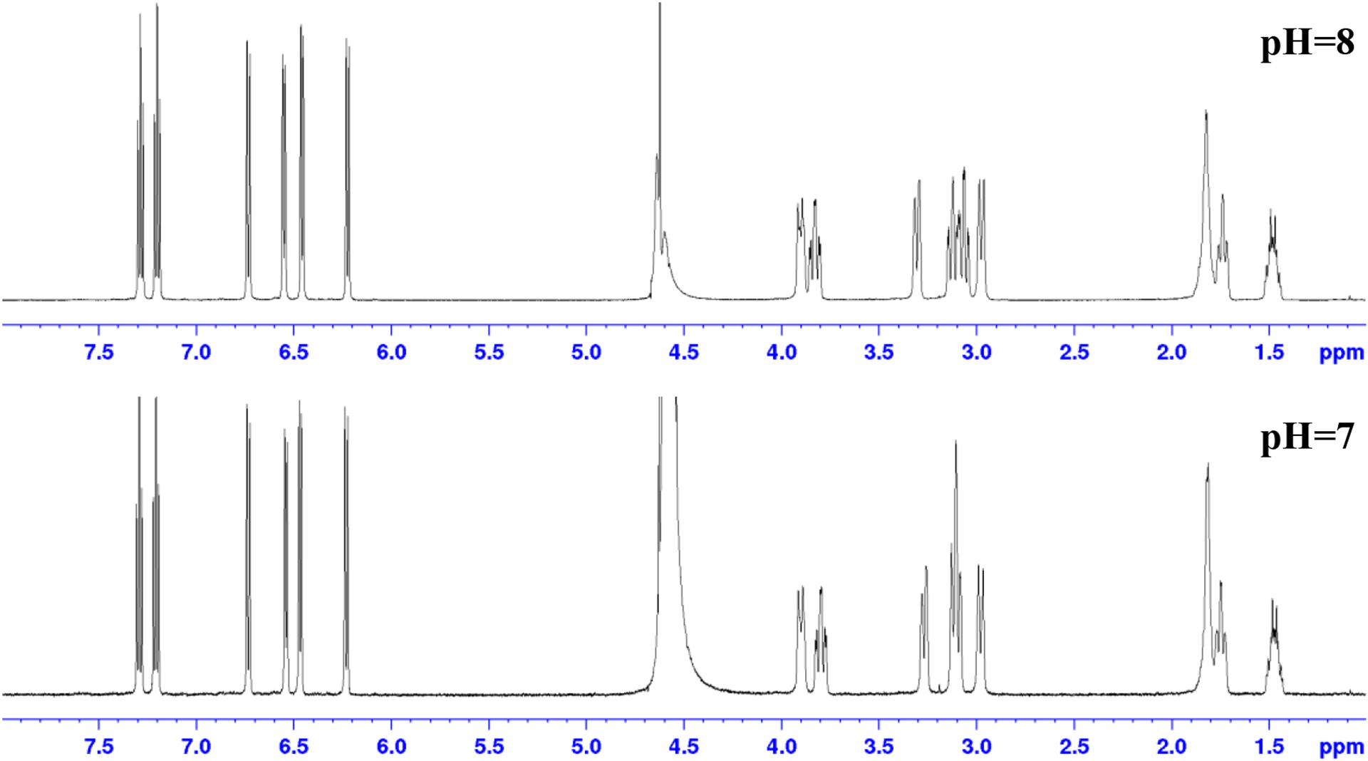
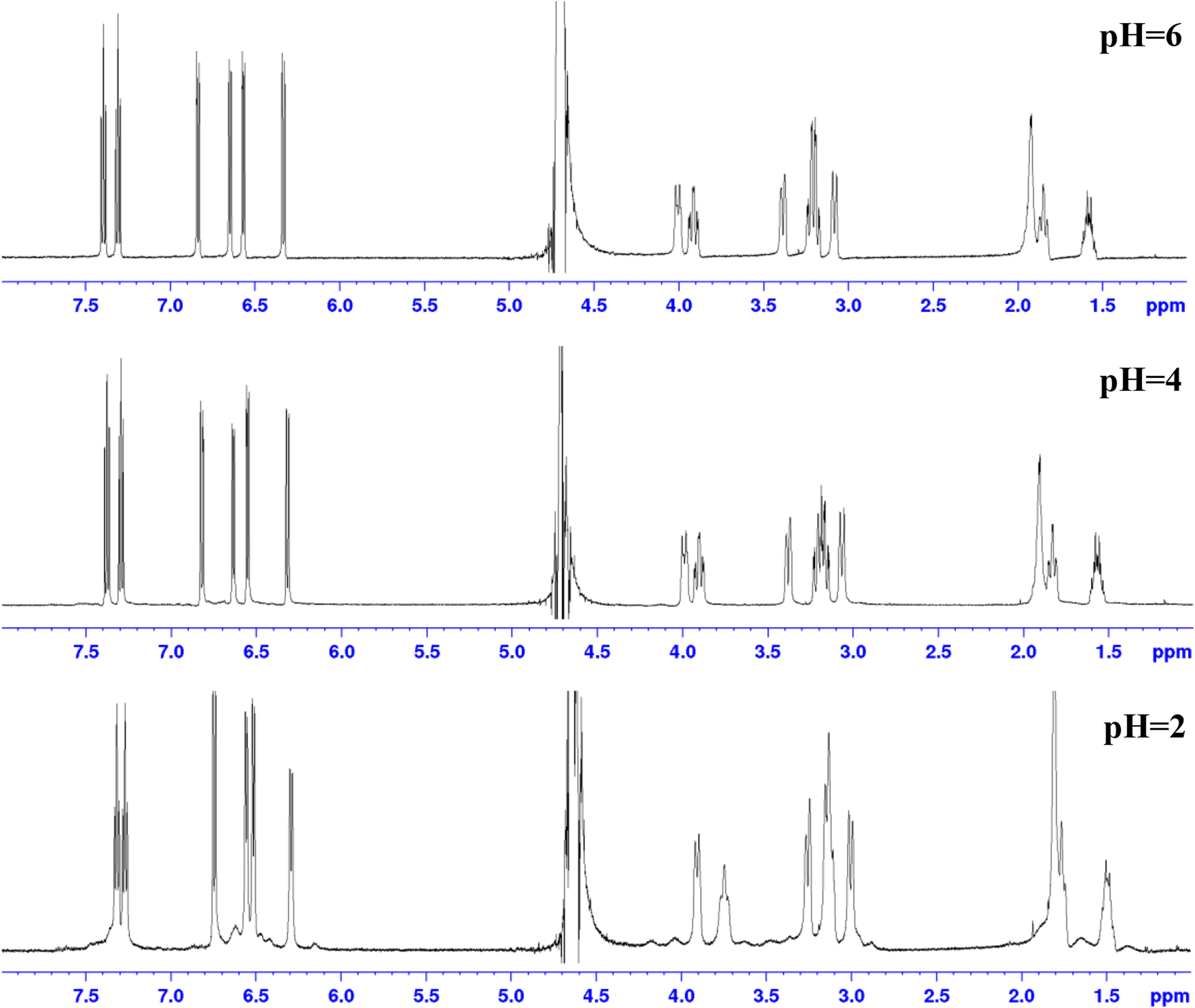
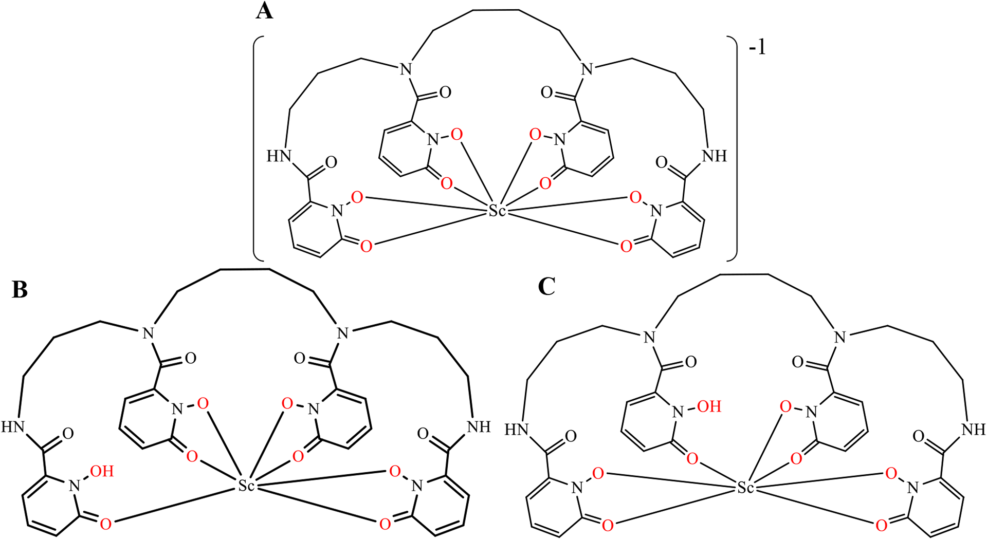
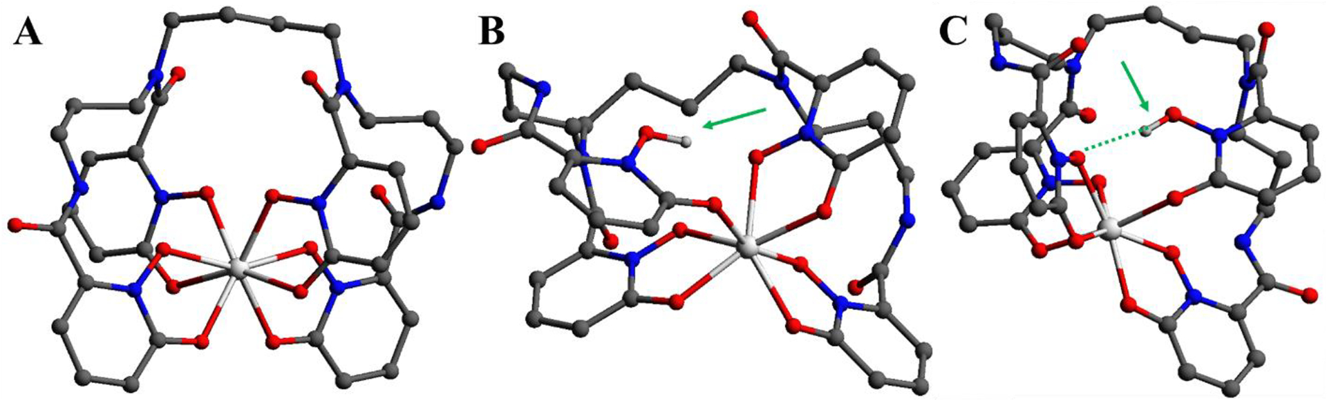
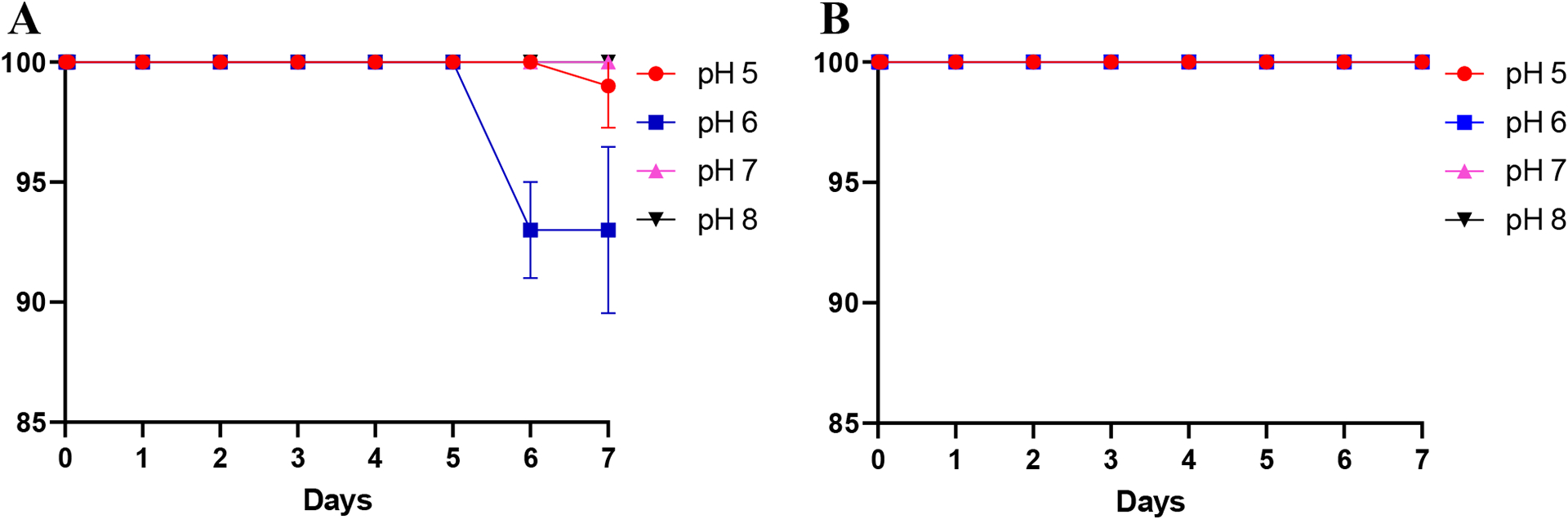
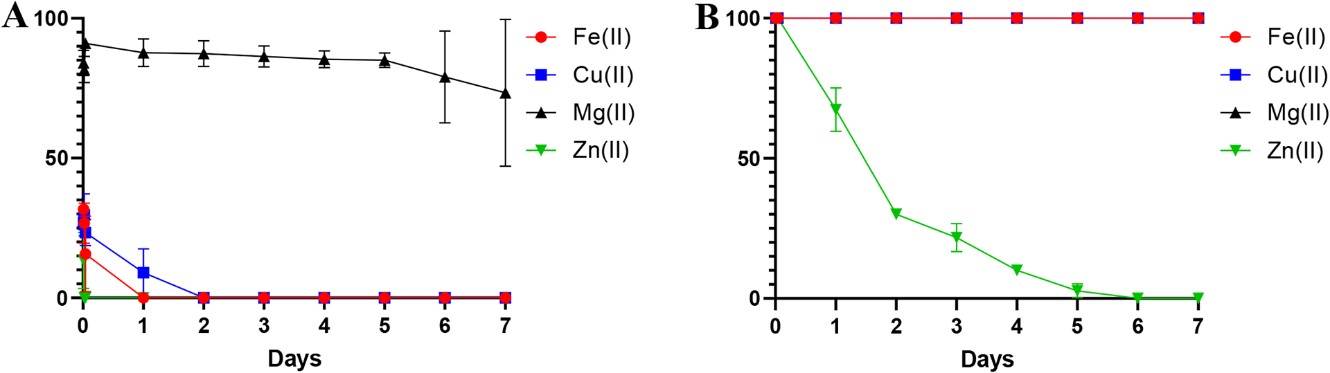
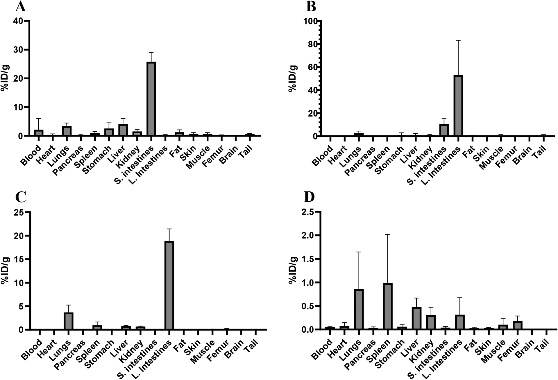
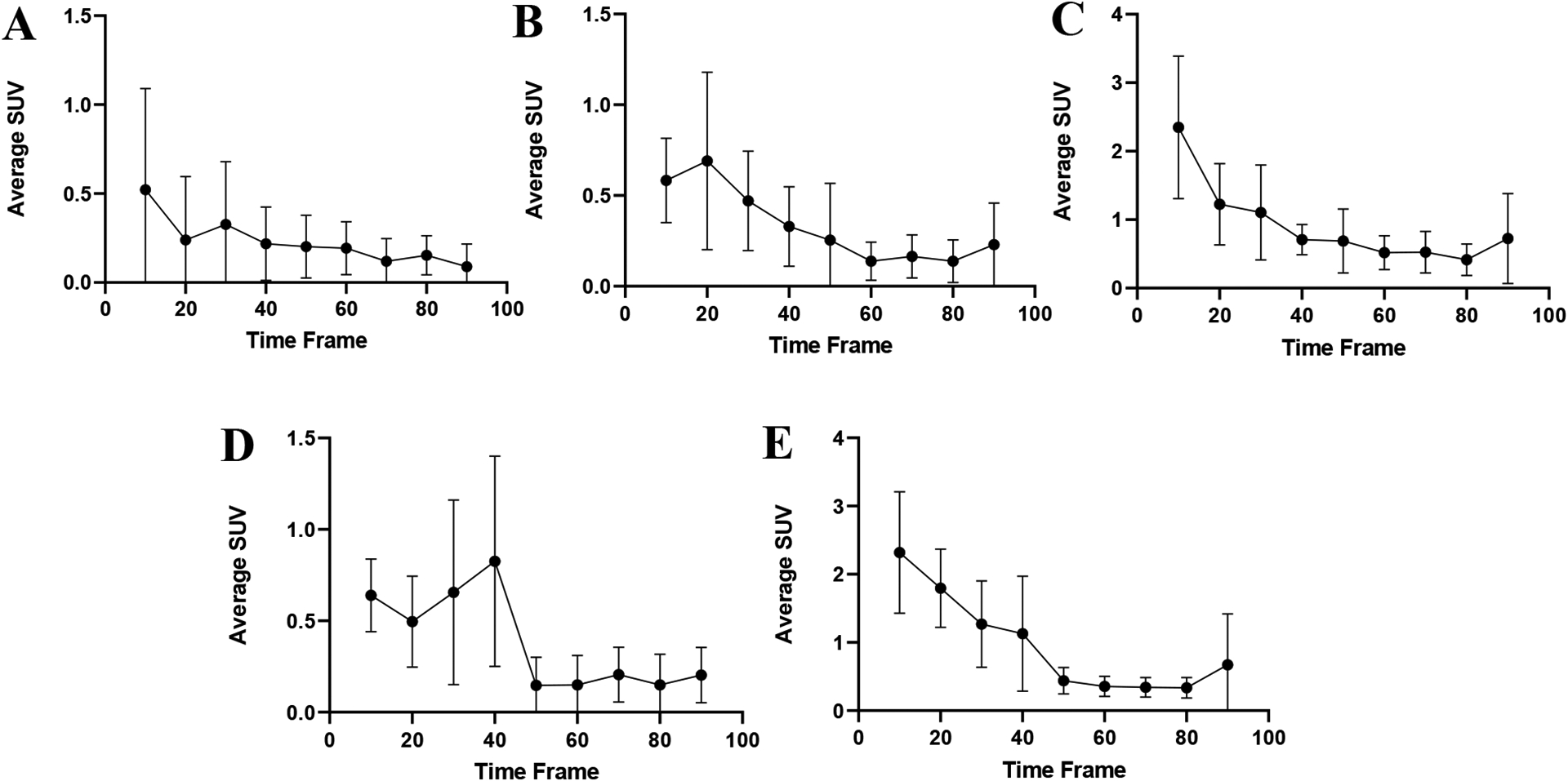
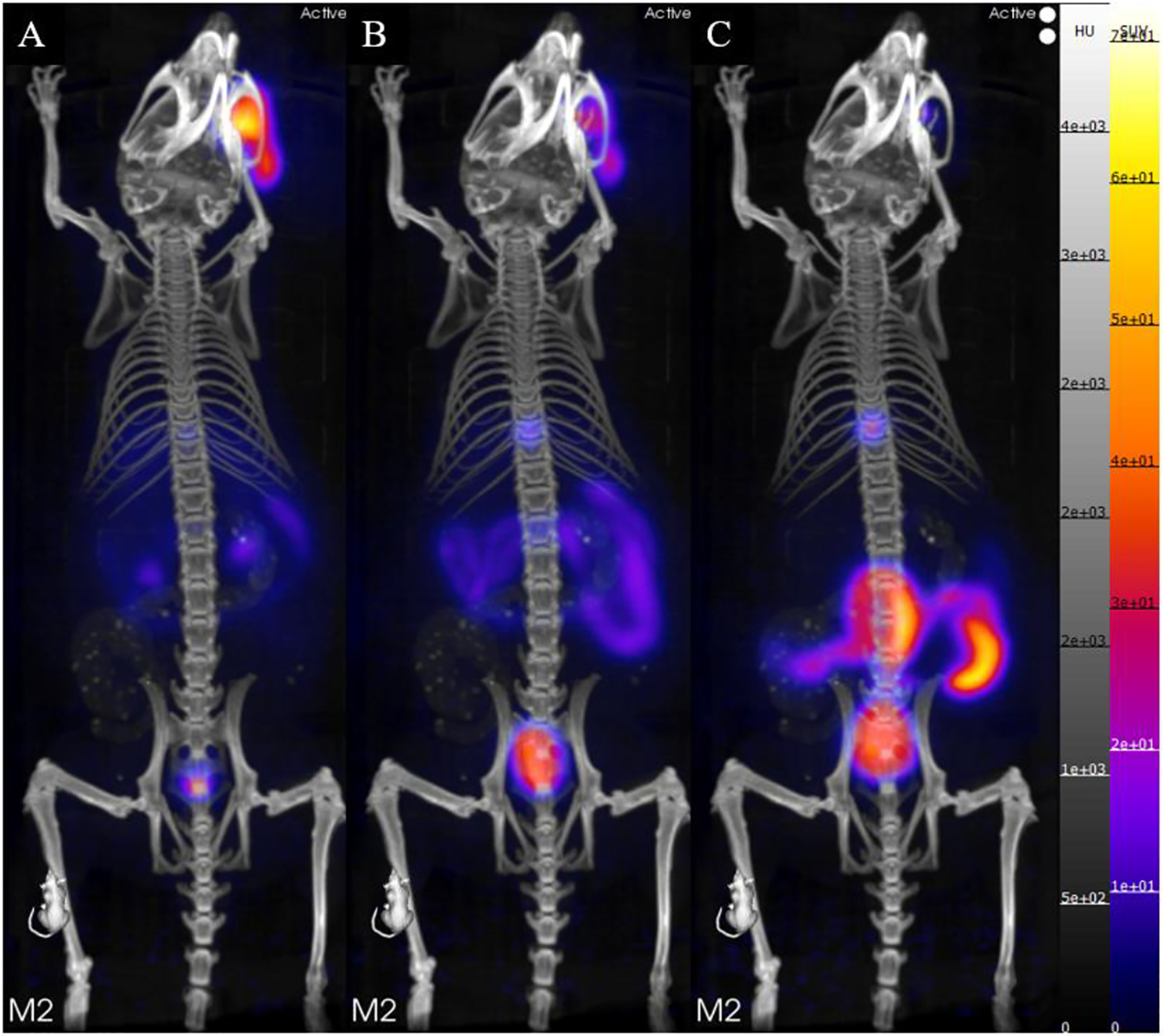
Three isotopes of scandium─43Sc, 44Sc, and 47Sc─have attracted increasing attention as potential candidates for use in imaging and therapy, respectively, as well as for possible theranostic use as an elementally matched pair. Here, we present the octadentate chelator 3,4,3-(LI-1,2-HOPO) (or HOPO), an effective chelator for hard cations, as a potential ligand for use in radioscandium constructs with simple radiolabeling under mild conditions. HOPO forms a 1:1 Sc-HOPO complex that was fully characterized, both experimentally and theoretically. [47Sc]Sc-HOPO exhibited good stability in chemical and biological challenges over 7 days. In healthy mice, [43,47Sc]Sc-HOPO cleared the body rapidly with no signs of demetalation. HOPO is a strong candidate for use in radioscandium-based radiopharmaceuticals.


Conflict of interest statement

Conflict of Interest statement

A patent on the bifunctional p-SCN-Bn-HOPO chelator has been filed with J.S.L., L.C.F, and M.A.D. as inventors.

Figures

Figure 1.
Figure 1.
Structural scheme for the HOPO ligand and the Sc-HOPO complex with the coordinating oxygen atoms shown in red.
Figure 2.
Figure 2.
1H NMR of HOPO and K[Sc(HOPO)] in D2O. Corresponding integral regions are highlighted by color.
Figure 3.
Figure 3.
45Sc NMR of K[Sc(HOPO)] in D2O. The broad peak (FWHM = 12414 Hz) is consistent with 45Sc NMR of other Sc chelates. 0.1 M ScCl3 in 0.01 M HCl was used as the reference standard for the NMR method.
Figure 4.
Figure 4.
Molecular diagram of the Sc-HOPO anion. Hydrogen atoms bound to carbon as well as disordered carbon atoms in the HOPO backbone are omitted for clarity. Atomic displacement parameters are displayed at the 30% probability level.
Figure 5.
Figure 5.
1H NMR of K[Sc(HOPO)] taken at different pH’s in D2O. pH was measured by pH probe, which introduced H2O to the sample. Although peak suppression for H2O was used in the NMR method, the quantity of water was too great for it to be effective. pH was adjusted with K2CO3 and DCl. The resultant increase of KCl concentration results in minor chemical shift changes for both the analyte and the solvent peak.
Figure 5.
Figure 5.
1H NMR of K[Sc(HOPO)] taken at different pH’s in D2O. pH was measured by pH probe, which introduced H2O to the sample. Although peak suppression for H2O was used in the NMR method, the quantity of water was too great for it to be effective. pH was adjusted with K2CO3 and DCl. The resultant increase of KCl concentration results in minor chemical shift changes for both the analyte and the solvent peak.
Figure 6.
Figure 6.
8-coordinate structural scheme of Sc-HOPO (A) and alternative 7-coordinate schemes for Sc-HOPOH in low pH conditions (B and C).
Figure 7.
Figure 7.
Optimized equilibrium structures for: A) 8-coordinate complex, B) 7-coordinate structure with outer group protonation, and C) 7-coordinate complex with inner group protonation. All hydrogens are omitted except for the protonated OH’s in complexes B and C, which are designated by green arrows. A dashed line indicates hydrogen bonding.
Figure 8.
Figure 8.
The five lowest occupied and five highest unoccupied molecular orbitals energies for the 8-coordinate A (1 in the x-axis) complex, 7-coordinate B (2 in the x-axis) and 7-coordinate C (3 in the x-axis) complexes. Frontier orbital representations for the LUMO and HOMO for each complex are shown.
Figure 9.
Figure 9.
Coelution of [47Sc]Sc-HOPO with macroscopic, characterized Sc-HOPO. As discussed above, both macroscopic and tracer levels show tailing caused by Sc-HOPOH.
Figure 10.
Figure 10.
Stability study of [47Sc]Sc-HOPO over the course of 7 days in the presence of 100 μM of EDTA at varying pH (A). Stability of [47Sc]Sc-DOTA in the presence of 100 μM EDTA at varying pH (B).
Figure 11.
Figure 11.
Stability study of [47Sc]Sc-HOPO and [47Sc]Sc-DOTA over the course of 7 days in the presence of metal ions. (A) [47Sc]Sc-HOPO in the presence of 100 μM of FeCl2, CuCl2. MgCl2 and ZnCl2. (B) Stability of [47Sc]Sc-DOTA in the presence of 100 μM of FeCl2, CuCl2. MgCl2 and ZnCl2.
Figure 12.
Figure 12.
Biodistribution of 10 μCi injections of [47Sc]Sc-HOPO in female Balb/c mice at four time points: 10 minutes (A), 1 hour (B), 4 hours (C) and 24 hours (D).
Figure 13:
Figure 13:
The standard uptake values for various organs of 100 μCi of [43Sc]Sc-HOPO were taken at 10 minute intervals over the course of the 90 minute scan and each organ was then plotted to create a time activity curve for each organ: Brain (A), Lung (B), Kidney (C), Heart (D) and Liver (E).
Figure 14.
Figure 14.
(A) PET/CT static image of a Balb/c mouse 0–10 minutes after injection of [43Sc]Sc-HOPO. (B) PET/CT static image of a Balb/c mouse 4–50 minutes after injection of [43Sc]Sc-HOPO. (C) PET/CT static image of a Balb/c mouse 80–90 minutes after injection of [43Sc]Sc-HOPO. Approximately 100 μCi of [43Sc]Sc-HOPO was injected.
Figure 15.
Figure 15.
General thermodynamics cycle used to calculate the change in thermodynamic properties in solution.

References

    1. . FDA-approved radiopharmaceuticals 2022. https://www.cardinalhealth.com/content/dam/corp/web/documents/fact-sheet... (accessed 10/19/2022).
    1. information extracted from the NuDat database. National Nuclear Data Center: 2021.
    1. Müller C; Bunka M; Haller S; Köster U; Groehn V; Bernhardt P; van der Meulen N; Türler A; Schibli R, Promising Prospects for 44Sc-/47Sc-Based Theragnostics: Application of 47Sc for Radionuclide Tumor Therapy in Mice. J. Nuc. Med 2014, 55 (10), 1658–1664. - PubMed
    1. Kerdjoudj R; Pniok M; Alliot C; Kubicek V; Havlickova J; Rosch F; Hermann P; Huclier-Markai S, Scandium(iii) complexes of monophosphorus acid DOTA analogues: a thermodynamic and radiolabelling study with 44Sc from cyclotron and from a 44Ti/44Sc generator. Dalton Trans. 2016, 45 (4), 1398–1409. - PubMed
    1. Domnanich KA; Müller C; Farkas R; Schmid RM; Ponsard B; Schibli R; Türler A; van der Meulen NP, (44)Sc for labeling of DOTA- and NODAGA-functionalized peptides: preclinical in vitro and in vivo investigations. EJNMMI radiopharmacy and chemistry 2017, 1 (1), 8–8. - PMC - PubMed

LinkOut - more resources