. 2023 Feb 7;120(6):e2219199120.
doi: 10.1073/pnas.2219199120. Epub 2023 Feb 1.

Losartan controls immune checkpoint blocker-induced edema and improves survival in glioblastoma mouse models

Affiliations

Losartan controls immune checkpoint blocker-induced edema and improves survival in glioblastoma mouse models

Meenal Datta et al. Proc Natl Acad Sci U S A. .

Abstract

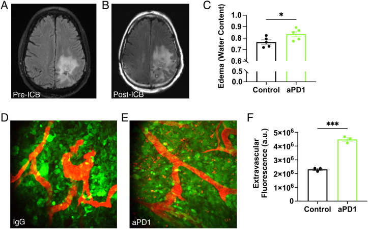
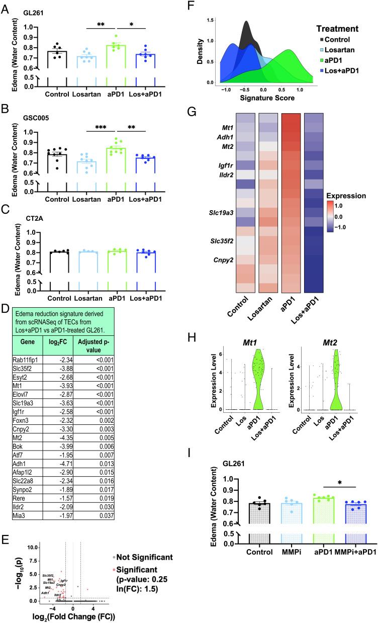
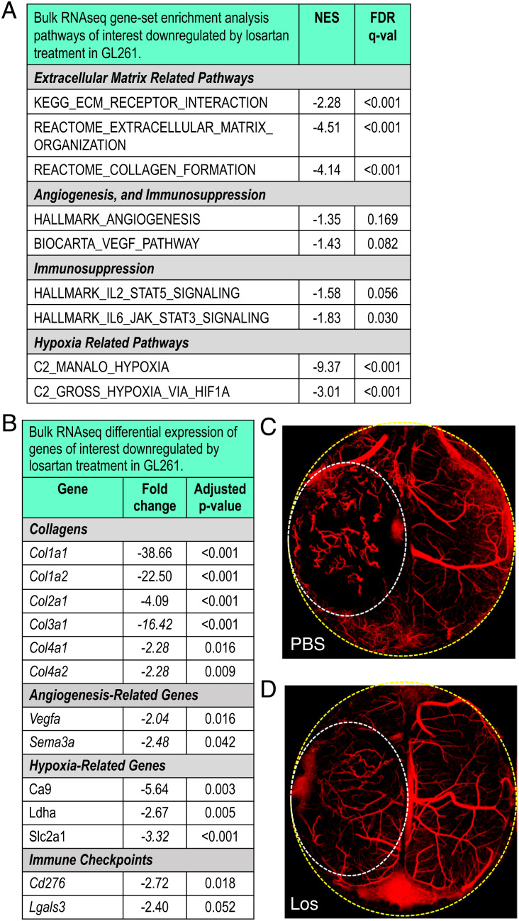
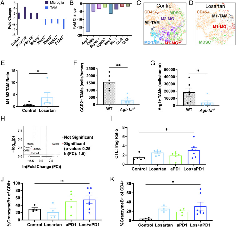
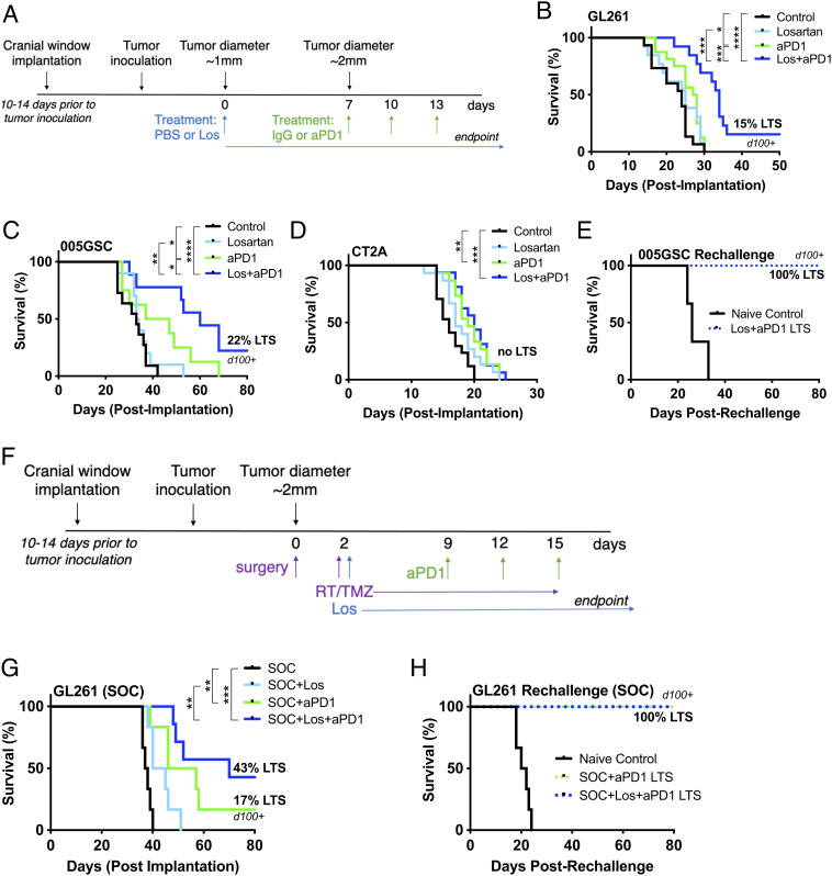
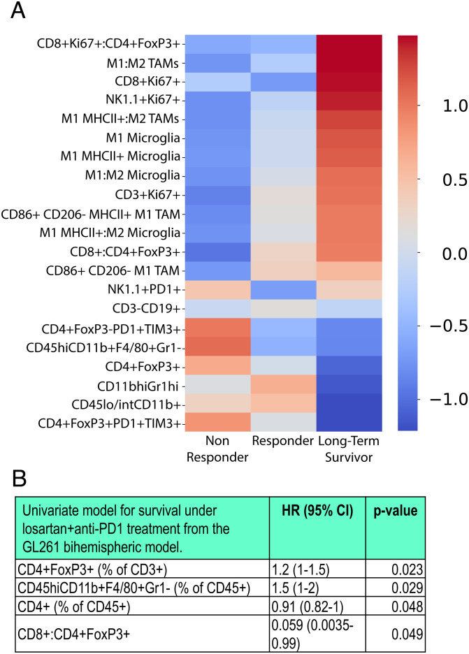
Immune checkpoint blockers (ICBs) have failed in all phase III glioblastoma trials. Here, we found that ICBs induce cerebral edema in some patients and mice with glioblastoma. Through single-cell RNA sequencing, intravital imaging, and CD8+ T cell blocking studies in mice, we demonstrated that this edema results from an inflammatory response following antiprogrammed death 1 (PD1) antibody treatment that disrupts the blood-tumor barrier. Used in lieu of immunosuppressive corticosteroids, the angiotensin receptor blocker losartan prevented this ICB-induced edema and reprogrammed the tumor microenvironment, curing 20% of mice which increased to 40% in combination with standard of care treatment. Using a bihemispheric tumor model, we identified a "hot" tumor immune signature prior to losartan+anti-PD1 therapy that predicted long-term survival. Our findings provide the rationale and associated biomarkers to test losartan with ICBs in glioblastoma patients.

Keywords: biomarkers; glioblastoma; immune checkpoint blockers; immune-related adverse events; tumor microenvironment.


Conflict of interest statement

The authors have organizational affiliations, stock ownership, and patent filings to disclose. S.C. is consultant at Guidepoint and Coleman Research. K.E.E. has intellectual property rights at NordicNeuroLab AS, Bergen, NO. M.C.S. is a current employee of GlaxoSmithKline and may own GSK stock. M.R.N. is a current employee of AbbVie and may own AbbVie stock. D.A.R. received research support from the following (paid to Dana-Farber Cancer Institute): Acerta Phamaceuticals, Agenus, Bristol-Myers Squibb, Celldex, EMD Serono, Enterome, Epitopoietic Research Coorporatioin, Incyte, Inovio, Insightec, Novartis, Omniox, and Tragara. D.A.R. received advisory/consultant fees from Abbvie; Advantagene; Agenus; Agios; Amgen; AnHeart Therapeutics; Bayer; Boston Biomedical; Boehringer Ingelheim; Bristol-Myers Squibb; Celldex; Deciphera; Del Mar Pharma; DNAtrix; Ellipses Pharma; EMD Serono; Genenta; Genentech/Roche; Hoffman-LaRoche, Ltd.; Imvax; Inovio; Kintara; Kiyatec; Medicenna Biopharma, Inc.; Merck; Merck KGaA; Monteris; Neuvogen; Novartis; Novocure; Oncorus; Oxigene; Regeneron; Stemline; Sumitono Dainippon Pharma; Pyramid; Taiho Oncology, Inc.; and Y-mabs Therapeutics. A.H.S. has patents/pending royalties on the PD-1 pathway from Roche and Novartis. A.H.S. is on advisory boards for Surface Oncology, SQZ Biotechnologies, Elpiscience, Selecta, Bicara, Monopteros, GlaxoSmithKline, and Janssen. A.H.S. has received research funding from Novartis, Roche, UCB, Ipsen, Merck, and AbbVie unrelated to this project. G.J.F. has patents/pending royalties on the PD-1/PD-L1 pathway from Roche, Merck MSD, Bristol-Myers-Squibb, Merck KGA, Boehringer-Ingelheim, AstraZeneca, Dako, Leica, Mayo Clinic, and Novartis. G.J.F. has served on advisory boards for Roche, Bristol-Myers-Squibb, Xios, Origimed, Triursus, iTeos, NextPoint, IgM, Jubilant, Trillium, GV20, IOME, and Geode. G.J.F. has equity in Nextpoint, Triursus, Xios, iTeos, IgM, Trillium, Invaria, GV20, and Geode. M.L.S. is an equity holder, scientific cofounder, and advisory board member of Immunitas Therapeutics. R.K.J. received consultant fees from Bristol Myers Squibb (BMS), Cur Therapeutics, Elpis, Innocoll, SPARC, and SynDevRx; owns equity in Accurius, Enlight, and SynDevRx; Board of Trustees of Tekla Healthcare Investors, Tekla Life Sciences Investors, Tekla Healthcare Opportunities Fund, Tekla World Healthcare Fund; and received research grants from Boehringer Ingelheim and Sanofi. No funding or reagents from these organizations were used in this study. M.Datta., L.X., M.L.S., and R.K.J. are coinventors of a patent application filed at the US Patent Office by Massachusetts General Hospital on, "Preventing immunotherapy-induced edema using angiotensin receptor blockers."

Figures

Fig. 1.
Fig. 1.
ICB increases GBM vascular leakage and induces brain edema. MR T2-weighted-Fluid-Attenuated Inversion Recovery (T2-FLAIR) images obtained from a recurrent GBM patient (A) before and (B) after 4 mo of anti-PD-L1 (MEDI4763; NCT02336165) treatment show increased edema after ICB treatment. In addition to ICB-induced inflammation, this change may be due in part to underlying tumor activity or growth. (C) In mice, anti-PD1 antibody (aPD1) treatment increases edema in GL261 tumors compared to IgG control [as measured by wet-dry weight (i.e., water content) evaluation of tumor tissue; n = 5]. Multiphoton visualization of the brain vasculature via injected tetramethylrhodamine (TAMRA) labeled albumin (red) imaged through transparent cranial windows in mice bearing GFP+ GL261 GBM (green) shows that compared to IgG controls (D) there is increased extravasation in anti-PD1-treated tumors after the third consecutive dose (E). (F) Quantification shows that more albumin in anti-PD1-treated mice has leaked outside of the tumor blood vessels (n = 3). (Bar plots: mean ± SEM; Student’s unpaired t test; *P < 0.05; ***P < 0.001.)
Fig. 2.
Fig. 2.
Losartan prevents ICB-induced edema by downregulating TEC MT-MMP-1 and -2 expression. Losartan decreases anti-PD1-induced edema in (A) GL261 and (B) 005 GSC models but not in (C) CT2A after 2 wk of treatment (n = 5 to 9). (D) scRNASeq of TECs reveals a set of downregulated genes that includes those related to metabolism (e.g., Adh1, Ildr2), angiogenesis/migration (e.g., Cnpy2, Igf1r), and solute carriers (e.g., Slc35f2, Slc19a3). When applied as an edema signature, this gene set is upregulated in anti-PD1-treated GL261 tumors compared to other treatment arms as visualized via (E) volcano plot, (F) density plot of edema signature scores (methods described in SI Appendix) by treatment and (G) mean gene expression heat map of edema signature genes. (H) Specialized MT-MMPs (Mt1, Mt2) are among these genes and are expressed in TECs only from the anti-PD1-treated tumors. (I) The MMP inhibitor Ilomastat (MMPi) controls anti-PD1-induced edema comparably to losartan in GL261 (n = 6). (Edema signaturegene expression units = ln(TP100k + 1); log2FC = fold changes > |2|; adjusted P value < 0.05. Bar plots: mean ± SEM; one-way ANOVA with Tukey’s post hoc test; *P < 0.05; **P < 0.01; ***P < 0.001.)
Fig. 3.
Fig. 3.
Losartan reprograms the GBM tumor microenvironment. (A) TME-related gene-set enrichment analysis pathways downregulated by losartan treatment compared to control in bulk RNASeq of GL261 tumors (n = 3). (B) Differential gene expression confirms these effects in matrix molecules such as collagen, hypoxia-related genes, and immune checkpoints. Intravital OCT imaging [to detect perfused vessels (red) vs. nonperfused areas (black)] shows that compared to PBS-treated controls (C), losartan (D) renders tumor blood vessels less tortuous and improves tumor perfusion (yellow dashed line—cranial window border; white dashed line—tumor area). (Sequencing: FDR, false discovery rate; all FDR q-values < 0.20; NES, normalized enrichment score; all adjusted P values < 0.05, FC > |2|. Bar plots: mean ± SEM; Student’s unpaired t test; *P < 0.05.)
Fig. 4.
Fig. 4.
Losartan promotes antitumor immunity in the GBM TME. Applying the human-derived signatures from our previous work (16), losartan is found to enrich microglia-like signatures and downregulate global (A) and M2-like (B) TAM signatures vs. controls as assessed in bulk RNASeq samples from GL261 (n = 3). t-distributed stochastic neighbor embedding (t-SNE) plots of flow cytometry data of myeloid populations reveal (C) a diverse and largely immunosuppressive (“M2”) microenvironment in GL261 controls that is (D) reprogrammed by losartan treatment to feature fewer myeloid cells that are polarized for anti-tumor (“M1”) activity (MG, microglia). (E) Losartan increases the ratio of anti- to pro-tumor TAMs, assessed via flow cytometry (n = 5 to 7). Highly suppressive TAM subsets (F) CCR2+ and (G) Arg1+ (of CD45hiCD11b+F4/80+) are downregulated in GL261 tumors implanted in Agtr1a−/− mice compared to those implanted in wild-type C57Bl/6 mice. (H) scRNASeq of CD8+ T cells reveals heightened Gzmb expression under combined treatment compared to anti-PD1 monotherapy. Losartan+anti-PD1 treatment increases (I) cytotoxic (CTL; CD45+CD3+CD8+GranzymeB+) to regulatory (Treg; CD45+CD3+CD4+FoxP3+) T cell ratios in the tumor, and effector Granzyme+ CD8 (J, not significant) and CD4 (K) T cells in the cervical lymph nodes. (Sequencing: all FDR q-values < 0.25, FC > |2|, adjusted P values < 0.05. Flow cytometry: Mann–Whitney unpaired t test or one-way ANOVA with Tukey’s post hoc test; *P < 0.05.)
Fig. 5.
Fig. 5.
Losartan improves survival under anti-PD1 treatment with and without the SOC. Losartan enhances the survival benefit of anti-PD1 therapy in (A) GL261 and (B) 005 GSC tumor models with 15% and 22% LTSs, respectively, with no detectable tumors via microultrasound imaging through transparent cranial windows for over 100 d (d100). In addition to lack of increased edema in the face of ICB treatment (Fig. 2C), (C) the CT2A model displays only a modest response to anti-PD1 therapy that does not result in LTSs nor is improved by the addition of losartan treatment. (D) Long-term surviving mice in the 005 GSC model reject a second tumor inoculation, suggesting the formation of an immune memory response. (E) The GL261 model subjected to SOC (F) therapy shows an improvement (G) in response to anti-PD1 (16% LTSs) that is tripled (43% LTSs) in combination with losartan. (H) Long-term surviving mice in the GL261 SOC model reject a second tumor rechallenge. (Log-rank Mantel–Cox test; *P < 0.05; **P < 0.01; ***P < 0.001; ****P < 0.0001.)
Fig. 6.
Fig. 6.
Bihemispheric model reveals predictors of response to losartan+anti-PD1 treatment. The bihemispheric mouse model can be used to resect one tumor for biomarker analysis prior to losartan+anti-PD1 treatment which has variable responses in GL261-bearing mice (n = 9). (A) Using flow cytometry, immune cells were profiled in individual mice under combinatorial therapy. As indicated by the heat-map z-scores (transformed relative populations of immune cell classes), LTSs have distinguished pretreatment biomarker signatures that indicate that strong antitumor immunity is present in the tumor prior to therapy. (B) The presence of CD4 T cells and higher ratios of CD8 to regulatory T cells in the GBM TME before therapy initiation are predictive of improved survival, while the presence of T regulatory cells and TAMs are associated with decreased survival, assessed via proportionate hazard models. (P values derived from univariate Cox regression model; HR, hazard ratio; CI, confidence interval.)

References

    1. Nayak L., et al. , Randomized phase II and biomarker study of pembrolizumab plus bevacizumab versus pembrolizumab alone for patients with recurrent glioblastoma. Clin. Cancer Res. 27, 1048–1057 (2021). - PMC - PubMed
    1. Qin L., et al. , Advanced MRI assessment to predict benefit of anti-programmed cell death 1 protein immunotherapy response in patients with recurrent glioblastoma. Neuroradiology 59, 135–145 (2017). - PMC - PubMed
    1. Arrieta O., et al. , Blockage of angiotensin II type I receptor decreases the synthesis of growth factors and induces apoptosis in C6 cultured cells and C6 rat glioma. Br J. Cancer 92, 1247–1252 (2005). - PMC - PubMed
    1. Carpentier A. F., et al. , Steroid-sparing effects of angiotensin-II inhibitors in glioblastoma patients. Eur. J. Neurol. 19, 1337–1342 (2012). - PubMed
    1. Kourilsky A., et al. , Impact of angiotensin-II receptor blockers on vasogenic edema in glioblastoma patients. J. Neurol. 263, 524–530 (2016). - PubMed

Publication types