

Review
doi: 10.1056/NEJMra2206346.
Humoral Innate Immunity and Acute-Phase Proteins
Affiliations
- PMID: 36724330
- PMCID: PMC9912245
- DOI: 10.1056/NEJMra2206346
Item in Clipboard
Review
Humoral Innate Immunity and Acute-Phase Proteins
N Engl J Med.
.
No abstract available
Figures
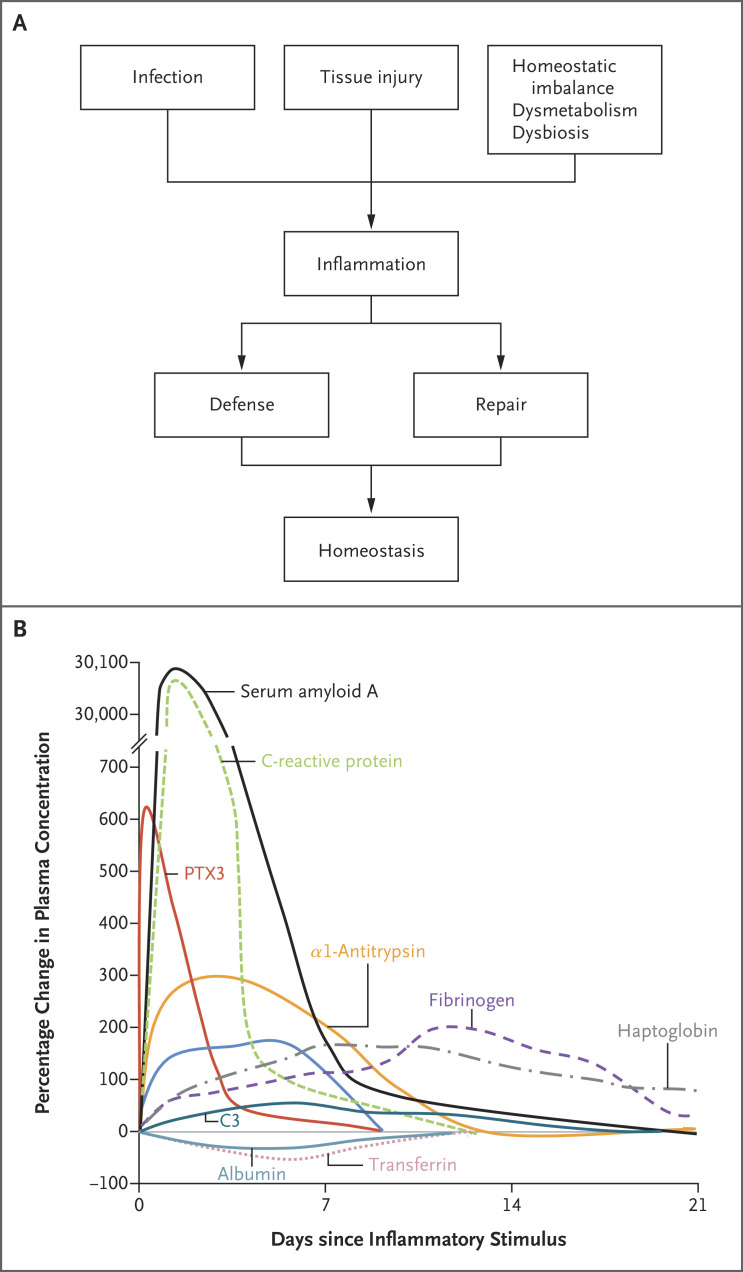
Panel B is adapted from Gitlin and Colten with permission from the publisher. PTX3 denotes pentraxin 3.
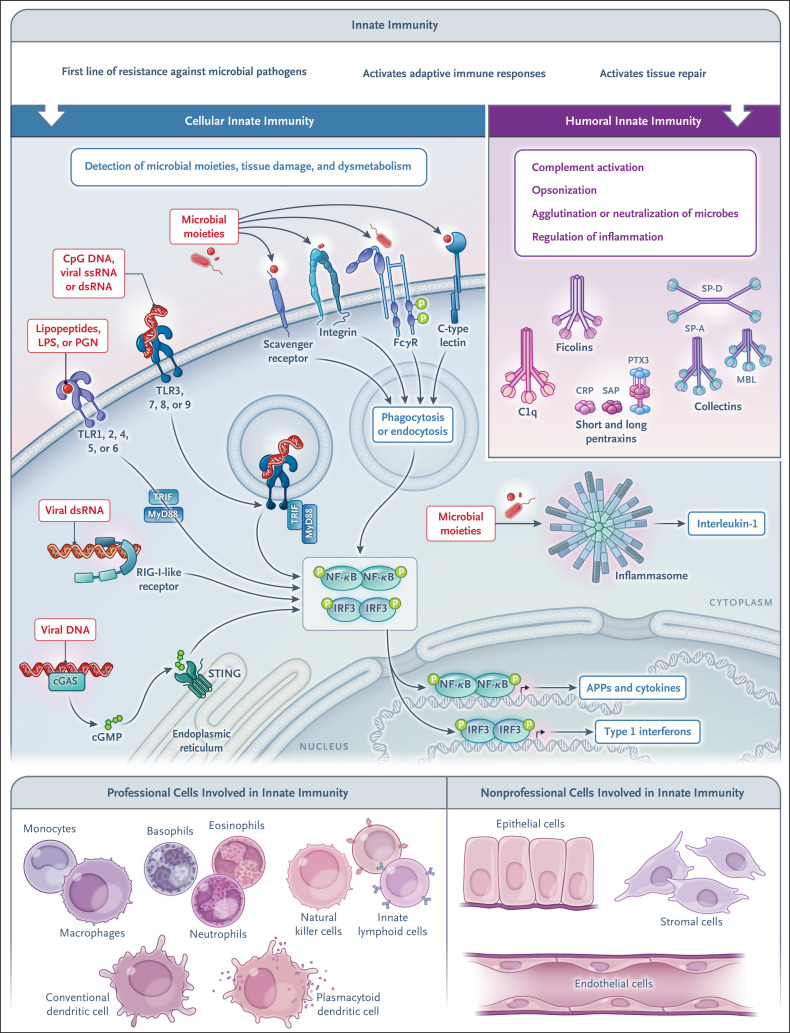
Cellular sensors of tissue damage, infection, and dysmetabolism are strategically localized on the cell surface, in the endosomal compartment, and in the cytoplasm, in both professional innate immune cells (i.e., those with innate immunity as their principal function) and nonprofessional innate immune cells (i.e., those with other principal functions). The latter include hepatocytes, a major source of acute-phase proteins. Under homeostatic conditions and in response to inflammation, components of the humoral arm of innate immunity are produced. These molecules serve complex functions, including immune resistance, by activating complement and having opsonic activity (ante-antibodies). APP denotes acute-phase protein, cGAS cyclic GMP–AMP synthase, cGMP cyclic guanosine monophosphate, CRP C-reactive protein, dsRNA double-stranded RNA, IRF interferon regulatory factor, LPS lipopolysaccharide, MBL mannose-binding lectin, MyD88 myeloid differentiation primary response 88, NF-κB nuclear factor kappa B, PGN peptidoglycan, RIG-I retinoid acid-inducible gene I, SAP serum amyloid P, SP-A surfactant protein A, SP-D surfactant protein D, ssRNA single-stranded RNA, STING stimulator of interferon genes, TLR toll-like receptor, TRIF toll/interleukin-1 receptor–domain–containing adapter-inducing interferon-β.
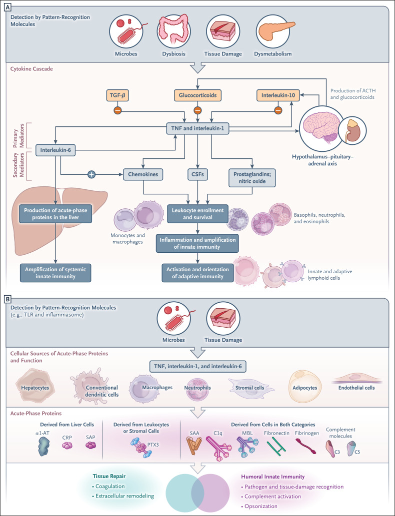
Panel A shows the cytokine cascade set in motion by pattern-recognition molecules; this process involves the production of primary and secondary mediators, activation of the acute-phase response, and promotion of leukocyte recruitment and leads to amplification of local and systemic innate immunity, as well as to the activation and orientation of adaptive immune responses. Inflammatory cytokines also promote the expression of negative regulators of inflammation (interleukin-10, transforming growth factor beta [TGF-β], and interleukin-1Ra) and the activation of the hypothalamus–pituitary–adrenal axis, which results in production of adrenocorticotropic hormone (ACTH) and glucocorticoid hormones. As shown in Panel B, in addition to hepatocytes, other cell types contribute to the synthesis of acute-phase proteins, which contribute to humoral innate immunity and tissue repair. α1-AT denotes α1-antitrypsin, CSF colony-stimulating factor, SAA serum amyloid A, and TNF tumor necrosis factor.
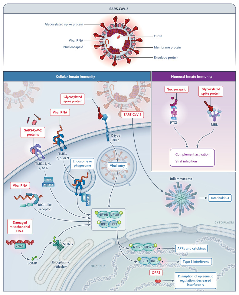
Severe acute respiratory syndrome coronavirus 2 (SARS-CoV-2) proteins and nucleic acids are recognized by cellular receptors involved in innate immunity, including C-type lectins, TLRs, cGAS–STING, and the inflammasome. Open reading frame 8 (ORF8) inhibits interferon production through epigenetic mechanisms. The humoral pattern-recognition molecules MBL and PTX3 bind glycosylated spike and the viral nucleocapsid, respectively.
Comment in
-
Humoral Innate Immunity and Acute-Phase Proteins.N Engl J Med. 2023 May 4;388(18):1725. doi: 10.1056/NEJMc2302460. N Engl J Med. 2023. PMID: 37133604 No abstract available.
-
Humoral Innate Immunity and Acute-Phase Proteins. Reply.N Engl J Med. 2023 May 4;388(18):1725-1726. doi: 10.1056/NEJMc2302460. N Engl J Med. 2023. PMID: 37133605 No abstract available.
References
-
- Medzhitov R. The spectrum of inflammatory responses. Science 2021;374:1070-1075. - PubMed
-
- Gitlin JD, Colten HR. Molecular biology of the acute phase plasma proteins. In: Pick E, Landy M, eds. Lymphokines. Vol. 14. San Diego: Academic Press, 1987:123-153.
-
- Gabay C, Kushner I. Acute-phase proteins and other systemic responses to inflammation. N Engl J Med 1999;340:448-454. - PubMed
-
- Dinarello CA. Interleukin-1 and the pathogenesis of the acute-phase response. N Engl J Med 1984;311:1413-1418. - PubMed
Publication types
MeSH terms
Substances
Associated data
LinkOut - more resources
Full Text Sources
Other Literature Sources
Medical