

Infants with Congenital Adrenal Hyperplasia Exhibit Thalamic Discrepancies in Early Brain Structure
- PMID: 36724764
- PMCID: PMC10505336
- DOI: 10.1159/000529403
Infants with Congenital Adrenal Hyperplasia Exhibit Thalamic Discrepancies in Early Brain Structure
Abstract
Introduction: Patients with classical congenital adrenal hyperplasia (CAH) have prenatal and postnatal hormonal imbalances. To characterize the ontogeny of reported brain and behavior changes in older children with CAH, we aimed to study the brain structure in infants with CAH compared to healthy controls.
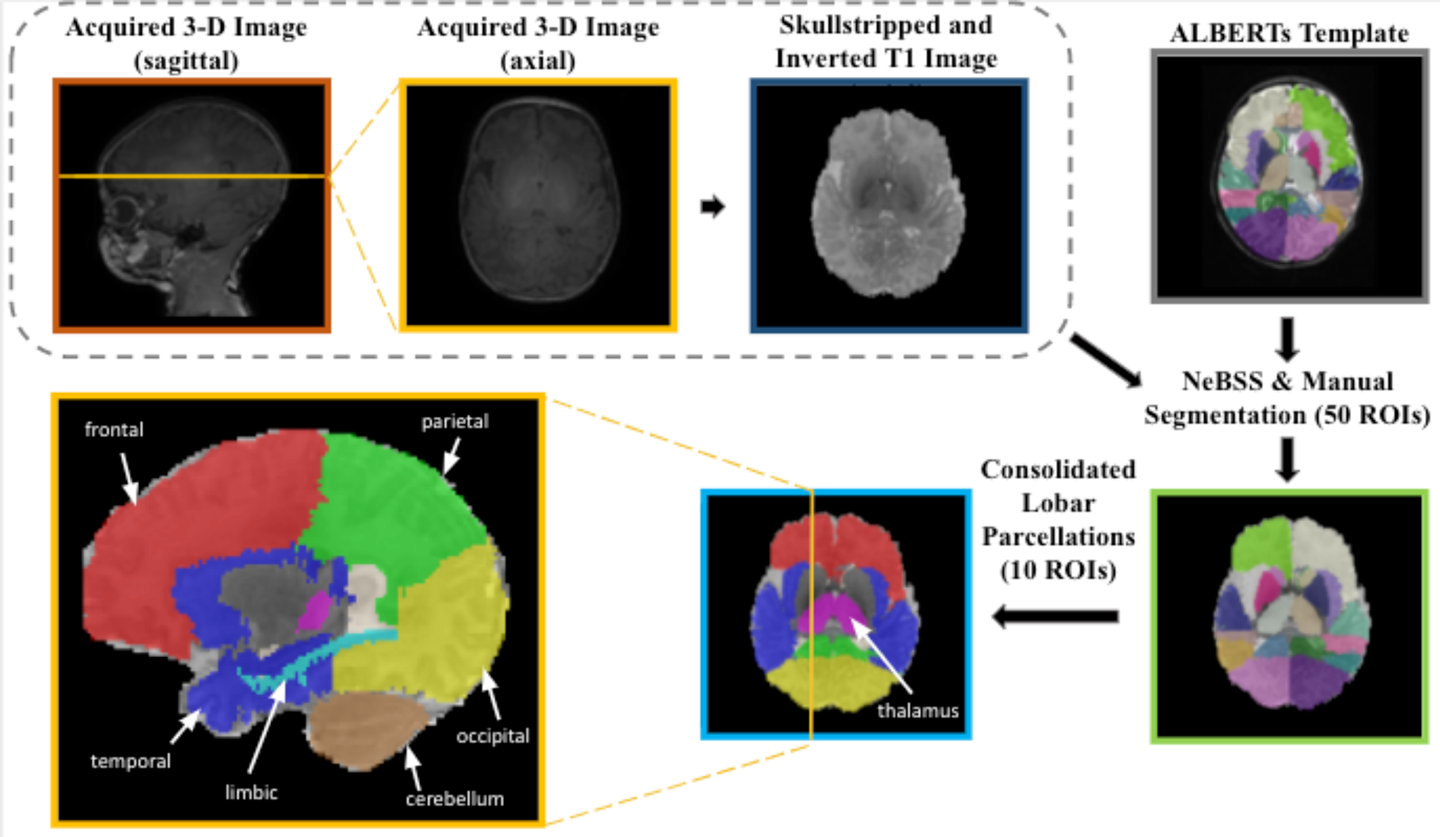
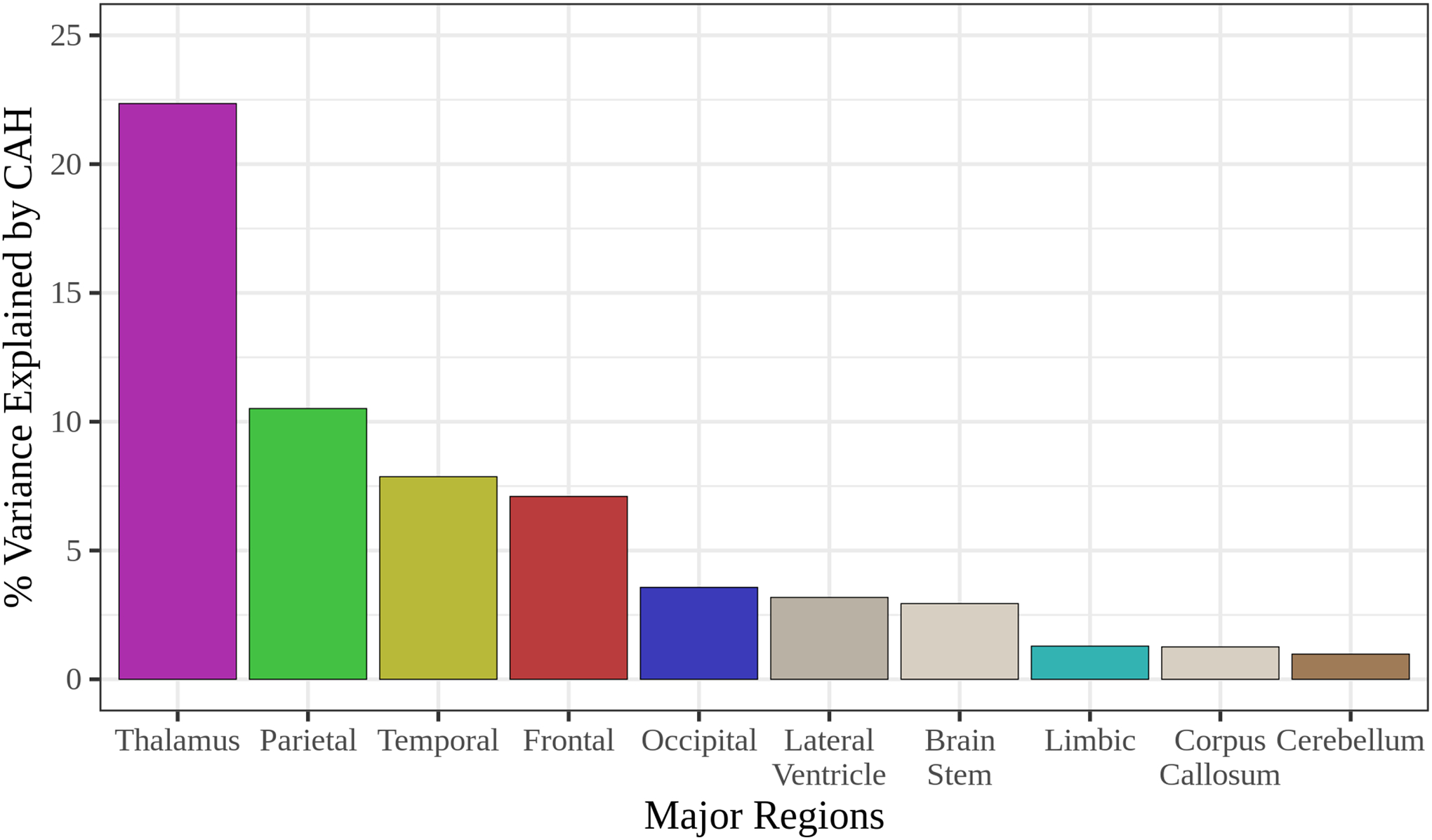
Methods: We performed neuroimaging in 16 infants with classical CAH due to 21-hydroxylase deficiency (8 males, gestational age 38.2 ± 1.7 weeks, post-conceptional age [PCA] 42.2 ± 3.0 weeks) and 14 control infants (9 males, gestational age 38.5 ± 1.8 weeks, PCA 42.5 ± 2.4 weeks) utilizing 3-Tesla magnetic resonance imaging. Regional brain volumes were adjusted for PCA and sex, along with an additional adjustment for total brain volume (TBV), for group comparisons by regression analyses (mean, 95% confidence interval [CI]). The degree to which each brain region was differentiated between CAH and control infants was examined by relaimpo analyses, adjusting for all other brain regions, PCA, and sex.
Results: Infants with CAH had significantly smaller thalamic volumes (8,606 mm3, 95% CI [8,209, 9,002]) compared to age-matched control infants (9,215 mm3, 95% CI [8,783, 9,647]; β = -609; p = 0.02) which remained smaller after further adjustment for TBV. Upon further adjustment for TBV, the temporal lobe was larger in infants with CAH (66,817 mm3, CI [65,957, 67,677]) compared to controls (65,616 mm3, CI [64,680, 66,551]; β = 1,202, p = 0.03). The brain regions most differentiated between CAH versus controls were the thalamus (22%) and parietal lobe (10%).
Conclusions: Infants with CAH exhibit smaller thalamic regions from early life, suggesting a prenatal influence on brain development in CAH. Thalamic emergence at 8-14 weeks makes the region particularly vulnerable to changes in the intrauterine environment, with potential implications for later maturing brain regions. These changes may take time to manifest, meriting longitudinal study through adolescence in CAH.
Keywords: 21-Hydroxylase deficiency; Brain development; Congenital adrenal hyperplasia; Magnetic resonance imaging; Newborn; Pediatrics.
© 2023 S. Karger AG, Basel.
Conflict of interest statement
M.S.K. receives unrelated research funding from Neurocrine Biosciences, Spruce Biosciences, Adrenas Therapeutics, and Diurnal. M.E.G. receives unrelated research support from Novo Nordisk, Adrenas Therapeutics, Neurocrine Biosciences, and Spruce Biosciences. M.E.G. serves on advisory boards or as a consultant for Adrenas Therapeutics, Ascendis, Eton Pharmaceuticals, Novo Nordisk, and Pfizer; serves on data safety monitoring boards for Ascendis; serves as an adjudication committee member for ICON Clinical Research, LLC/Aeterna Zentaris; and receives royalties from McGraw-Hill and UpToDate.
Figures
References
-
- Faa G, et al., Fetal Programming of Neuropsychiatric Disorders. Birth Defects Res C Embryo Today, 2016. 108(3): p. 207–223. - PubMed
-
- Godfrey KM and Barker DJ, Fetal Programming and Adult Health. Public Health Nutr, 2001. 4(2B): p. 611–24. - PubMed
-
- Athanasiadis L, Psychological Evaluation of Patients with Congenital Adrenal Hyperplasia (CAH), in Fertility and Reproductive Outcomes in Different Forms of Congenital Adrenal Hyperplasia. 2021, Springer, Cham. p. 141–155.
MeSH terms
Grants and funding
LinkOut - more resources
Full Text Sources
Medical
