Review
. 2023 Mar;56(2):145-152.
doi: 10.5483/BMBRep.2023-0002.

Mechanosensitive ion channels in apoptosis and ferroptosis: focusing on the role of Piezo1

Affiliations
Review

Mechanosensitive ion channels in apoptosis and ferroptosis: focusing on the role of Piezo1

Yong-Jae Kim et al. BMB Rep. 2023 Mar.

Abstract

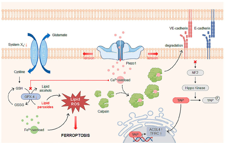
Mechanosensitive ion channels sense mechanical stimuli applied directly to the cellular membranes or indirectly through their tethered components, provoking cellular mechanoresponses. Among others, Piezo1 mechanosensitive ion channel is a relatively novel Ca2+-permeable channel that is primarily present in non-sensory tissues. Recent studies have demonstrated that Piezo1 plays an important role in Ca2+-dependent cell death, including apoptosis and ferroptosis, in the presence of mechanical stimuli. It has also been proven that cancer cells are sensitive to mechanical stresses due to higher expression levels of Piezo1 compared to normal cells. In this review, we discuss Piezo1-mediated cell death mechanisms and therapeutic strategies to inhibit or induce cell death by modulating the activity of Piezo1 with pharmacological drugs or mechanical perturbations induced by stretch and ultrasound. [BMB Reports 2023; 56(3): 145-152].


Conflict of interest statement

CONFLICTS OF INTEREST

The authors have no conflicting interests.

Figures

Fig. 1
Fig. 1
Piezo1-mediated Ca2+-dependent apoptosis mechanism. Piezo1, a mechanosensitive ion channel, opens in response to mechanical (tensile, compressive, shear) forces and physical cues in the extracellular matrix (ECM) that induce membrane tension, allowing Ca2+ to enter the cytosol. Mitochondrial damage caused by Ca2+ overload increases the levels of reactive oxygen species (ROS) that cause endoplasmic reticulum (ER) stress, indicated by upregulation of GRP78 and CHOP. Ca2+ is then released from the stressed ER via ryanodine receptors (RyRs) and inositol-1,4,5-trisphosphate receptors (Ins(1,4,5)P3Rs) and taken up by mitochondria via mitochondrial calcium uniporters (MCU). In addition, cytosolic Ca2+ activates the calpain protease to cleave and translocate p53 and Bcl-2-associated X (BAX) proteins to the mitochondria. Once Ca2+ binds to cardiolipin at the inner mitochondrial membrane, cytochrome c (Cyt c) is dissociated from cardiolipin and the BAX protein weakens mitochondrial outer membrane integrity, then releases Cyt c into the cytosol through the mitochondrial permeability transition pore (MPTP). Subsequently, Cyt c forms an apoptosome complex with apoptotic protease activating factor-1 (APAF-1) and procaspase-9 to promote the activation of caspase-9 and caspase-3 to initiate apoptosis. Piezo1-mediated apoptosis under mechanical stress can be inhibited by treatment with Gd3+ (an inhibitor of mechanically gated cation channels), GsMTx4 (a peptide blocker of Piezo1), or RNA interference (RNAi) to Piezo1, whereas the Piezo1 agonist Yoda1 promotes Piezo1-induced apoptosis by lowering the mechanical activation threshold of Piezo1.
Fig. 2
Fig. 2
Piezo1-mediated Ca2+-dependent ferroptosis mechanism. Piezo1 is activated upon mechanical stimulation and increases the intracellular Ca2+ level, increasing the activity of the calpain protease that degrades (V)E-cadherin. Cells with loss of (V)E-cadherin and subsequent inactivation of NF2 and hippo kinase are susceptible to ferroptosis due to upregulation of multiple regulators of ferroptosis including ACSL4 and TFRC through YAP activation. The cystine/glutamate antiporter system Xc-(consisting of light chain SLC7A11 and heavy chain SLC3A2) mediates uptake of cystine for the production of cysteine and glutathione (GSH), which is converted to glutathione disulfide (GSSG) by glutathione peroxidase-4 (GPX-4), an antioxidant enzyme that metabolizes lipid peroxides into non-toxic lipid alcohols. However, Ca2+ suppresses the expression of GPX-4, which promotes accumulation of lipid peroxides that increase lipid ROS, consequently causing ferroptosis. Otherwise, Fe2+ binds to reactive oxygen species (ROS) to participate in iron-dependent lipid peroxidation and finally induce ferroptosis. Abbreviations: ACSL4, Acyl-CoA Synthetase Long Chain Family Member 4; TFRC, Transferrin Receptor 1.
Fig. 3
Fig. 3
Cancer treatment strategies through the induction of mechanosensitive ion channel-mediated Ca2+-dependent cell death by mechanical perturbations. (A) Microdiscs biofunctionalized with an antibody (Ab) that captures cancer cell surface marker vibrate (i.e., oscillation) under a magnetic field to deliver cytotoxic levels of mechanical force to cancer cells. Interfacing with these microdiscs disrupts membrane integrity and increases intracellular Ca2+ levels, presumably through activation of mechanosensitive (MS) ion channels, eventually leading to programmed cell death in cancer cells. (B) Schematic diagram highlighting the different expression of mechanosensing proteins including Piezo1, tropomyosin 2.1 (Tpm2.1) and myosin IIA between normal and cancer cells. Cancer cells have higher levels of Piezo1 than normal cells, making them more susceptible to apoptosis due to the increase in intracellular Ca2+ levels to the extent of cytotoxicity upon mechanical stimulation. Cyclic stretching stimulates normal cell growth while slowing growth and eventually causing apoptosis in cancer cells. Stretch-induced apoptosis in cancer cells occurs through the Piezo1-mediated Ca2+-dependent mitochondrial apoptosis pathway. Tpm2.1 is eliminated from cancer cells, allowing them to grow anchorage-independently. Cancer cells recovered with Tpm2.1 survive and proliferate upon cyclic stretching, whereas normal cells depleted of Tpm2.1 become susceptible to stretch-induced apoptosis. In addition, restoration of myosin IIA inhibits cancer cell growth, which can induce apoptosis through induction of Piezo1 activation (see Fig. 3C). (C) Piezo1 is activated by ultrasound (US)-mediated mechanical force, triggering Ca2+ influx to activate calpain proteases, which activates the mitochondrial apoptosis pathway and induces microtubule disassembly. Destabilized microtubules enhance myosin IIA contractility through activation of GEF-H1, a Rho guanine nucleotide exchange factor that can increase RhoA activity. Increased contractility, in turn, promotes Piezo1 expression and localization to peripheral adhesions where Piezo1 channel opening allows Ca2+ influx as a positive feedback loop. (D) Cancer cells in contact with nanogels carrying the MscL plasmid and polyethylenamine (PEI) as a transfection reagent are induced to express and activate MscL MS ion channels upon mechanical US waves, resulting in sustained Ca2+ entry to cause apoptosis when ultrasound was applied. Apoptotic cell fragments act as tumor-associated antigens to promote dendritic cell (DC) maturation and subsequent anti-cancer CD8+ T cell activation. Schematic illustrations were recomposed based on key finding in refs. (51) Kim et al. Nat Mater (2010) for (A), (54) Tijore et al. Biomaterials (2021) for (B), (61) Singh et al. Bioeng Transl Med (2021) for (C) and (65) He et al. Chem Eng J (2021) for (D).

Similar articles

Cited by

References

    1. Barnes JM, Przybyla L, Weaver VM. Tissue mechanics regulate brain development, homeostasis and disease. J Cell Sci. 2017;130:71–82. doi: 10.1242/jcs.191742. - DOI - PMC - PubMed
    1. Zhang Y, Su SA, Li W, et al. Piezo1-mediated mechanotransduction promotes cardiac hypertrophy by impairing calcium homeostasis to activate calpain/calcineurin signaling. Hypertension. 2021;78:647–660. doi: 10.1161/HYPERTENSIONAHA.121.17177. - DOI - PubMed
    1. Bonakdar N, Gerum R, Kuhn M, et al. Mechanical plasticity of cells. Nat Mater. 2016;15:1090–1094. doi: 10.1038/nmat4689. - DOI - PubMed
    1. Rafiq NBM, Nishimura Y, Plotnikov SV, et al. A mechano-signalling network linking microtubules, myosin IIA filaments and integrin-based adhesions. Nat Mater. 2019;18:638–649. doi: 10.1038/s41563-019-0371-y. - DOI - PubMed
    1. Sanghvi-Shah R, Weber GF. Intermediate filaments at the junction of mechanotransduction, migration, and development. Front Cell Dev Biol. 2017;5:81. doi: 10.3389/fcell.2017.00081.2b040d4f48e4405a890b6cdf32e0a66c - DOI - PMC - PubMed