

Attenuated Familial Adenomatous Polyposis
- PMID: 36725040
- PMCID: PMC10569909
- DOI: 10.2169/internalmedicine.1101-22
Attenuated Familial Adenomatous Polyposis
Abstract
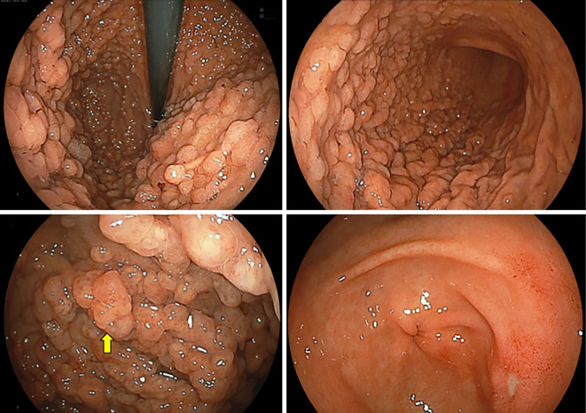
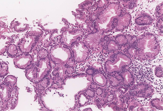
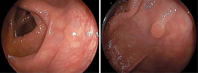
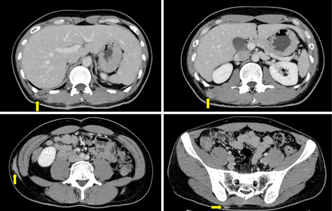
A 36-year-old man was diagnosed with multiple gastric polyps by esophagogastroduodenoscopy. Subsequent colonoscopy identified two tubular adenomas, and computed tomography revealed subcutaneous tumors. Based on these findings, we suspected that gastric polyposis was associated with the APC gene, either attenuated familial adenomatous polyposis (AFAP) or gastric adenocarcinoma and proximal polyposis of the stomach (GAPPS). A genetic analysis demonstrated that he had a frameshift variant at codon 1928 of APC, suggesting AFAP. In this era of less Helicobacter pylori infection and frequent use of proton pump inhibitors, diagnoses of AFAP and GAPPS should be considered in patients with prominent gastric fundic gland polyposis.
Keywords: APC-associated polyposis; attenuated familial adenomatous polyposis; desmoid tumor; gastric adenocarcinoma and proximal polyposis of the stomach.
Conflict of interest statement
Figures
References
-
- Genta RMG. Fundic gland polyps. WHO Classification of Tumours. In: Digestive System Tumours. 5th ed. Lyon, 2019: 67-68.
-
- Burt R. Gastric fundic gland polyps. Gastroenterology 125: 1462-1469, 2003. - PubMed
-
- Jalving M, Koornstra JJ, Wesseling J, Boezen HM, De Jong S, Kleibeuker JH. Increased risk of fundic gland polyps during long‐term proton pump inhibitor therapy. Aliment Pharmacol Ther 24: 1341-1348, 2006. - PubMed