. 2023 Apr;10(10):e2206155.
doi: 10.1002/advs.202206155. Epub 2023 Feb 1.

Engineered Sensory Nerve Guides Self-Adaptive Bone Healing via NGF-TrkA Signaling Pathway

Affiliations

Engineered Sensory Nerve Guides Self-Adaptive Bone Healing via NGF-TrkA Signaling Pathway

Zengjie Zhang et al. Adv Sci (Weinh). 2023 Apr.

Abstract

The upstream role of sensory innervation during bone homeostasis is widely underestimated in bone repairing strategies. Herein, a neuromodulation approach is proposed to orchestrate bone defect healing by constructing engineered sensory nerves (eSN) in situ to leverage the adaptation feature of SN during tissue formation. NGF liberated from ECM-constructed eSN effectively promotes sensory neuron differentiation and enhances CGRP secretion, which lead to improved RAOECs mobility and osteogenic differentiation of BMSC. In turn, such eSN effectively drives ossification in vivo via NGF-TrkA signaling pathway, which substantially accelerates critical size bone defect healing. More importantly, eSN also adaptively suppresses excessive bone formation and promotes bone remodeling by activating osteoclasts via CGRP-dependent mechanism when combined with BMP-2 delivery, which ingeniously alleviates side effects of BMP-2. In sum, this eSN approach offers a valuable avenue to harness the adaptive role of neural system to optimize bone homeostasis under various clinical scenario.

Keywords: BMP-2; extracellular matrix; nerve growth factor; osteogenesis; sensory nerve.


Conflict of interest statement

The authors declare no conflict of interest.

Figures

Scheme 1
Scheme 1
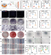
Engineered sensory nerve guides self‐adaptive bone healing via NGF‐TrkA signaling pathway: The adsorption capacity of the acellular scaffold was leveraged to construct a sustained release system of NGF (NGF@S), which promoted the sensory nerve reinnervation at the site of bone tissue injury, and then promoted bone repair. Engineered sensory nerve system sustained‐release NGF to promote sensory nerve reinnervation through NGF‐TrkA signaling pathway and reinnervated sensory nerve secretes CGRP to promote MSC osteogenic differentiation and vascular regeneration in tissues. In addition, sensory nerve regulates osteoblast and osteoclasts to participate in bone remodeling and guides self‐adaptive bone healing.
Figure 1
Figure 1
Active bone formation is highly co‐localized with NGF‐mediated sensory nerve innervation. A,B) Representative tile scans of H&E‐stained images and Masson‐stained images of cranial defect. C,G) IHC was performed on a fracture callus on day 3 after injury, including staining for TUBB, CGRP, and NGF; Semiquantitative analysis of D) TUBB3, E) CGRP, F) CGRP/TUBB3, and H) NGF expression on day 15 after cranial defect. I) Schematic of hypothesis that macrophage secreted NGF to promote sensory nerve re‐innervation, which in turn secrete CGRP to regulate bone regeneration. n = 3 animals per group. Data are represented as mean ± SD.
Figure 2
Figure 2
The construction of NGF@S and their effects for nerve regeneration in vitro. A) Schematic illustration of the construction of the NGF@S scaffold; B) HE‐stained images of ECM and decellularized ECM; C,D) Representative scanning electron microscopy (SEM) of ECM@S and NGF@S; E) NGF release curve of NGF@S in SBF; F) Cell proliferation of Schwann cells co‐cultured with different scaffolds were evaluated by Edu kit. G) Immunofluorescence staining of Tuj‐1 and NF‐200 images on Schwann cells after co‐culture with different scaffolds. I) Immunofluorescence staining of TUBB3 images on Dorsal root ganglion cells after co‐culture with different scaffolds. J,K) CGRP expression and CGRP secretion of Dorsal root ganglion cells with different scaffolds were detected respectively by WB and ELISA. n = 3 times per experiments. Data are represented as mean ± SD.
Figure 3
Figure 3
DRGs secret CGRP to regulate BMSC osteogenic differentiation co‐cultured with NGF@S. A) Schematic illustration of DRGs/scaffolds co‐culture with BMSCs. BMSCs or RAOECs were co‐cultured with DRGs and different scaffolds with or without Olcegepant. B) The proliferation of BMSCs with DRGs/Scaffold system were evaluated by Edu kit. C) Quantification analysis of Edu evaluation. D,E) ALP staining images of BMSCs with DRGs/scaffold system after 7 days co‐culture. F) Quantification analysis of ALP staining. G,H) ARS staining images of BMSCs with DRGs/scaffold system after 14 days and quantification analysis of ARS staining. Osteogenic differentiation related genes mRNA expression of I) OSX, J) RUNX‐2, K) Collagen I, L) ALP, with DRGs/scaffold system after 14 days. M) Representative optical microscopic images of RAOECs tube formation assay at 6 h and 12 h, respectively. N) Quantification analysis of RAOECs tube formation. O) Representative optical microscopic images of scratch line at 0, 5, and 8 h, respectively. P) Quantification analysis of scratch test. n = 3 times per experiments. Data are represented as mean ± SD.
Figure 4
Figure 4
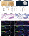
NGF@S promoted scaffold mineralization under subcutaneous. A) Photograph of a scaffold subcutaneous implantation experiment in Balb/c mice. B) 3D reconstruction of micro‐CT images with multiple metrics of µCT quantitative including C) BV, D) BV/TV, E) TB/N. F,G) HE‐stained and Masson‐stained images of subcutaneous tissue in different groups (scale bar:400 µm,Red circle:implaned materials). H) TUBB3 and CGRP immunofluorescence staining of subcutaneous tissues on 30 days after implantation (scale bar: 200 µm or 100 µm). n = 3 animals per time point and per group, each animal was embedded with two scaffolds. Data are represented as mean ± SD.
Figure 5
Figure 5
NGF@S promoted critical size bone defect healing. A) Photograph of a scaffold cranial defect implantation experiment in rats. B) HE‐stained and Masson‐stained images of cranial defect in different groups at 28 days or 56 days after implantation (scale bar: 100 µm), and 3D reconstruction of micro‐CT images with multiple metrics of µCT quantitative including C) BV, D) BV/TV, E) TB/N. F) CD31 immunofluorescence staining of cranial defect in G) different groups and quantified; n = 3 animals with 6 bone defects per time point and per group. Data are represented as mean ± SD.
Figure 6
Figure 6
NGF@S promoted critical size bone defect healing through sensory nerve reinnervation. A) Representative fluorescence images showing TUBB3+ nerve fibers (Green) and CGRP+ nerve fibers (Red) in the cranial bone after scaffold implantation for 15 days. Semiquantitative analysis of B) the fluorescence distribution CGRP, C) TUBB3, and D) ratio of CGRP/TUBB. E) Representative fluorescence of NGF (Green) and nucleus (DAPI, Blue) in cranial bone after scaffold implantation for 28 or 56 days and F,G) quantified. n = 3 animals with 6 bone defects per time point and per group. Data are represented as mean ± SD.
Figure 7
Figure 7
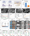
NGF@S optimized BMP‐2 induced bone healing. A) Schematic illustration of the construction of NGF and BMP2 sequential sustained release system (NGF@S/BMP‐2). B) Binding efficiency of mineralized coated microparticles (MCM) with BMP2. C,D) Representative Scanning electron microscope image for MCM and NGF@S/BMP‐2. White scale bar as shown in picture. The characteristic of constructed system was measured, including E) FITR, F) calcium release, G) BMP‐2 release curve, and H) BMP2 and NGF release cumulative release curve. n = 3 times experiments. Data are represented as mean ± SD. I) Schematic illustration of animal experiments. J) Representative photograph and reconstruction µCT images of cranial bone in each group at 28‐ or 56‐days post‐surgery. The bone volume (BV), bone volume fraction (BV/TV), trabecular number (Tb.N), and trabecular separation (Tb.sp) of the different groups. n = 3 animals with 6 bone defects per time point and per group. Data are represented as mean ± SD.
Figure 8
Figure 8
NGF@S optimized BMP‐2 induced bone healing were associated with sensory nerve re‐innervation. A) Representative images of trap staining of cranial bone in different groups and B) quantification of trap positive cells in each group. C) Representative fluorescence images showing TUBB3+ nerve fibers (Green) and CGRP+ nerve fibers (Red) in the cranial bone after scaffold implantation for 30 days. D) 3D reconstruction of micro‐CT images of scaffold implanted subcutaneous with multiple metrics of µCT quantitative including E) BV, F) BV/TV, G) TB/N. H) TUBB3 and CGRP immunofluorescence staining of subcutaneous tissues on 30 days after implantation. n = 3 animals per group. Data are represented as mean ± SD.

References

    1. a) Rupp M., Biehl C., Budak M., Thormann U., Heiss C., Alt V., Int. Orthop. 2018, 42, 247; - PubMed
    2. b) Stewart S. K., Malays Orthop. J. 2019, 13, 1.
    1. a) Dimitriou R., Jones E., McGonagle D., Giannoudis P. V., BMC Med. 2011, 9, 66; - PMC - PubMed
    2. b) Filipowska J., Tomaszewski K. A., Niedźwiedzki Ł., Walocha J. A., Niedźwiedzki T., Angiogenesis 2017, 20, 291. - PMC - PubMed
    1. Freeman F. E., Pitacco P., Dommelen L. H. A., Nulty J., Browe D. C., Shin J.‐Y., Alsberg E., Kelly D. J., Sci. Adv. 2020, 6, eabb5093. - PMC - PubMed
    1. a) Cai B., Lin D., Li Y., Wang L., Xie J., Dai T., Liu F., Tang M., Tian L., Yuan Y., Kong L., Shen S. G. F., Adv. Sci. 2021, 8, 2100584; - PMC - PubMed
    2. b) Mahon O. R., Browe D. C., Gonzalez‐Fernandez T., Pitacco P., Whelan I. T., Euw S. V., Hobbs C., Nicolosi V., Cunningham K. T., Mills K. H. G., Kelly D. J., Dunne A., Biomaterials 2020, 239, 119833; - PubMed
    3. c) Jin S.‐S., He D.‐Q., Luo D., Wang Y., Yu M., Guan B., Fu Y., Li Z.‐X., Zhang T., Zhou Y.‐H., Wang C.‐Y., Liu Y., ACS Nano 2019, 13, 6581; - PubMed
    4. d) Zhao D. W., Zuo K. Q., Wang K., Sun Z. Y., Lu Y. P., Cheng L., Xiao G. Y., Liu C., Mater. Sci. Eng., C 2021, 118, 111512. - PMC - PubMed
    1. a) Abeynayake N., Arthur A., Gronthos S., Bone 2021, 142, 115645; - PubMed
    2. b) Hu C.‐H., Sui B.‐D., Liu J., Dang L., Chen J., Zheng C.‐X., Shi S., Zhao N., Dang M.‐Y., He X.‐N., Zhang L.‐Q., Gao P.‐P., Chen N., Kuang H.‐J., Chen K., Xu X.‐L., Yu X.‐R., Zhang G., Jin Y., Small Methods 2022, 6, 2100763; - PubMed
    3. c) Chen H., Hu B., Lv X., Zhu S., Zhen G., Wan M., Jain A., Gao B., Chai Y., Yang M., Wang X., Deng R., Wang L., Cao Y., Ni S., Liu S., Yuan W., Chen H., Dong X., Guan Y., Yang H., Cao X., Nat. Commun. 2019, 10, 181; - PMC - PubMed
    4. d) Kondo H., Kondo M., Hayashi K., Kusafuka S., Hamamura K., Tanaka K., Kodama D., Hirai T., Sato T., Ariji Y., Miyazawa K., Ariji E., Goto S., Togari A., Eur. J. Orthod. 2022, 44, 404. - PubMed

Publication types

MeSH terms

LinkOut - more resources