. 2023 Feb 1;14(2):75.
doi: 10.1038/s41419-023-05574-w.

SARS-CoV-2 infection induces persistent adipose tissue damage in aged golden Syrian hamsters

Affiliations

SARS-CoV-2 infection induces persistent adipose tissue damage in aged golden Syrian hamsters

Gemma Bogard et al. Cell Death Dis. .

Abstract

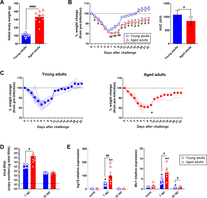
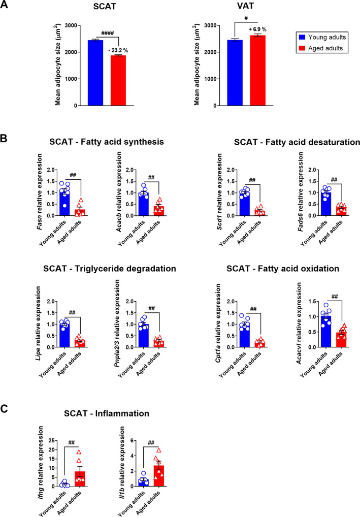
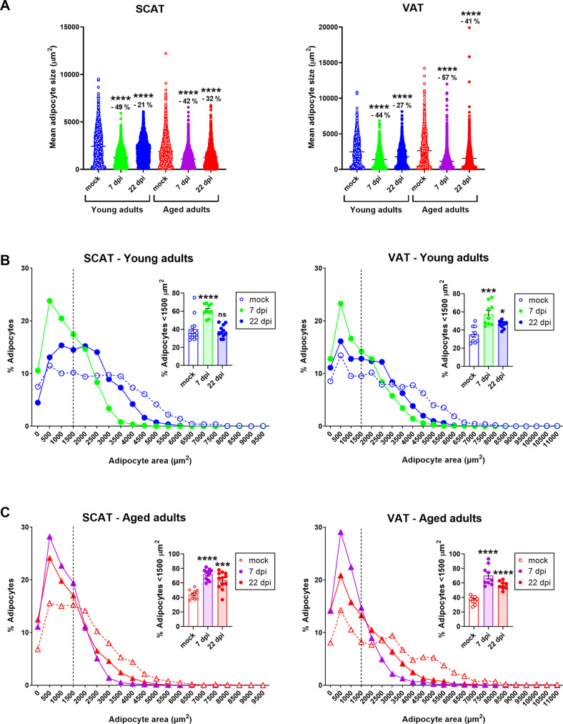
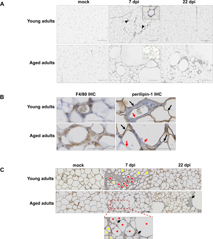
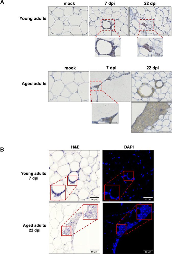
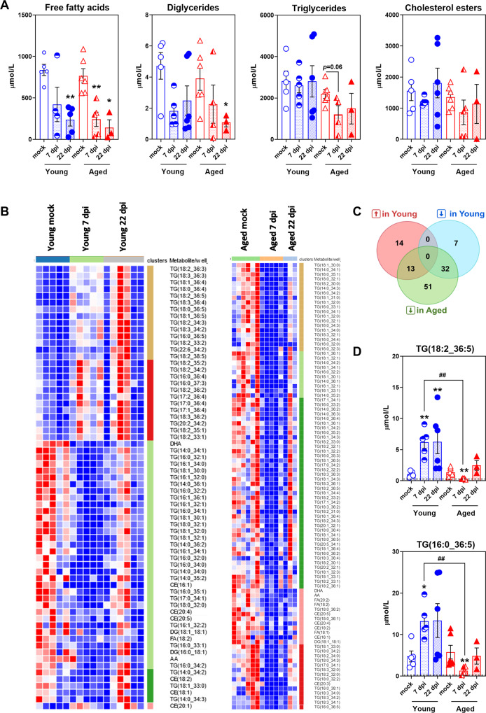
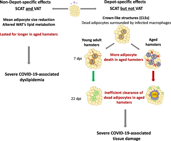
Coronavirus disease 2019 (COVID-19, caused by severe acute respiratory syndrome-coronavirus 2 (SARS-CoV-2)) is primarily a respiratory illness. However, various extrapulmonary manifestations have been reported in patients with severe forms of COVID-19. Notably, SARS-CoV-2 was shown to directly trigger white adipose tissue (WAT) dysfunction, which in turn drives insulin resistance, dyslipidemia, and other adverse outcomes in patients with COVID-19. Although advanced age is the greatest risk factor for COVID-19 severity, published data on the impact of SARS-CoV-2 infection on WAT in aged individuals are scarce. Here, we characterized the response of subcutaneous and visceral WAT depots to SARS-CoV-2 infection in young adult and aged golden hamsters. In both age groups, infection was associated with a decrease in adipocyte size in the two WAT depots; this effect was partly due to changes in tissue's lipid metabolism and persisted for longer in aged hamsters than in young-adult hamsters. In contrast, only the subcutaneous WAT depot contained crown-like structures (CLSs) in which dead adipocytes were surrounded by SARS-CoV-2-infected macrophages, some of them forming syncytial multinucleated cells. Importantly, older age predisposed to a unique manifestation of viral disease in the subcutaneous WAT depot during SARS-CoV-2 infection; the persistence of very large CLSs was indicative of an age-associated defect in the clearance of dead adipocytes by macrophages. Moreover, we uncovered age-related differences in plasma lipid profiles during SARS-CoV-2 infection. These data suggest that the WAT's abnormal response to SARS-CoV-2 infection may contribute to the greater severity of COVID-19 observed in elderly patients.


Conflict of interest statement

The authors declare no competing interests.

Figures

Fig. 1
Fig. 1. SARS-CoV-2 infection is more severe in aged golden hamsters.
Golden (Syrian) hamsters aged 2 months (the “young adults” group) or 22 months (the “aged adults” group) were treated intranasally with a SARS-CoV-2 inoculate (n = 12 young adults and n = 10 aged adults) or with DMEM (mock infection) (n = 6 young adults and n = 6 aged adults). A Body weight (g) on the day of infection. Data are expressed as the mean ± SEM, and individual replicates are shown (n = 12 young adults and n = 10 aged adults). B Percentage body weight change after infection (left). Data are expressed as the mean ± SEM (n = 12 young adults and n = 10 aged adults). The corresponding area under the curve (AUC) is shown (right). Data are expressed in arbitrary units (AU). C Percentage body weight change after infection in individual animals. Median weight losses are depicted as plain circles. Asterisks indicate death. D Quantification of SARS-CoV-2 RNA in the lungs of young adult and aged adult hamsters, using an RT-qPCR assay. Viral RNA levels are expressed as the mean ± SEM copy number/μg of total RNA, and individual replicates are shown (mock and 7 days post infection (dpi): n = 6 animals per group, 22 dpi: n = 6 young adults and n = 3 aged adults). The dashed line represents the assay’s limit of detection. E mRNA expression levels (RT-qPCR assay) of the Isg15 and Mx1 genes in the lungs of young adult and aged adult hamsters, presented according to the 2−ΔΔCT method (housekeeping gene: Actg1, coding for gamma (γ) actin). Data are expressed as the mean ± SEM, and individual replicates are shown (mock and 7 dpi: n = 6 animals per group, 22 dpi: n = 6 young adults and n = 3 aged adults). Groups were compared in a two-sided Mann–Whitney test; # indicates the P values for the comparison of young adults and aged adults (the effect of age: #P < 0.05, ##P < 0.01 and ####P < 0.0001), and * indicates the P values for the comparison of mock-treated and SARS-CoV-2-infected groups (the effect of infection: *P < 0.05 and **P < 0.01). For intergroup differences, the threshold for statistical significance was set to P < 0.05.
Fig. 2
Fig. 2. Aging has a regional impact on WAT in golden hamsters.
A Mean ± SEM adipocyte size (μm2) in the (inguinal) SCAT and (epididymal) VAT of mock-treated young adult and aged hamsters. For SCAT, 2052 adipocytes in young adults and 2621 in aged adults were measured. For VAT, 1531 adipocytes in young adults and 1412 in aged adults were measured. Percentage differences in adipocyte size between young adult and aged hamsters are indicated. B mRNA expression levels (RT-qPCR assay) of Fasn and Acacb (involved in fatty acid synthesis), Scd1 and Fasd6 (involved in fatty acid desaturation), Lipe and Pnpla2/3 (involved in triglyceride degradation), and Cpt1a and Acadvl (involved in fatty acid oxidation) in the SCAT of mock-treated young adult and aged hamsters. C mRNA expression levels (RT-qPCR assay) of the inflammatory genes Ifng and Il1b in the SCAT of mock-treated young adult and aged hamsters. B, C Relative expression is presented as 2−ΔΔCT (housekeeping gene: GusB, coding for glucuronidase beta). Data are expressed as the mean ± SEM, and individual replicates are shown (n = 6 animals per group). Groups were compared in a two-sided Mann–Whitney test; # indicates the P values for the comparison of young adults and aged adults (the effect of age: #P < 0.05, ##P < 0.01 and ####P < 0.0001). For intergroup differences, the threshold for statistical significance was set to P < 0.05.
Fig. 3
Fig. 3. SARS-CoV-2 infection induces a persistent decrease in adipocyte size in SCAT and VAT depots in young adult and aged golden hamsters.
Adipocyte size and distribution in the (inguinal) SCAT and (epididymal) VAT were determined by quantitative histomorphometry, at 0 (mock), 7, and 22 dpi in both age groups. A Superplots showing the size of individual adipocytes, as well as the mean values (μm2), in the SCAT (left) and the VAT (right) of young adult and aged hamsters at 0 (mock), 7, and 22 dpi. Percentage differences in adipocyte size are indicated. B Adipocyte size distribution (%) in the SCAT (left) and VAT (right) of young-adult hamsters. The relative frequency of adipocytes <1500 μm2 is shown in the insert (mean ± SEM, and individual replicates are shown). C Adipocyte size distribution (%) in the SCAT (left) and VAT (right) of aged hamsters. The relative frequency of adipocytes <1500 μm2 is shown in the insert (mean ± SEM, and individual replicates are shown). AC n = 3 animals per group, and a mean (range) of 1035 (649–1500) adipocytes per tissue sample were analyzed. Groups were compared in a two-sided Mann–Whitney test; # indicates the P values for the comparison of young adults and aged adults (the effect of age: #P < 0.05, ##P < 0.01 and ###P < 0.001) and * indicates the P values for the comparison of mock-treated and SARS-CoV-2-infected groups (the effect of infection: *P < 0.05 and **P < 0.01). For intergroup differences, the threshold for statistical significance was set to P < 0.05.
Fig. 4
Fig. 4. SARS-CoV-2 infection induces adipocyte death in the SCAT, and dead adipocytes are not cleared in aged golden hamsters.
A Representative microscopy images of the (inguinal) SCAT of young adult and aged hamsters at 0 (mock), 7, and 22 dpi (H&E staining). At 7 dpi, one can note the presence of CLSs in the SCAT of young-adult animals, and the presence of fibrosis in the SCAT of the aged animals (inserts: higher magnification). B Representative immunochemical (IHC) staining for F4/80 (left panels) highlighted the presence of macrophages (in brown) clustered around adipocytes. Representative IHC staining for lipid-droplet-specific perilipin-1 (right panels) highlighted live (stained) adipocytes (in brown, black arrows) and damaged/dead (non-stained, red arrows) adipocytes in the SCAT of young adults and aged adults at 7 dpi. C Representative images of dead adipocytes in the SCAT of young adult and aged hamsters at 0 (mock), 7, and 22 dpi. IHC staining for perilipin-1 shows live (stained) adipocytes. A higher magnification image is shown for aged adult animals at 7 dpi. Live adipocytes are indicated by yellow stars, and damaged/dead adipocytes are indicated by red stars. Lipid spillovers are indicated by black arrows. AC scale bars = 100 μm. B, C Slides were counterstained with Mayer’s hematoxylin.
Fig. 5
Fig. 5. In the SCAT of golden hamsters, the macrophages around dead adipocytes are infected with SARS-CoV-2 and formed syncytia.
A Representative IHC staining for SARS-CoV-2 spike protein (brown precipitate) in the (inguinal) SCAT of young-adult and aged hamsters at 0 (mock), 7 and 22 dpi (scale bars = 100 μm). Higher magnification images of CLSs are shown (scale bars = 50 μm). Slides were counterstained with Mayer’s hematoxylin. B The same CLSs found in the (inguinal) SCAT of young-adult and aged hamsters at, respectively, 7 and 22 dpi, were H&E-stained (left panels) or DAPI-stained (right panels) (scale bars = 50 μm). In left panels, the blue fluorescence indicates DAPI staining for cell nuclei, and higher magnifications are shown that revealed the presence of multinucleated cells.
Fig. 6
Fig. 6. SARS-CoV-2 infection differently disturbs plasma lipid metabolism in young-adult and aged golden hamsters.
Plasma levels of free fatty acids (FFAs), diglycerides (DGs), cholesterol esters (CEs) and triglycerides (TGs) were quantified by ultrahigh-pressure liquid chromatography-tandem mass spectrometry (UPLC-MS/MS) in young-adult and aged hamsters at 0 (mock), 7 and 22 dpi. A Plasma levels of FFAs (n = 5 molecules), DGs (n = 4 molecules), TGs (n = 154 molecules) and CEs (n = 16 molecules) at days 0 (mock), 7 and 22 post-SARS-CoV-2 infection. Data are expressed as mean ± SEM μmol/L, and individual replicates are shown. B Heatmaps of significantly changed lipid species (fold change >1.5, P < 0.05) at 7 dpi in young-adult hamsters and aged hamsters (Young adults: n = 66 lipid species (27 increased, 39 decreased), and aged adults: n = 96 lipid species (all decreased)). Differential intensities in red and blue colors denote increased or reduced levels, respectively (less concentrated: dark blue, most concentrated: dark red). C Venn diagrams (https://bioinformatics.psb.ugent.be/) showing the number of distinct or overlapping lipid species that are increased (↑) or decreased (↓) upon SARS-CoV-2 infection (7 dpi) in young-adult and aged hamsters. D Plasma levels of TG(18:2_36:5) and TG(16:0_36:5) in young-adult and aged hamsters at days 0 (mock), 7 and 22 post-SARS-CoV-2 infection. Data are expressed as mean ± SEM mmol/L, and individual replicates are shown. Dotted lines indicate the limit of detection (LOD) of the assay for each lipid species. AD n = 5 young adults mock, n = 6 aged adults mock, n = 5 young adults 7 dpi, n = 5 aged adults 7 dpi, n = 6 young adults 22 dpi, and n = 3 aged adults 22 dpi. Young = young-adult hamsters, Aged = aged adult hamsters. A, D Groups were compared in a two-sided Mann–Whitney test; # indicates the P values for the comparison of young adults and aged adults (the effect of age: ##P < 0.01) and * indicates the P values for the comparison of mock-treated and SARS-CoV-2-infected groups (the effect of infection: *P < 0.05 and **P < 0.01). For intergroup differences, the threshold for statistical significance was set to P < 0.05.
Fig. 7
Fig. 7. Age-related differences in WAT’s response to SARS-CoV-2 infection in golden hamsters.
Schematic diagram showing the non-depot-specific (left) and the depot-specific (right) effects of a SARS-CoV-2 infection on WAT in young-adult vs. aged golden hamsters. The viral infection perturbed lipid metabolism in both the (inguinal) SCAT and the (epididymal) VAT but induced CLS formation—indicative of infection-induced adipocyte death—in SCAT only. Importantly, massive adipose death was evident in the SCAT of aged hamsters. The repair phase post infection-induced injury was effective in young adults but not in aged animals. We hypothesize that the accumulation of tissue damage in the SCAT of aged animals results from both the defective clearance of dead adipocytes by surrounding macrophages having age-related compromised efferocytic capacity, and/or the absence of replacement of dead adipocytes by newly formed adipocytes due to age-related impaired adipogenesis. CLSs crown-like structures. Preadipocytes are shown in red, and newly formed adipocytes are shown in green. Both non-depot-specific and depot-specific WAT remodeling upon SARS-CoV-2 infection might participate to COVID-19 severity in aged individuals. Created with BioRender.com.

References

    1. Wiersinga WJ, Rhodes A, Cheng AC, Peacock SJ, Prescott HC. Pathophysiology, transmission, diagnosis, and treatment of coronavirus disease 2019 (COVID-19): a review. J Am Med Assoc. 2020;324:782–93. - PubMed
    1. Zaim S, Chong JH, Sankaranarayanan V, Harky A. COVID-19 and multiorgan response. Curr Probl Cardiol. 2020;45:100618. - PMC - PubMed
    1. Robba C, Battaglini D, Pelosi P, Rocco PRM. Multiple organ dysfunction in SARS-CoV-2: MODS-CoV-2. Expert Rev Respir Med. 2020;14:865–8. - PMC - PubMed
    1. Sia SF, Yan LM, Chin AWH, Fung K, Choy KT, Wong AYL, et al. Pathogenesis and transmission of SARS-CoV-2 in golden hamsters. Nature. 2020;583:834–8. - PMC - PubMed
    1. Chan JFW, Zhang AJ, Yuan S, Poon VKM, Chan CCS, Lee ACY, et al. Simulation of the clinical and pathological manifestations of Coronavirus Disease 2019 (COVID-19) in golden Syrian hamster model: implications for disease pathogenesis and transmissibility. Clin Infect Dis Publ Infect Dis Soc Am [Internet] 2020;71:2428–46. - PMC - PubMed

Publication types