Review
. 2023 Jul;23(7):415-432.
doi: 10.1038/s41577-022-00821-1. Epub 2023 Feb 1.

The therapeutic age of the neonatal Fc receptor

Affiliations
Review

The therapeutic age of the neonatal Fc receptor

Michal Pyzik et al. Nat Rev Immunol. 2023 Jul.

Abstract

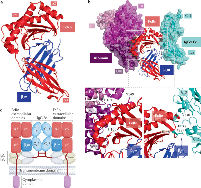
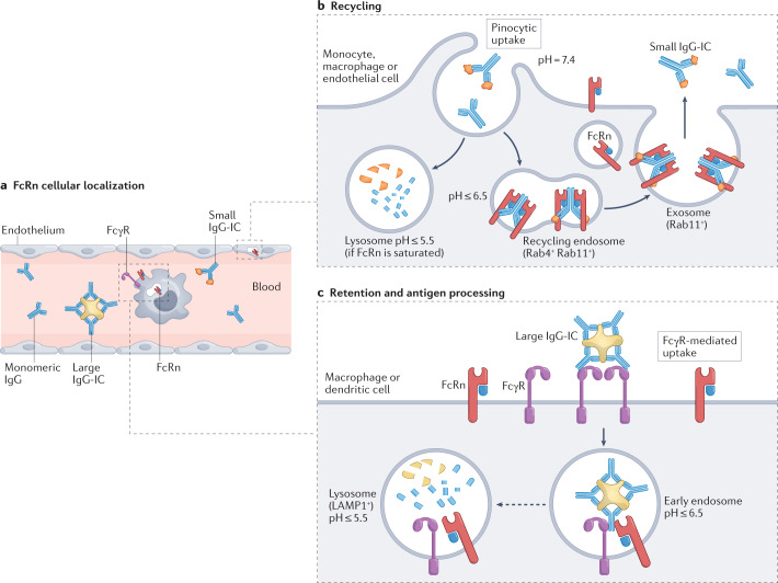
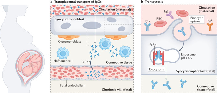
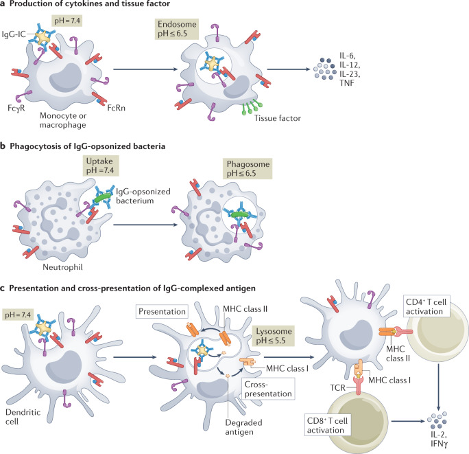
IgGs are essential soluble components of the adaptive immune response that evolved to protect the body from infection. Compared with other immunoglobulins, the role of IgGs is distinguished and enhanced by their high circulating levels, long half-life and ability to transfer from mother to offspring, properties that are conferred by interactions with neonatal Fc receptor (FcRn). FcRn binds to the Fc portion of IgGs in a pH-dependent manner and protects them from intracellular degradation. It also allows their transport across polarized cells that separate tissue compartments, such as the endothelium and epithelium. Further, it is becoming apparent that FcRn functions to potentiate cellular immune responses when IgGs, bound to their antigens, form IgG immune complexes. Besides the protective role of IgG, IgG autoantibodies are associated with numerous pathological conditions. As such, FcRn blockade is a novel and effective strategy to reduce circulating levels of pathogenic IgG autoantibodies and curtail IgG-mediated diseases, with several FcRn-blocking strategies on the path to therapeutic use. Here, we describe the current state of knowledge of FcRn-IgG immunobiology, with an emphasis on the functional and pathological aspects, and an overview of FcRn-targeted therapy development.


Conflict of interest statement

R.S.B. had equity interests in Syntimmune Inc., a company developing therapeutic agents to target FcRn. Syntimmune Inc. was a wholly owned subsidiary of Alexion Pharmaceuticals Inc., which was acquired by AstraZeneca. M.P., L.K.K. and A.G. declare no competing interests.

Figures

Fig. 1
Fig. 1. Structure of FcRn and its ligands.
a, Ribbon diagram of the human neonatal Fc receptor (FcRn) heavy chain (red) with indicated α1, α2 and α3 domains, and β2-microglobulin (β2m; blue) light chain (Protein Data Bank (PDB) ID 1EXU). b, Ternary complex between human FcRn, human albumin (purple) and portion of human IgG1 Fc fragment (cyan) (PDB ID 4N0U). Albumin and half of the IgG1 Fc in ribbon and surface diagrams are shown with the indicated domains: albumin domains I (DI, purple), II (DII, light purple) and III (DIII, dark purple), and IgG1 Fc domains CH2 and CH3. Bottom-right inset box shows FcRn residues E115, E116, D130 and E133 binding with Fc residues H310 and H435 (not shown). Bottom-left inset box shows that, at acidic pH, the presence of H166 leads to intramolecular hydrogen bonding, which facilitates optimal binding of human FcRn residues W53 and W59 with albumin. Human FcRn residues S58 (not shown), N149 and H161 make various hydrogen-bonded interactions with albumin DI whereas FcRn residues W53 and W59 engage albumin DIII. c, Cartoon representation of two FcRn–β2m heterodimers engaging monomeric IgG (in a T conformation), which presumably occurs on the membrane of acidified recycling endosomes. A short stalk, the transmembrane domain and the cytoplasmic domain are also depicted. Fab, antigen-binding fragment.
None
a, Cryogenic electron microscopy structures of human FcRn in complex with echovirus 6 nucleocapsid (Protein Data Bank IDs 6ILL, 6ILM). The icosahedral nucleocapsid consisting of viral protein 1 (VP1, green) and viral protein 2 (VP2, light green) are shown by surface and ribbon representation. Human FcRn α2 domain binds to echovirus 6 at a site between VP1 and VP2. b, FcRn α2 domain residues Q139, R140, Q142, Q143, D145, K146 and N149 mediate most of the interactions with VP1 residues.
Fig. 2
Fig. 2. FcRn-mediated recycling provides a long half-life to monomeric IgG or small IgG immune complexes whereas multimeric IgG immune complexes are degraded.
a, IgG recycling occurs within macrophages, monocytes and endothelial cells, based on mouse studies. b, Monomeric IgGs and small IgG immune complexes (IgG-ICs) consisting of a single antibody engaging two antigens are taken up by pinocytosis as they can not bind to neonatal Fc receptor (FcRn) at the neutral pH of the cell surface. Following the formation of an early endosome and its acidification, FcRn can bind to IgGs, which are diverted into recycling endosomes (Rab4+Rab11+) and exocytosed within Rab11+ exosomes, where the IgGs or small IgG-ICs are released from FcRn at neutral pH, extending their half-life in the body, while FcRn is recovered for another round of recycling. When FcRn is saturated, the excess unbound antibody is degraded in the lysosome. c, FcRn retains and regulates the degradation of multimeric IgG-ICs. Multimeric IgG-ICs bind Fc receptors for IgG (FcγRs) on the cell surface (neutral pH), where they are taken up into endosomes by receptor-mediated endocytosis allowing for co-engagement with FcRn and FcγRs at acidic pH. The multimeric IgG-ICs are retained in a FcRn+LAMP1+ lysosomal compartment (pH ~5.5) enriched in antigen presentation machinery (dashed arrow). It is not well understood how FcRn differentiates between the recycling and retention pathways. LAMP1, lysosomal-associated membrane protein 1.
Fig. 3
Fig. 3. FcRn-mediated transplacental transfer of maternal IgG to the offspring.
a, In humans, maternal and fetal circulations are separated by a single layer of polarized epithelium called the syncytiotrophoblast, which the antibody must intially pass to reach the fetus. b, Transport of IgGs is largely mediated through the presence of neonatal Fc receptor (FcRn) at this site. The initial step of internalization of maternal IgGs is thought to occur via fluid-phase pinocytosis. The formed vesicles containing IgGs then fuse with endosomes, where at a mildly acidic pH, the interaction with FcRn allows transport of the antibody to the basolateral membrane and release into the stroma at neutral pH. From there, IgG is hypothesized to passively diffuse and reach fetal endothelial cells; whether a similar FcRn-dependent transcytosis of IgG across fetal endothelium occurs in vivo is currently unknown. RBC, red blood cell.
Fig. 4
Fig. 4. Active immune functions of FcRn in innate immune cells.
a, IgG immune complexes (IgG-ICs) bind Fc receptors for IgG (FcγRs) on the cell surface at neutral pH. As the IgG-ICs are taken up into endosomes, they engage neonatal Fc receptor (FcRn) at a slightly acidic pH. In response to IgG-ICs, FcRn actively induces the production of pro-inflammatory cytokines IL-12, IL-23, tumour necrosis factor (TNF) and IL-6 by neutrophils, monocytes, macrophages and dendritic cells and the induction of tissue factor expression in monocytes and macrophages, through co-operation with FcγRs. b, FcRn promotes phagocytosis of IgG-coated Streptococcus pneumoniae in neutrophils. The IgG-opsonized bacteria bind to the neutrophil cell surface at neutral pH, likely by FcγRs, whereas FcRn binds to the IgGs at a slightly acidic pH and actively enhances phagocytosis. c, Once IgG-ICs are taken up by FcγR-expressing cells, FcRn enhances both antigen presentation to CD4+ T cells by dendritic cells and macrophages, and antigen cross-presentation to CD8+ T cells by dendritic cells. FcRn-associated antigen presentation by MHC class II occurs via degradation and peptide loading within the acidic endosome, whereas antigen cross-presentation by MHC class I occurs via a separate cytoplasmic and proteasomal processing pathway. The active participation of FcRn in these antigen-presenting pathways results in greatly enhanced T cell activation, expansion and production of IL-2 and interferon-γ (IFNγ) by the interacting immune cells, which probably further amplifies the immune response. TCR, T cell receptor.

References

    1. Akula S, Mohammadamin S, Hellman L. Fc receptors for immunoglobulins and their appearance during vertebrate evolution. PLoS ONE. 2014;9:e96903. doi: 10.1371/journal.pone.0096903. - DOI - PMC - PubMed
    1. Delidakis G, Kim JE, George K, Georgiou G. Improving antibody therapeutics by manipulating the Fc domain: immunological and structural considerations. Annu. Rev. Biomed. Eng. 2022;24:249–274. doi: 10.1146/annurev-bioeng-082721-024500. - DOI - PMC - PubMed
    1. Ward ES, et al. Clinical significance of serum albumin and implications of FcRn inhibitor treatment in IgG-mediated autoimmune disorders. Front. Immunol. 2022;13:892534. doi: 10.3389/fimmu.2022.892534. - DOI - PMC - PubMed
    1. Bern M, Sand KM, Nilsen J, Sandlie I, Andersen JT. The role of albumin receptors in regulation of albumin homeostasis: implications for drug delivery. J. Control. Rel. 2015;211:144–162. doi: 10.1016/j.jconrel.2015.06.006. - DOI - PubMed
    1. Kandil E, et al. The human gene encoding the heavy chain of the major histocompatibility complex class I-like Fc receptor (FCGRT) maps to 19q13.3. Cytogenetics Cell Genet. 1996;73:97–98. doi: 10.1159/000134316. - DOI - PubMed

Publication types