

A human T-lymphotropic virus-1 carrier who developed progressive multifocal leukoencephalopathy following immunotherapy for sarcoidosis: a case report
- PMID: 36726087
- PMCID: PMC9893603
- DOI: 10.1186/s12883-023-03094-w
A human T-lymphotropic virus-1 carrier who developed progressive multifocal leukoencephalopathy following immunotherapy for sarcoidosis: a case report
Abstract
Background: Progressive multifocal leukoencephalopathy (PML) is a devastating demyelinating disorder of the central nervous system caused by opportunistic infection of the JC virus (JCV).
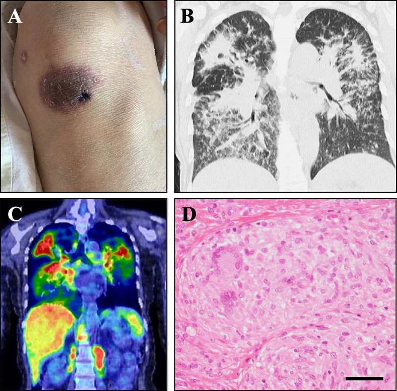
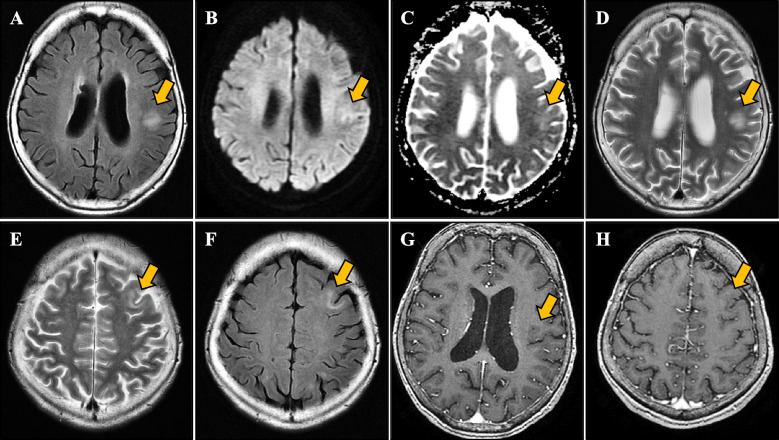
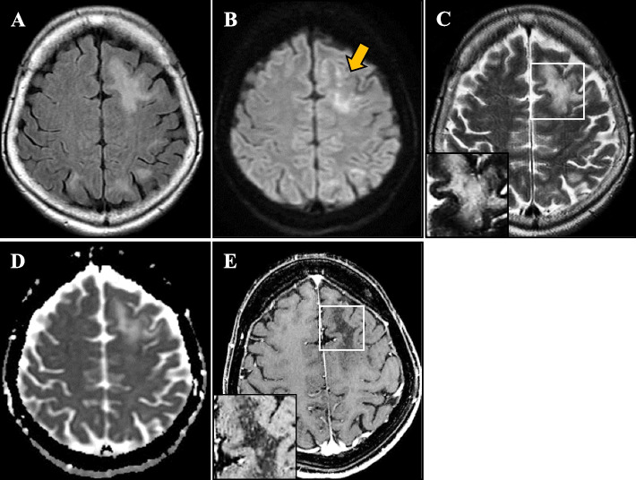
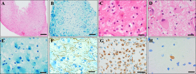
Case presentation: A 58-year-old Japanese woman was admitted to our hospital for aphasia. She had a 5-year history of untreated sarcoidosis and was a human T cell lymphotropic virus-1 (HTLV-1) carrier. Serum angiotensin-converting enzyme, soluble interleukin-2 receptor, lysozyme, and calcium levels were elevated. JCV-DNA was not detected in cerebrospinal fluid by PCR testing. Skin biopsy revealed noncaseating granuloma formation. Bilateral multiple nodular lesions were present on chest X-ray. Brain magnetic resonance imaging showed left frontal and temporal lesions without gadolinium enhancement. As we suspected that systemic sarcoidosis had developed into neurosarcoidosis, we started steroid and infliximab administration. After treatment, the chest X-ray and serum abnormalities ameliorated, but the neurological deficits remained. At 1 month after immunotherapy, she developed right hemiparesis. Cerebrospinal fluid was positive for prototype (PML-type) JCV on repeated PCR testing. Brain biopsy revealed demyelinating lesions with macrophage infiltration, atypical astrocytes, and JCV antigen-positive cells. We diagnosed her with PML and started mefloquine, leading to partial remission.
Conclusions: Sarcoidosis and HTLV-1 infection both affect T cell function, especially CD4+ T cells, and may developped the patient's PML. The comorbidity of sarcoidosis, PML, and HTLV-1 infection has not been reported, and this is the world's first report of PML associated with HTLV-1 infection and sarcoidosis.
Keywords: Case report; Demyelination; Human T-lymphotropic virus-1 (HTLV-1); Progressive multifocal leukoencephalopathy (PML); Sarcoidosis.
© 2023. The Author(s).
Conflict of interest statement
The authors declare that they have no competing interests.
Figures
Similar articles
-
Inflammatory progressive multifocal leukoencephalopathy with human T-cell lymphotropic virus-1 coinfection.BMJ Case Rep. 2024 Apr 30;17(4):e257805. doi: 10.1136/bcr-2023-257805. BMJ Case Rep. 2024. PMID: 38688573
-
[A case of progressive multifocal leukoencephalopathy presenting white matter MRI lesions extending over the cerebral cortex and a marked decrease in cerebral blood flow on SPECT, and associated with HTLV-I infection].Rinsho Shinkeigaku. 2005 Jun;45(6):426-30. Rinsho Shinkeigaku. 2005. PMID: 16022467 Review. Japanese.
-
Clearance of cerebrospinal fluid JCV DNA with mirtazapine in a patient with progressive multifocal leukoencephalopathy and sarcoidosis.Antivir Ther. 2016;21(7):633-635. doi: 10.3851/IMP3032. Epub 2016 Feb 9. Antivir Ther. 2016. PMID: 26857363
-
Case report: Progressive multifocal leukoencephalopathy co-occurring with neurosarcoidosis: early brain biopsy and appropriate therapy for PML resulted in a favorable prognosis.Front Immunol. 2024 Oct 11;15:1447992. doi: 10.3389/fimmu.2024.1447992. eCollection 2024. Front Immunol. 2024. PMID: 39464878 Free PMC article.
-
Probable progressive multifocal leukoencephalopathy-immune reconstitution inflammatory syndrome with immunosuppressant dose reduction following lung transplantation: a case report and literature review.BMC Neurol. 2019 Oct 31;19(1):263. doi: 10.1186/s12883-019-1493-1. BMC Neurol. 2019. PMID: 31672142 Free PMC article. Review.
Cited by
-
Inflammatory progressive multifocal leukoencephalopathy with human T-cell lymphotropic virus-1 coinfection.BMJ Case Rep. 2024 Apr 30;17(4):e257805. doi: 10.1136/bcr-2023-257805. BMJ Case Rep. 2024. PMID: 38688573
References
Publication types
MeSH terms
Substances
Grants and funding
LinkOut - more resources
Full Text Sources
Medical
Research Materials
