Review
. 2023 Apr;102(4):364-375.
doi: 10.1177/00220345221145555. Epub 2023 Feb 1.

The Application of Organs-on-a-Chip in Dental, Oral, and Craniofacial Research

Affiliations
Review

The Application of Organs-on-a-Chip in Dental, Oral, and Craniofacial Research

C Huang et al. J Dent Res. 2023 Apr.

Abstract

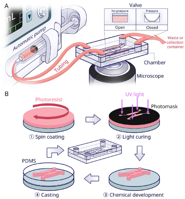
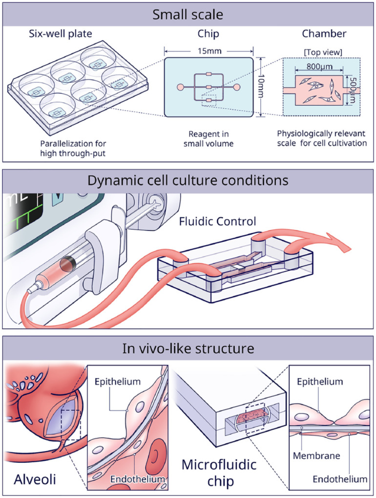
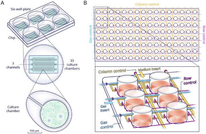
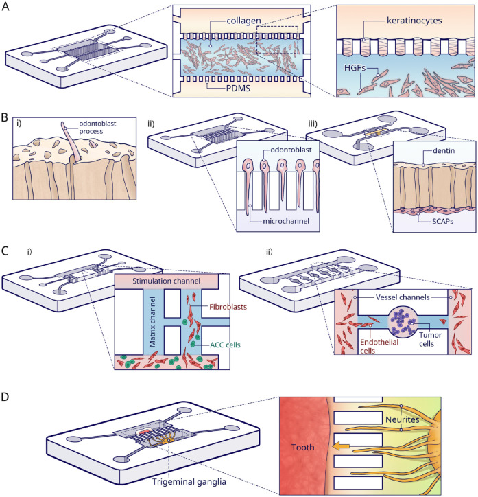
The current development of microfluidics-based microphysiological systems (MPSs) will rapidly lead to a paradigm shift from traditional static 2-dimensional cell cultivation towards organized tissue culture within a dynamic cellular milieu. Especially organs-on-a-chip (OoCs) can very precisely re-create the mechanical and unique anatomical structures of the oral environment. This review provides an introduction to such technology, from commonly used chip materials and fabrication methods to the application of OoC in in vitro culture. OoCs are advantageous because of their small-scaled culture environment, the highly controlled dynamic experimental conditions, and the likeness to the in vivo structure. We specifically focus on current chip designs in dental, oral, and craniofacial (DOC) research. Also, future perspectives are discussed, like model standardization and the development of integrated platforms with advanced read-out functionality. By doing so, it will be possible for OoCs to serve as an alternative for animal testing and to develop highly predictive human models for clinical experiments and even personalized medicine.

Keywords: biofilm(s); dentin; mineralized tissue/development; mucosal immunity; odontoblast(s); pulp biology.


Conflict of interest statement

The authors declared no potential conflicts of interest with respect to the research, authorship, and/or publication of this article.

Figures

Figure 1.
Figure 1.
Overview of microfluidic chip technology. (A) Schematic of an automated microfluidic chip with monitoring microscope. The basic chip components include an inlet and outlet, cell culture chamber, and media transportation channel. A pump and valve are used for fluidic control in the chip. The valve is usually controlled by pressure. (B) Fabrication flow of a polydimethylsiloxane (PDMS) chip (adapted from Scott and Ali 2021). The so-called soft lithography production method, first, a photoresist material (pink), is spin-coated on a (usually silicon) substrate (gray). By UV irradiation through a photomask (black), the desired pattern is transferred onto the photoresist-coated substrate. The exposed part is subsequently cured and the non-cross-linked resist is removed. Thus, a master mold is fabricated. From the mold, PDMS casting leads to the correct microfluidic architecture. Finally, after sealing the channels and chambers by a cover, the PDMS chip is completed.
Figure 2.
Figure 2.
The 3 main advantages of using microfluidic chips for in vitro culture are the small scale of the models, the high control over dynamic cell culture conditions, and the possibility to efficiently construct in vivo–like structures.
Figure 3.
Figure 3.
Multiarray chips. (A) Schematic view of a multiarray chip with 99 chambers distributed in 3 independent channels evenly. By adding 1 chip to each well of a 6-well culture plate, 18 different conditions are supported. Lower panel shows that bacterial exclusion zones were assessed in the small-scale chambers when coculturing different bacteria. (B) Schematic of a high-throughput platform with 128 chambers (8 rows × 16 columns). The 8 chambers in each column are connected by media-transporting channels (yellow). The column control and row valves (purple) are intended to control the media insertion in each individual chamber. In addition, dissolved oxygen conditions can be controlled through a gas insert (blue) in combination with control valves.
Figure 4.
Figure 4.
Parallel-chamber chips. (A) In the mucosa-on-a-chip, fibroblasts were seeded in collagen in the central channel, and keratinocytes were grown on top. The upper channel was used for the insertion of dental materials, and the bottom channel was for the media. (B) Dentin-on-a-chip and tooth-on-a-chip models. i) Illustration of a functional dentin–pulp complex. ii) For the dentin-on-a-chip system, microchannels were made to induce odontoblast processes. iii) Tooth-on-a-chip with a native dentin disc inserted in between 2 channels. Pulp cells were seeded in 1 channel and adhered to the dentin. The opposite channel was used to provide exogenous oral components. (C) Adenoid cystic carcinoma (ACC)-on-a-chip model. i) ACC-related fibroblasts were cocultured with ACC cells in the bottom channel. The media in the upper channel induced cells to migrate through the vertical channel. The invasion pattern in the vertical channel showed that fibroblasts (red) localized at the invasion front and ACC cells (green) following behind. ii) Another ACC-on-a-chip for the investigation of tumor-induced angiogenesis. The ACC cells were seeded in the round chamber and the Human umbilical vein endothelial cells (HUVECs) were seeded in the vessel channels. The angiogenesis was then tracked in the side channels. (D) Tooth innervation on a microfluidic chip. Trigeminal ganglia and tooth tissue were cultured in parallel chambers in different media. The neurites (yellow) are growing toward the tooth (red) through the microgrooves.
Figure 5.
Figure 5.
Serial-chamber chips. (A) Schematic top and cross-section view of the immunosystem-on-a-chip. Media (M) flow direction was from reconstructed human gingiva (RHG) to reconstructed human skin with Langerhans cells (RHS-LC). After culture in the chip, the RHG were exposed to nickel sulfate (NiSO4) and then used for the analysis. (B) Representation of the digestive-track-on-chip. This chip consists of a mouth, stomach, and intestine chamber. In each chamber, the flow was mixed with different digestive juices in physiologically relevant ratios. Connected to the outlet of each compartment, tubing loops were used for incubation. Samples can be collected at different time points in these loops to closely assess the whole digestive progress.

References

    1. Ahadian S., Civitarese R., Bannerman D., Mohammadi M. H., Lu R., Wang E., Davenport-Huyer L., Lai B., Zhang B., Zhao Y., Mandla S., Korolj A., Radisic M., Adv. Healthcare Mater. 2018, 7, 1700506. - PubMed
    1. Ai X, Lu W, Zeng K, Li C, Jiang Y, Tu P. 2018. Microfluidic coculture device for monitoring of inflammation-induced myocardial injury dynamics. Anal Chem. 90(7):4485–4494. - PubMed
    1. Alvarez MMP, Moura GE, Machado MFM, Viana GM, Costa CAD, Tjaderhane L, Nader HB, Tersariol ILS, Nascimento FD. 2017. Par-1 and par-2 expression is enhanced in inflamed odontoblast cells. J Dent Res. 96(13):1518–1525. - PubMed
    1. Atif AR, Pujari-Palmer M, Tenje M, Mestres G. 2021. A microfluidics-based method for culturing osteoblasts on biomimetic hydroxyapatite. Acta Biomater. 127:327–337. - PubMed
    1. Cao X, Ashfaq R, Cheng F, Maharjan S, Li J, Ying G, Hassan S, Xiao H, Yue K, Zhang YS. 2019. A tumor-on-a-chip system with bioprinted blood and lymphatic vessel pair. Adv Funct Mater. 29(31):1807173. - PMC - PubMed

Publication types

LinkOut - more resources