

Reorganization, specialization, and degradation of oocyte maternal components for early development
- PMID: 36726596
- PMCID: PMC9884333
- DOI: 10.1002/rmb2.12505
Reorganization, specialization, and degradation of oocyte maternal components for early development
Abstract
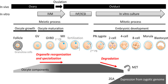
Background: Oocyte components are maternally provided, solely determine oocyte quality, and coordinately determine embryo quality with zygotic gene expression. During oocyte maturation, maternal organelles are drastically reorganized and specialized to support oocyte characteristics. A large number of maternal components are actively degraded after fertilization and gradually replaced by zygotic gene products. The molecular basis and the significance of these processes on oocyte/embryo quality are not fully understood.
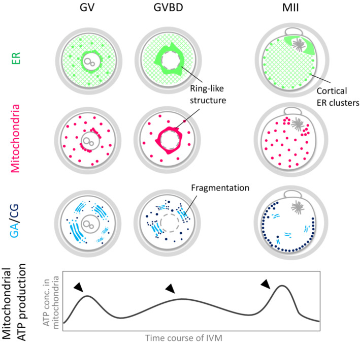
Methods: Firstly, recent findings in organelle characteristics of other cells or oocytes from model organisms are introduced for further understanding of oocyte organelle reorganization/specialization. Secondly, recent progress in studies on maternal components degradation and their molecular mechanisms are introduced. Finally, future applications of these advancements for predicting mammalian oocyte/embryo quality are discussed.
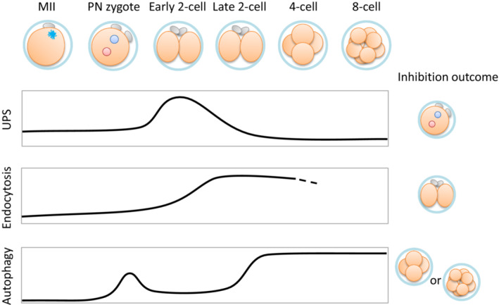
Main findings: The significance of cellular surface protein degradation via endocytosis for embryonic development, and involvement of biogenesis of lipid droplets in embryonic quality, were recently reported using mammalian model organisms.
Conclusion: Identifying key oocyte component characteristics and understanding their dynamics may lead to new applications in oocyte/embryo quality prediction and improvement. To implement these multidimensional concepts, development of new technical approaches that allow us to address the complexity and efficient studies using model organisms are required.
Keywords: autophagy; endocytosis; oocyte organelles; oocyte quality; ubiquitin‐proteasome system.
© 2023 The Authors. Reproductive Medicine and Biology published by John Wiley & Sons Australia, Ltd on behalf of Japan Society for Reproductive Medicine.
Conflict of interest statement
The authors declare no conflict of interest.
Figures
References
-
- Vazquez‐Diez C, FitzHarris G. Causes and consequences of chromosome segregation error in preimplantation embryos. Reproduction. 2018;155(1):R63–76. - PubMed
-
- Hoffmann EK, Lambert IH, Pedersen SF. Physiology of cell volume regulation in vertebrates. Physiol Rev. 2009;89(1):193–277. - PubMed
-
- Tscherner AK, Macaulay AD, Ortman CS, Baltz JM. Initiation of cell volume regulation and unique cell volume regulatory mechanisms in mammalian oocytes and embryos. J Cell Physiol. 2021;236(10):7117–33. - PubMed
-
- Yaniz JL, Soler C, Santolaria P. Computer assisted sperm morphometry in mammals: a review. Anim Reprod Sci. 2015;156:1–12. - PubMed
Publication types
LinkOut - more resources
Full Text Sources