Peripheral blood cellular profile at pre-lymphodepletion is associated with CD19-targeted CAR-T cell-associated neurotoxicity
- PMID: 36726971
- PMCID: PMC9886226
- DOI: 10.3389/fimmu.2022.1058126
Peripheral blood cellular profile at pre-lymphodepletion is associated with CD19-targeted CAR-T cell-associated neurotoxicity
Abstract
Background: Infusion of second generation autologous CD19-targeted chimeric antigen receptor (CAR) T cells in patients with R/R relapsed/refractory B-cell lymphoma (BCL) is affected by inflammatory complications, such as Immune Effector Cell-Associated Neurotoxicity Syndrome (ICANS). Current literature suggests that the immune profile prior to CAR-T infusion modifies the chance to develop ICANS.
Methods: This is a monocenter prospective study on 53 patients receiving approved CAR T-cell products (29 axi-cel, 24 tisa-cel) for R/R-BCL. Clinical, biochemical, and hematological variables were analyzed at the time of pre-lymphodepletion (pre-LD). In a subset of 21 patients whose fresh peripheral blood sample was available, we performed cytofluorimetric analysis of leukocytes and extracellular vesicles (EVs). Moreover, we assessed a panel of soluble plasma biomarkers (IL-6/IL-10/GDF-15/IL-15/CXCL9/NfL) and microRNAs (miR-146a-5p, miR-21-5p, miR-126-3p, miR-150-5p) which are associated with senescence and inflammation.
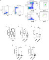
Results: Multivariate analysis at the pre-LD time-point in the entire cohort (n=53) showed that a lower percentage of CD3+CD8+ lymphocytes (38.6% vs 46.8%, OR=0.937 [95% CI: 0.882-0.996], p=0.035) and higher levels of serum C-reactive protein (CRP, 4.52 mg/dl vs 1.00 mg/dl, OR=7.133 [95% CI: 1.796-28], p=0.005) are associated with ICANS. In the pre-LD samples of 21 patients, a significant increase in the percentage of CD8+CD45RA+CD57+ senescent cells (median % value: 16.50% vs 9.10%, p=0.009) and monocytic-myeloid derived suppressor cells (M-MDSC, median % value: 4.4 vs 1.8, p=0.020) was found in ICANS patients. These latter also showed increased levels of EVs carrying CD14+ and CD45+ myeloid markers, of the myeloid chemokine CXCL-9, as well of the MDSC-secreted cytokine IL-10. Notably, the serum levels of circulating neurofilament light chain, a marker of neuroaxonal injury, were positively correlated with the levels of senescent CD8+ T cells, M-MDSC, IL-10 and CXCL-9. No variation in the levels of the selected miRNAs was observed between ICANS and no-ICANS patients.
Discussion: Our data support the notion that pre-CAR-T systemic inflammation is associated with ICANS. Higher proportion of senescence CD8+ T cells and M-MDSC correlate with early signs of neuroaxonal injury at pre-LD time-point, suggesting that ICANS may be the final event of a process that begins before CAR-T infusion, consequence to patient clinical history.
Trial registration: ClinicalTrials.gov NCT04892433.
Keywords: chimeric antigen receptor; inflammation; myeloid activation; neurotoxicity; senescence.
Copyright © 2023 De Matteis, Dicataldo, Casadei, Storci, Laprovitera, Arpinati, Maffini, Cortelli, Guarino, Vaglio, Naddeo, Sinigaglia, Zazzeroni, Guadagnuolo, Tomassini, Bertuccio, Messelodi, Ferracin, Bonafè, Zinzani and Bonifazi.
Conflict of interest statement
PLZ: scientific advisory boards: Secura Bio BIO, Celltrion, Gilead, Janssen-Cilag, BS, Servier, Sandoz, MSD, TG Therap., Takeda, Roche, EUSA Pharma, Kiowa Kirin, Novartis, ADC Therap., Incyte, Beigene; consultancy: EUSA Pharma, MSD, Novartis; speaker’s bureau: Celltrion, Gilead, Janssen-Cilag, BMS, Servier, MSD, TG Therap., Takeda, Roche, EUSA Pharma, Kiowa Kirin, Novartis, Incyte, Beigene. FB: scientific advisory boards and speaker fees: NEOVII, NOVARTIS, KITE, GILEAD, PFIZER, CELGENE, MSD. MB: Research Grant from NEOVII. The remaining authors declare that the research was conducted in the absence of any commercial or financial relationships that could be construed as a potential conflict of interest. The handling editor GL declared a past collaboration with the author FB.
Figures


References
Publication types
MeSH terms
Substances
Associated data
LinkOut - more resources
Full Text Sources
Medical
Research Materials
Miscellaneous

