

Activatable G-quadruplex based catalases for signal transduction in biosensing
- PMID: 36727464
- PMCID: PMC9976883
- DOI: 10.1093/nar/gkad031
Activatable G-quadruplex based catalases for signal transduction in biosensing
Abstract
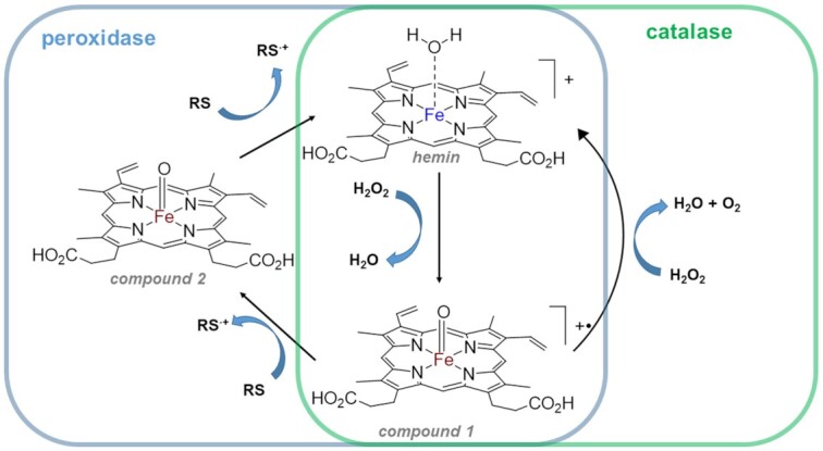
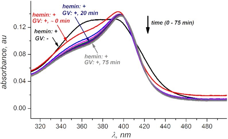
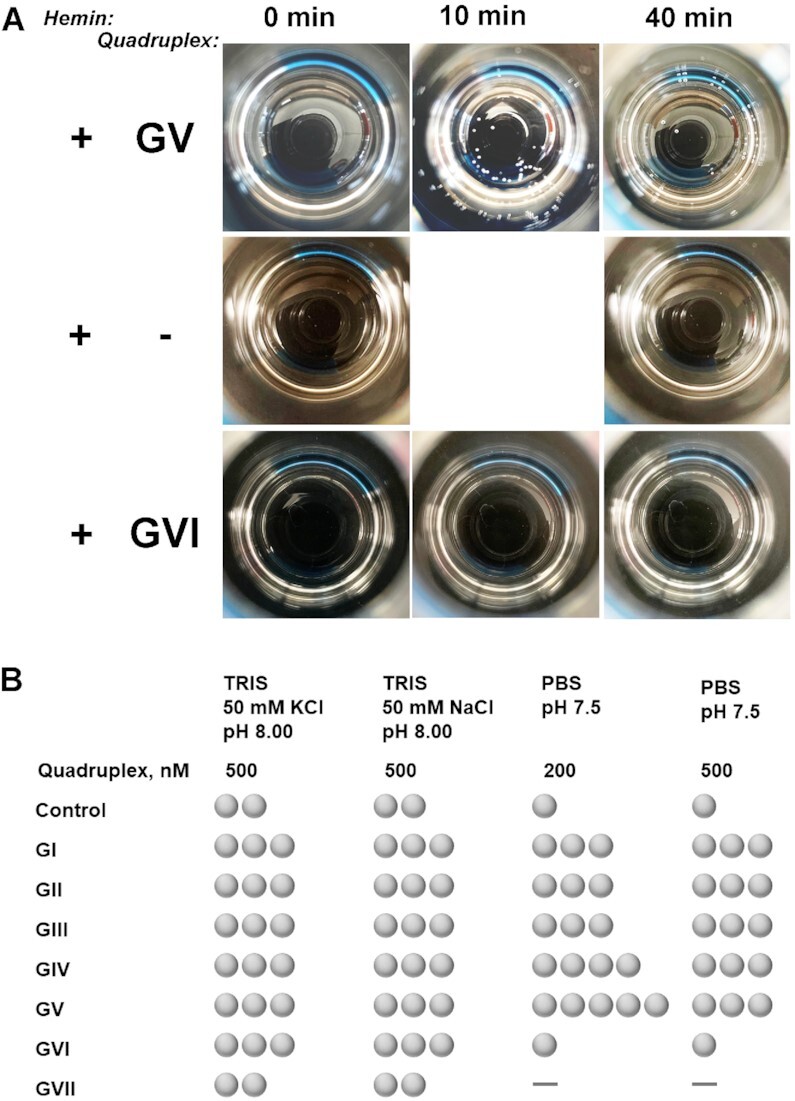
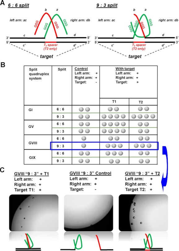
Discovery of oxidative catalysis with G-quadruplex•hemin constructs prompted a range of exciting developments in the field of biosensor design. Thus, G-quadruplex based DNAzymes with peroxidase activity found a niche as signal transduction modules in a wide range of analytical applications. The ability of nucleic acid scaffolds to recognise a variety of practically meaningful markers and to translate the recognition events into conformational changes powers numerous sensor design possibilities. In this work, we establish a catalase activity of G-quadruplex•hemin scaffolds. Catalase activated hydrogen peroxide decomposition generates molecular oxygen that forms bubbles. Observation of bubbles is a truly equipment free signal readout platform that is highly desirable in limited resources or do-it-yourself environments. We take a preliminary insight into a G-quadruplex structure-folding topology-catalase activity correlation and establish efficient operating conditions. Further, we demonstrate the platform's potential as a signal transduction modality for reporting on biomolecular recognition using an oligonucleotide as a proof-of-concept target. Ultimately, activatable catalases based on G-quadruplex•hemin scaffolds promise to become valuable contributors towards accessible molecular diagnostics applications.
© The Author(s) 2023. Published by Oxford University Press on behalf of Nucleic Acids Research.
Figures
Similar articles
-
Highly Sensitive Biosensing Applications of a Magnetically Immobilizable Covalent G-Quadruplex-Hemin DNAzyme Catalytic System.Anal Chem. 2022 Feb 1;94(4):2212-2219. doi: 10.1021/acs.analchem.1c04842. Epub 2022 Jan 20. Anal Chem. 2022. PMID: 35050586
-
Positive effects of ATP on G-quadruplex-hemin DNAzyme-mediated reactions.Anal Chem. 2010 Jul 15;82(14):6148-53. doi: 10.1021/ac100940v. Anal Chem. 2010. PMID: 20552961
-
Exploring the catalytic mechanism of multivalent G-quadruplex/hemin DNAzymes by modulating the position and spatial orientation of connected G-quadruplexes.Anal Chim Acta. 2022 Aug 15;1221:340105. doi: 10.1016/j.aca.2022.340105. Epub 2022 Jun 21. Anal Chim Acta. 2022. PMID: 35934350
-
G-quadruplex based probes for visual detection and sensing.Curr Pharm Des. 2012;18(14):2048-57. doi: 10.2174/138161212799958341. Curr Pharm Des. 2012. PMID: 22380516 Review.
-
Hemin/G-Quadruplex Horseradish Peroxidase-Mimicking DNAzyme: Principle and Biosensing Application.Adv Biochem Eng Biotechnol. 2020;170:85-106. doi: 10.1007/10_2017_37. Adv Biochem Eng Biotechnol. 2020. PMID: 29143069 Review.
Cited by
-
Protocellular Heme and Iron-Sulfur Clusters.Acc Chem Res. 2024 Aug 20;57(16):2293-2302. doi: 10.1021/acs.accounts.4c00254. Epub 2024 Aug 5. Acc Chem Res. 2024. PMID: 39099316 Free PMC article. Review.
References
-
- Peng H.Y., Newbigging A.M., Wang Z.X., Tao J., Deng W.C., Le X.C., Zhang H.Q.. DNAzyme-mediated assays for amplified detection of nucleic acids and proteins. Anal. Chem. 2018; 90:190–207. - PubMed
-
- McConnell E.M., Cozma I., Morrison D., Li Y.. Biosensors made of synthetic functional nucleic acids toward better human health. Anal. Chem. 2020; 92:327–344. - PubMed
-
- Peeling R.W.W., Mabey D.. Point-of-care tests to reduce the burden of sexually transmitted infections. Lancet Infect. Dis. 2019; 19:570–571. - PubMed
Publication types
MeSH terms
Substances
Grants and funding
LinkOut - more resources
Full Text Sources
