. 2023 Feb 28;14(1):e0339322.
doi: 10.1128/mbio.03393-22. Epub 2023 Feb 2.

Siglec-9 Restrains Antibody-Dependent Natural Killer Cell Cytotoxicity against SARS-CoV-2

Affiliations

Siglec-9 Restrains Antibody-Dependent Natural Killer Cell Cytotoxicity against SARS-CoV-2

Pratima Saini et al. mBio. .

Abstract

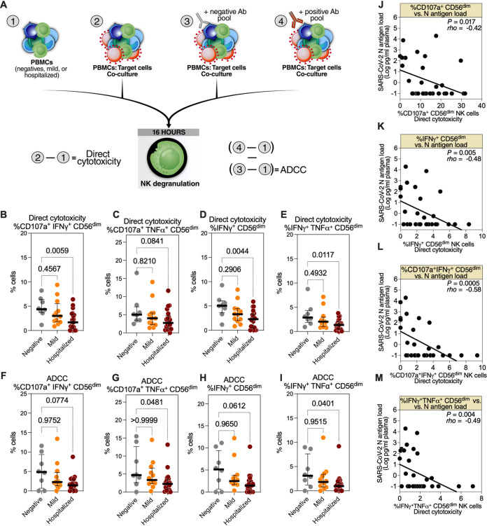
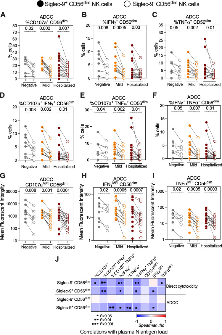
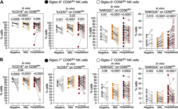
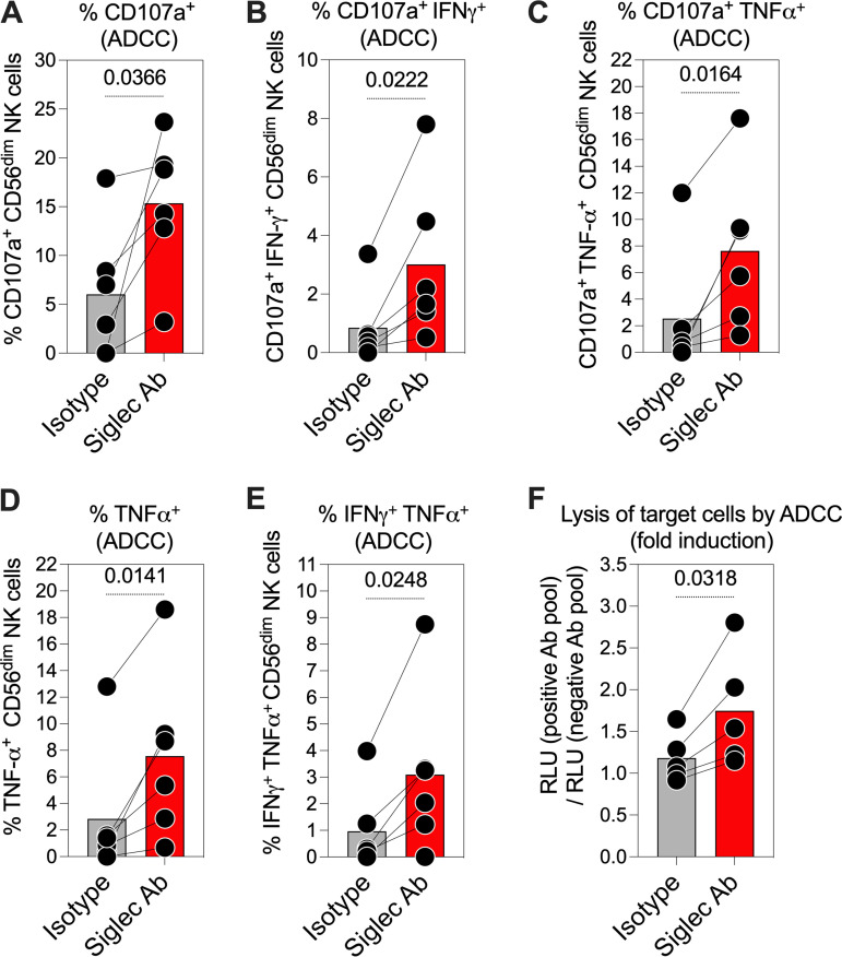
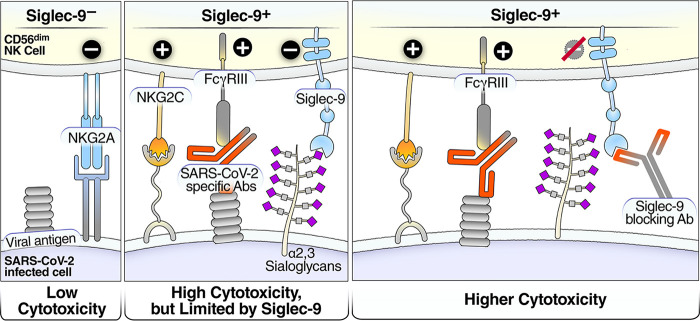
Severe acute respiratory syndrome coronavirus 2 (SARS-CoV-2) infection alters the immunological profiles of natural killer (NK) cells. However, whether NK antiviral functions are impaired during severe coronavirus disease 2019 (COVID-19) and what host factors modulate these functions remain unclear. We found that NK cells from hospitalized COVID-19 patients degranulate less against SARS-CoV-2 antigen-expressing cells (in direct cytolytic and antibody-dependent cell cytotoxicity [ADCC] assays) than NK cells from mild COVID-19 patients or negative controls. The lower NK degranulation was associated with higher plasma levels of SARS-CoV-2 nucleocapsid antigen. Phenotypic and functional analyses showed that NK cells expressing the glyco-immune checkpoint Siglec-9 elicited higher ADCC than Siglec-9- NK cells. Consistently, Siglec-9+ NK cells exhibit an activated and mature phenotype with higher expression of CD16 (FcγRIII; mediator of ADCC), CD57 (maturation marker), and NKG2C (activating receptor), along with lower expression of the inhibitory receptor NKG2A, than Siglec-9- CD56dim NK cells. These data are consistent with the concept that the NK cell subpopulation expressing Siglec-9 is highly activated and cytotoxic. However, the Siglec-9 molecule itself is an inhibitory receptor that restrains NK cytotoxicity during cancer and other viral infections. Indeed, blocking Siglec-9 significantly enhanced the ADCC-mediated NK degranulation and lysis of SARS-CoV-2-antigen-positive target cells. These data support a model in which the Siglec-9+ CD56dim NK subpopulation is cytotoxic even while it is restrained by the inhibitory effects of Siglec-9. Alleviating the Siglec-9-mediated restriction on NK cytotoxicity may further improve NK immune surveillance and presents an opportunity to develop novel immunotherapeutic tools against SARS-CoV-2 infected cells. IMPORTANCE One mechanism that cancer cells use to evade natural killer cell immune surveillance is by expressing high levels of sialoglycans, which bind to Siglec-9, a glyco-immune checkpoint molecule on NK cells. This binding inhibits NK cell cytotoxicity. Several viruses, such as hepatitis B virus (HBV) and HIV, also use a similar mechanism to evade NK surveillance. We found that NK cells from SARS-CoV-2-hospitalized patients are less able to function against cells expressing SARS-CoV-2 Spike protein than NK cells from SARS-CoV-2 mild patients or uninfected controls. We also found that the cytotoxicity of the Siglec-9+ NK subpopulation is indeed restrained by the inhibitory nature of the Siglec-9 molecule and that blocking Siglec-9 can enhance the ability of NK cells to target cells expressing SARS-CoV-2 antigens. Our results suggest that a targetable glyco-immune checkpoint mechanism, Siglec-9/sialoglycan interaction, may contribute to the ability of SARS-CoV-2 to evade NK immune surveillance.

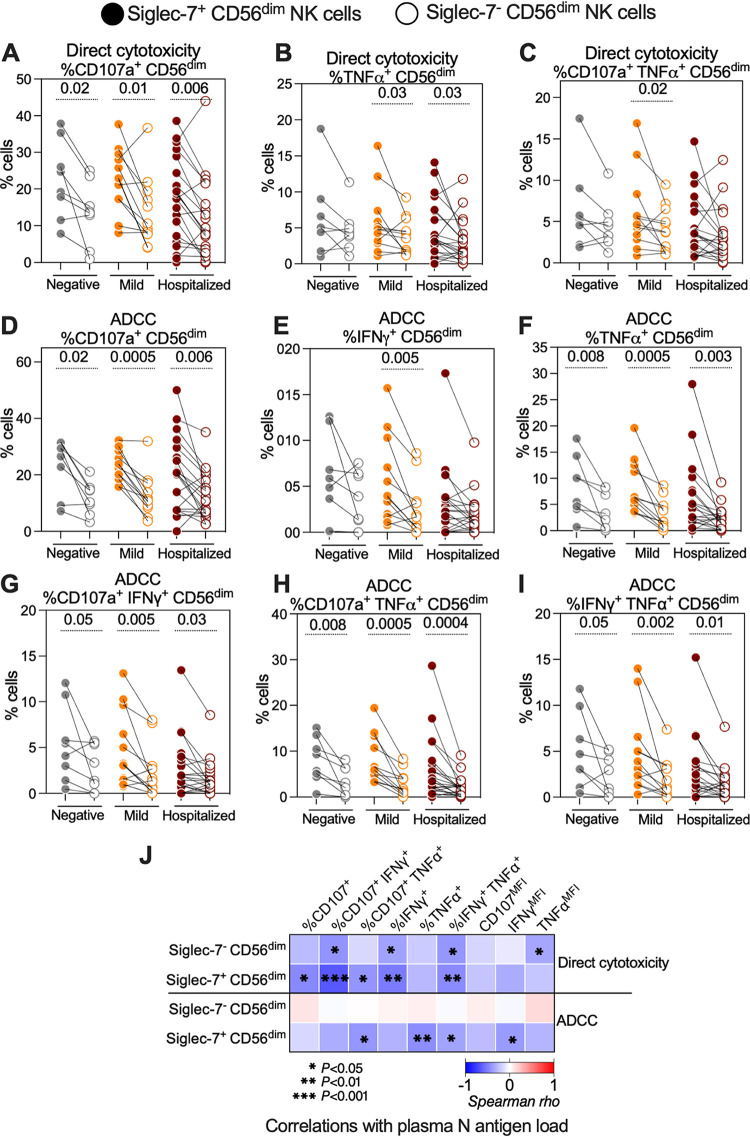
Keywords: COVID-19; SARS-CoV-2; Siglec-7; Siglec-9; antibody-dependent cell cytotoxicity; natural killer cells.


Conflict of interest statement

The authors declare no conflict of interest.

Figures

FIG 1
FIG 1
Hospitalized coronavirus disease 2019 (COVID-19) is associated with reduced CD56dim natural killer (NK) cell degranulation against severe acute respiratory syndrome coronavirus 2 (SARS-CoV-2) Spike-expressing target cells. (A) Schematic overview of the experiments to evaluate the direct cytotoxicity- and antibody-dependent cell cytotoxicity (ADCC)-mediated degranulation of CD56dim NK cells during different severities of COVID-19. To examine direct cytotoxicity-mediated degranulation, peripheral blood mononuclear cells (PBMCs) from each donor, from three COVID-19 status groups (n = 8 SARS-CoV-2 negative, n = 12 mild COVID-19, and n = 21 hospitalized COVID-19), were cocultured (2) or not (1) with SARS-CoV-2 Spike-expressing 293T target cells. To examine ADCC-mediated degranulation, identical cocultures were performed in the presence of the negative (3) or positive (4) antibody pool. Direct cytotoxicity was assessed by subtracting (1) from (2). 10:1 effector:target [E:T] ratio. ADCC was assessed by subtracting the results of subtracting (1) from (3) from the results of subtracting (1) from (4). (B to E) Direct cytotoxicity-mediated degranulation and cytokine production of the CD56dim NK population was assessed as the percentage of (B) CD107a+ interferon γ (IFN-γ)+, (C) CD107a+ tumor necrosis factor α (TNF-α)+, (D) IFN-γ+, and (E) IFN-γ+ TNF-α+ cells. Medians and interquartile ranges (IQR) are displayed. Kruskal-Wallis tests with Dunn’s multiple comparisons correction were used for statistical analyses. (F to I) ADCC-mediated degranulation and cytokine production of the CD56dim NK population was assessed as the percentage of (F) CD107a+ IFN-γ+, (G) CD107a+ TNF-α+, (H) IFN-γ+, or (I) IFN-γ+ TNF-α+ cells. Median and IQR are shown. Kruskal-Wallis tests with Dunn’s multiple-comparisons correction were used for statistical analyses. (J to M) Spearman’s rank-order correlations between plasma N-antigen load and the percentage of CD56dim NK cells expressing (J) CD107a+, (K) IFN-γ+, (L) CD107a+ IFN-γ+, and (M) IFN-γ+ TNF-α+ during direct cytotoxicity assays; only samples from COVID-19-positive donors were used (n = 33).
FIG 2
FIG 2
The Siglec-9+ CD56dim NK subpopulation exhibits a higher SARS-CoV-2 specific ADCC than the Siglec-9 CD56dim NK subpopulation. (A to I) ADCC-mediated degranulation and/or cytokine production of the Siglec-9+ and Siglec-9 CD56dim NK cells within each COVID-19 status group as assessed by the percentage of cells expressing (A) CD107a, (B) IFN-γ, (C) TNF-α, (D) CD107a and IFN-γ, (E) CD107a and TNF-α, and (F) IFN-γ and TNF-α. The mean fluorescence intensity (MFI) was also evaluated for (G) CD107a, (H) IFN-γ, and (I) TNF-α. Wilcoxon’s signed-rank tests were used to compare the Siglec-9+ and Siglec-9 CD56dim NK cells within each COVID-19 status group. (J) Spearman’s correlation heat-map showing associations between direct cytotoxicity and ADCC-mediated NK degranulation of the Siglec-9+ and Siglec-9 CD56dim NK cells and plasma N-antigen load. Square color represents correlation strength, with blue representing negative correlations and red representing positive correlations; only samples from COVID-19-positive donors were used (n = 33).
FIG 3
FIG 3
The Siglec-7+ CD56dim NK subpopulation exhibits higher SARS-CoV-2-specific direct cytolytic and ADCC activity than does the Siglec-7 CD56dim NK subpopulation. (A to C) Direct cytotoxicity-mediated degranulation and/or cytokine production of the Siglec-7+ and Siglec-7 CD56dim NK cells within each COVID-19 status group as assessed by the percentage of cells expressing (A) CD107a, (B) TNF-α, and (C) CD107a and TNF-α. (D to I) ADCC-mediated degranulation and/or cytokine production of the Siglec-7+ and Siglec-7 CD56dim NK cells within each COVID-19 status group as assessed by the percentage of cells expressing (D) CD107a, (E) IFN-γ, (F) TNF-α, (G) CD107a and IFN-γ, (H) CD107a and TNF-α, or (I) IFN-γ and TNF-α. Wilcoxon’s signed-rank tests were used to compare the Siglec-7+ and Siglec-7 CD56dim NK cells within each disease group. (J) Spearman’s correlation heat-map showing associations between direct cytotoxicity- and ADCC-mediated NK degranulation of the Siglec-7+ and Siglec-7 CD56dim NK cells and plasma N-antigen load. Square color represents correlation strength, with blue representing negative correlations and red representing positive correlations; only samples from COVID-19-positive donors were used (n = 33).
FIG 4
FIG 4
Siglec-9 marks activated and mature CD56dim NK subpopulation in vivo. (A) Comparisons of the expression of CD16 (FcγRIII; mediator of ADCC), CD57 (maturation marker), NKG2C (activating receptor), and NKG2A (inhibitory receptor) on Siglec-9+ and Siglec-9 CD56dim NK cells obtained from 79 individuals with three COVID-19 disease states (negative; n = 12, mild; n = 26, and hospitalized; n = 41). Siglec-9+ cells exhibit higher levels of CD16, CD57, NKG2C, and lower levels of NKG2A compared to Siglec-9 cells. Wilcoxon’s signed-rank tests were used to compare the Siglec-9+ and Siglec-9 CD56dim NK cells within each COVID-19 disease state group. Mann-Whitney U tests were used to compare disease state groups. (B) Comparisons of CD16, CD57, NKG2C, and NKG2A expression on Siglec-7+ and Siglec-7 CD56dim NK cells. Siglec-7+ cells exhibited higher levels of CD16, NKG2C, and NKG2A than Siglec-7 cells. No differences were observed in CD57 expression between the Siglec-7+ and Siglec-7 cells. As in panel A, Wilcoxon’s signed-rank tests were used to compare the Siglec-7+ and Siglec-7 CD56dim NK cells within each COVID-19 status group, and Mann-Whitney U tests were used to compare the status groups.
FIG 5
FIG 5
Siglec-9, but not Siglec-7, marks CD56dim NK cells with high ADCC activity against SARS-CoV-2. (A) Percentage of each Siglec-expressing cell subpopulation (Siglec-7 Siglec-9, Siglec-7+ Siglec-9, Siglec-7 Siglec-9+, or Siglec-7+ Siglec-9+) within the CD56dim NK cells from all donors (n = 79). (B to E) In vivo expression of (B) CD16, (C) CD57, (D) NKG2C, and (E) NKG2A on the indicated cell subpopulations (n = 79). Median and IQR are shown. Friedman tests with Dunn’s multiple-comparisons correction were used for statistical analyses. (F to H) The ADCC-mediated degranulation of the indicated NK cell subpopulations (n = 41) as assessed by (F) the percentage of cells expressing CD107a, (G) the MFI of CD107a, or (H) the percentage of cells expressing CD107a and IFN-γ. Median and IQR are displayed. Friedman tests with Dunn’s multiple-comparisons correction were used for statistical analyses.
FIG 6
FIG 6
Blocking Siglec-9 interactions, using a Siglec-9-blocking antibody, enhances the anti-SARS-CoV-2 ADCC of CD56dim NK cells. (A to E) The impact of the Siglec-9-blocking antibody, compared to an isotype control, on ADCC-mediated NK degranulation and/or cytokine production against SARS-CoV-2. PBMCs from six healthy controls were used as effector cells, and SARS-CoV-2 Spike-expressing 293T cells were used as target cells. Cells were cocultured at a 10:1 (E:T) ratio for 12 h. NK degranulation and/or cytokine production was assessed as the percentage of cells expressing (A) CD107a, (B) CD107a and IFN-γ, (C) CD107a and TNF-α, (D) TNF-α, (E) and IFN-γ and TNF-α. Paired t tests were used for statistical analysis. (F) The impact of the Siglec-9-blocking antibody, compared to an isotype control, on the ADCC-mediated lysis of SARS-CoV-2 target cells. Purified NK cells isolated from the PBMCs of five healthy donors were used as effector cells, and SARS-CoV-2 S CHO-K1 cells were used as target cells. Cells were cocultured at a 5:1 E:T ratio for 5 h. The SARS-CoV-2 S CHO-K1 cells stably express the SARS-CoV-2 Spike (S) protein and a HaloTag-HiBiT protein. When the target cells are lysed by ADCC, the intracellular HaloTag-HiBiT protein interacts with the extracellular detection reagent to generate a luminescence signal that can quantitatively measure the degree of target cell lysis. A paired t test was used for statistical analysis.
FIG 7
FIG 7
Model of how a Siglec-9-blocking antibody increases the cytotoxicity of Siglec-9+ NK cells. Left panel: Siglec-9 cells have low cytotoxicity. Middle panel: the Siglec-9+ CD56dim NK subset has high ADCC activity, possibly due to elevated expression of CD16 (FcγRIII; a mediator of ADCC activity), CD57 (maturation marker), and NKG2C (activating receptor), and reduced expression of the inhibitory receptor NKG2A, compared to Siglec-9 CD56dim NK cells. However, the Siglec-9 molecule itself is an inhibitory receptor which restrains the cytolytic ability of these highly cytotoxic Siglec-9+ CD56dim NK cells by binding to Sialic acid on the surface of target cells. Right panel: blocking the inhibitory receptor, Siglec-9, using a blocking antibody can unleash a higher ADCC potential of the Siglec-9+ CD56dim subpopulation.

References

    1. Guan WJ, Ni ZY, Hu Y, Liang WH, Ou CQ, He JX, Liu L, Shan H, Lei CL, Hui DSC, Du B, Li LJ, Zeng G, Yuen KY, Chen RC, Tang CL, Wang T, Chen PY, Xiang J, Li SY, Wang JL, Liang ZJ, Peng YX, Wei L, Liu Y, Hu YH, Peng P, Wang JM, Liu JY, Chen Z, Li G, Zheng ZJ, Qiu SQ, Luo J, Ye CJ, Zhu SY, Zhong NS, China Medical Treatment Expert Group for Covid-19 . 2020. Clinical characteristics of coronavirus disease 2019 in China. N Engl J Med 382:1708–1720. doi:10.1056/NEJMoa2002032. - DOI - PMC - PubMed
    1. Gandhi RT, Lynch JB, Del Rio C. 2020. Mild or moderate COVID-19. N Engl J Med 383:1757–1766. doi:10.1056/NEJMcp2009249. - DOI - PubMed
    1. Schulien I, Kemming J, Oberhardt V, Wild K, Seidel LM, Killmer S, Sagar Daul F, Salvat Lago M, Decker A, Luxenburger H, Binder B, Bettinger D, Sogukpinar O, Rieg S, Panning M, Huzly D, Schwemmle M, Kochs G, Waller CF, Nieters A, Duerschmied D, Emmerich F, Mei HE, Schulz AR, Llewellyn-Lacey S, Price DA, Boettler T, Bengsch B, Thimme R, Hofmann M, Neumann-Haefelin C. 2021. Characterization of pre-existing and induced SARS-CoV-2-specific CD8+ T cells. Nat Med 27:78–85. doi:10.1038/s41591-020-01143-2. - DOI - PubMed
    1. Stephenson E, Reynolds G, Botting RA, Calero-Nieto FJ, Morgan MD, Tuong ZK, Bach K, Sungnak W, Worlock KB, Yoshida M, Kumasaka N, Kania K, Engelbert J, Olabi B, Spegarova JS, Wilson NK, Mende N, Jardine L, Gardner LCS, Goh I, Horsfall D, McGrath J, Webb S, Mather MW, Lindeboom RGH, Dann E, Huang N, Polanski K, Prigmore E, Gothe F, Scott J, Payne RP, Baker KF, Hanrath AT, Schim van der Loeff ICD, Barr AS, Sanchez-Gonzalez A, Bergamaschi L, Mescia F, Barnes JL, Kilich E, de Wilton A, Saigal A, Saleh A, Janes SM, Smith CM, Gopee N, Wilson C, Coupland P, Coxhead JM, Cambridge Institute of Therapeutic Immunology and Infectious Disease-National Institute of Health Research (CITIID-NIHR) COVID-19 BioResource Collaboration , et al. 2021. Single-cell multi-omics analysis of the immune response in COVID-19. Nat Med 27:904–916. doi:10.1038/s41591-021-01329-2. - DOI - PMC - PubMed
    1. Leem G, Cheon S, Lee H, Choi SJ, Jeong S, Kim ES, Jeong HW, Jeong H, Park SH, Kim YS, Shin EC. 2021. Abnormality in the NK-cell population is prolonged in severe COVID-19 patients. J Allergy Clin Immunol 148:996–1006 e18. doi:10.1016/j.jaci.2021.07.022. - DOI - PMC - PubMed

Publication types

MeSH terms

Substances