. 2023 May;137(3):733-742.
doi: 10.1007/s00414-023-02958-7. Epub 2023 Feb 2.

Automated localization of the medial clavicular epiphyseal cartilages using an object detection network: a step towards deep learning-based forensic age assessment

Affiliations

Automated localization of the medial clavicular epiphyseal cartilages using an object detection network: a step towards deep learning-based forensic age assessment

Philipp Wesp et al. Int J Legal Med. 2023 May.

Abstract

Background: Deep learning is a promising technique to improve radiological age assessment. However, expensive manual annotation by experts poses a bottleneck for creating large datasets to appropriately train deep neural networks. We propose an object detection approach to automatically annotate the medial clavicular epiphyseal cartilages in computed tomography (CT) scans.

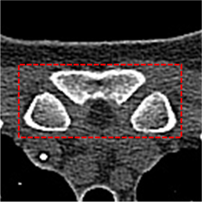
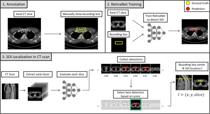
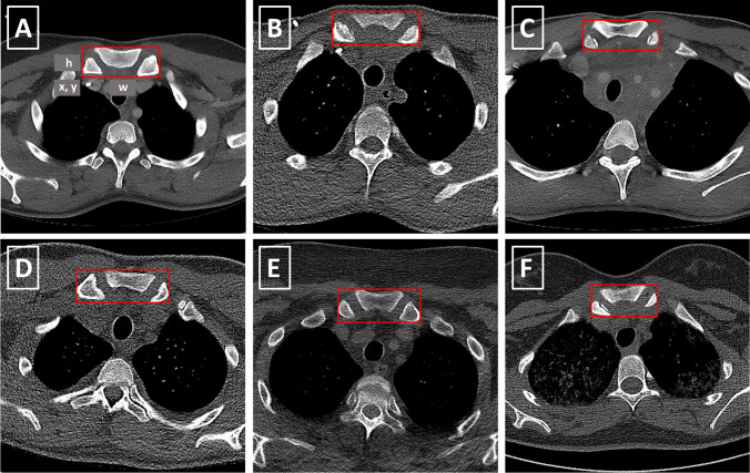
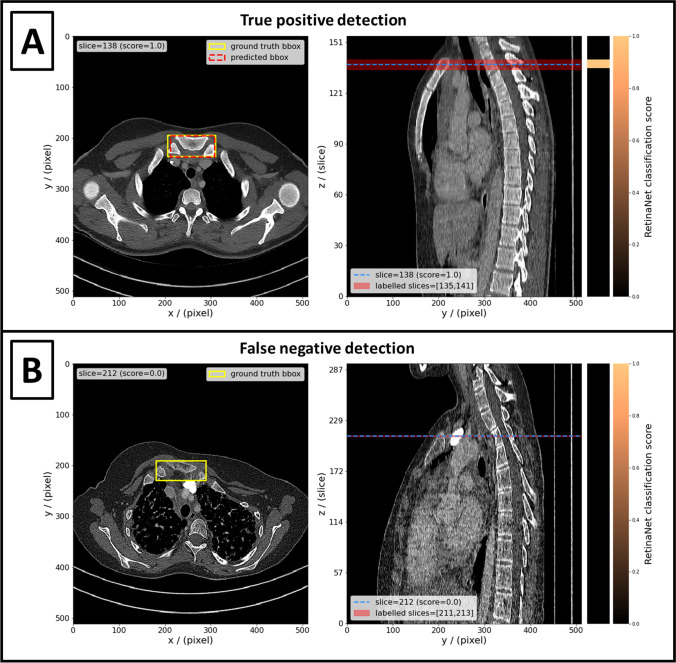
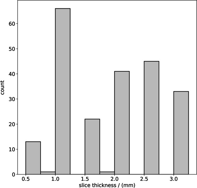
Methods: The sternoclavicular joints were selected as structure-of-interest (SOI) in chest CT scans and served as an easy-to-identify proxy for the actual medial clavicular epiphyseal cartilages. CT slices containing the SOI were manually annotated with bounding boxes around the SOI. All slices in the training set were used to train the object detection network RetinaNet. Afterwards, the network was applied individually to all slices of the test scans for SOI detection. Bounding box and slice position of the detection with the highest classification score were used as the location estimate for the medial clavicular epiphyseal cartilages inside the CT scan.

Results: From 100 CT scans of 82 patients, 29,656 slices were used for training and 30,846 slices from 110 CT scans of 110 different patients for testing the object detection network. The location estimate from the deep learning approach for the SOI was in a correct slice in 97/110 (88%), misplaced by one slice in 5/110 (5%), and missing in 8/110 (7%) test scans. No estimate was misplaced by more than one slice.

Conclusions: We demonstrated a robust automated approach for annotating the medial clavicular epiphyseal cartilages. This enables training and testing of deep neural networks for age assessment.

Keywords: Age assessment; Anatomic landmark detection; Deep learning; Medial clavicular epiphyseal cartilages; Object detection.


Conflict of interest statement

The authors declare no competing interests.

Figures

Fig. 1
Fig. 1
The structure-of-interest (SOI), defined as the sternoclavicular joints, together with their contributing portions of the sternum and the medial clavicles
Fig. 2
Fig. 2
Schematic workflow diagram of the proposed medial clavicular epiphyseal cartilage localization. 1. Annotation: CT images are manually annotated with two-dimensional ground-truth bounding boxes in axial slices around the structure-of-interest (SOI). The SOI is an easy-to-identify proxy structure for the actual medial clavicular epiphyseal cartilage. 2. RetinaNet Training: A RetinaNet is trained to detect the SOI in axial slices and predict bounding boxes. 3: Localization in CT scan: The SOI can be localized in an unknown CT scan of the upper body. For this purpose, the trained RetinaNet is applied to each slice in a CT scan and all positive detections are collected. Afterwards, the center of the bounding box which corresponds to the best detection (highest classification score) is used as the predicted location for the SOI
Fig. 3
Fig. 3
(AF) Bounding boxes around the SOI in different CT scans after preprocessing. The SOI is defined as the sternoclavicular joints, together with their contributing portions of the sternum and the medial clavicles. In addition, (A) illustrates the 4 bounding box location parameters x, y, width (w), and height (h)
Fig. 4
Fig. 4
The left panels show axial CT slices with ground-truth bounding boxes around the SOI (yellow boxes) and detections (if predicted by network) (red boxes). The right panels show the central sagittal slice of the respective CT. The position of the axial slice in the left panel is indicated by the dashed blue line in the right panel. The red area in the right panel indicates the positions of all axial slices which contain the SOI and have ground-truth bounding boxes annotated. The heatmaps next to the right panels show the classification score returned by the RetinaNet for each axial slice (light orange = 1.0; black = 0.0). Detections made by the RetinaNet are true positive, if the axial slice has a ground-truth bounding box (red area) and the classification score is > 0.05 (e.g., light orange). A Shows an example of a true-positive localization of the SOI; i.e., the highest classification score was returned for a slice which indeed contains the SOI. B shows an example of a false-negative localization; i.e., the RetinaNet returned only classification scores < 0.05 even though the SOI is present in one or more slices
Fig. 5
Fig. 5
Slice thickness distribution of the 222 chest CT scans

References

    1. United Nations (1989) The convention on the rights of the child. https://www.ohchr.org/en/instruments-mechanisms/instruments/convention-r.... Accessed 31 Jan 2023
    1. The European Parliament and The Council Of The European Union (2013) Directive 2013/33/EU of the European Parliament and of the Council of 26 June 2013 laying down standards for the reception of applicants for international protection (recast). https://eur-lex.europa.eu/eli/dir/2013/33/oj. Accessed 31 Jan 2023
    1. European Asylum Support Office (2018) EASO Practical guide on age assessment. Publications Office. 10.2847/292263
    1. Schmeling A, Dettmeyer R, Rudolf E, et al. Forensic age estimation: methods, certainty, and the law. Dtsch Arzteblatt Int. 2016;113:44–50. doi: 10.3238/arztebl.2016.0044. - DOI - PMC - PubMed
    1. Schmeling A, Schulz R, Reisinger W, et al. Studies on the time frame for ossification of the medial clavicular epiphyseal cartilage in conventional radiography. Int J Legal Med. 2004;118:5–8. doi: 10.1007/s00414-003-0404-5. - DOI - PubMed

LinkOut - more resources