Review
. 2023 Jul;41(7):703-711.
doi: 10.1007/s11604-023-01395-1. Epub 2023 Feb 2.

The diagnostic challenge of non-traumatic bladder rupture: a pictorial essay

Affiliations
Review

The diagnostic challenge of non-traumatic bladder rupture: a pictorial essay

Hideki Kunichika et al. Jpn J Radiol. 2023 Jul.

Abstract

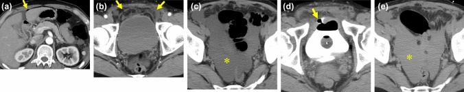
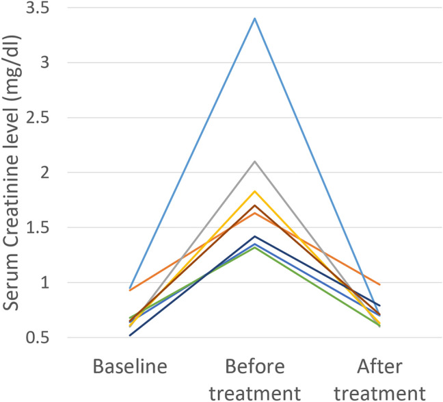
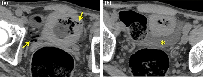
Non-traumatic bladder rupture (NTBR) is relative rare pathology including spontaneous rupture and iatrogenic injury. As increasing the medical intervention for the pelvic malignancy or elderly population, NTBR will be encountered more frequently. There are few previous studies summarizing the imaging features of NTBR. We reviewed imaging characteristics of 18 previous cases of NTBR experienced. In addition, 3 presentative cases that can be a pitfall to differentiate from NTBR. The aim of this article is to clarify the key CT findings of NTBR and its pitfalls.

Keywords: Bladder rupture; Computed tomography; Non-traumatic; Pseudo-renal failure.


Conflict of interest statement

The authors declare that they have no conflict of interest.

Figures

Fig. 1
Fig. 1
60-Year-old woman with bladder rupture complained abdominal pain the day after TURBT. a, b The axial a and sagittal b images of CECT show the tract-like gas at the bladder dome (arrows), retroperitoneal fluid collection with gas (in the paravesical space and Retzius cavity) (*) and moderate amount of ascites. c T2-weighted MR image before TURBT shows bladder tumor matching with the ruptured point (arrow)
Fig. 2
Fig. 2
60-Year-old woman with bladder rupture who also had bacteremia, lung abscess, and epidural abscess of the lumbar spine. The cause of this rupture was unknown. a b The axial a and MPR. b CECT images demonstrate bladder wall (arrowheads) and its discontinuity (arrows). Small amount of uremic ascites (*) leaks through the ruptured site. c. Bilateral hydronephrosis is also observed as the indirect finding
Fig. 3
Fig. 3
40-Year-old male with bladder rupture due to bladder hemangioma associated with Klippel-Trenaunay-Weber syndrome. He presented with lower abdominal pain and pseudo-renal failure. a CECT shows massive ascites (*) and lymphangioma of subcutaneous and spleen (arrows) associated with Klippel–Trenaunay–Weber syndrome. b The coronal image shows the discontinuity of bladder wall (arrow) and low dense uremic ascites. c Small amount of contrast medium has reached to the bladder (arrow) in delayed phase (180 s after administration of contrast medium) but the uremic ascites showed still low density (*). It is difficult to identify the leakage of contrast medium. d T2-weighted MR image shows bladder hemangioma as the cause of bladder hemorrhagic tamponade in the past (arrow). The fragility of the bladder wall may be caused by the history of tamponade and several placements of catheter
Fig. 4
Fig. 4
80-Year-old woman with bladder rupture, who presented lower abdominal pain and abdominal distension. She had the history of radiation therapy for cervical cancer 27 years ago. a b CECT in axial a and coronal b images shows thickened bladder wall without any discontinuity. Retroperitoneal fluid (arrow) and a small amount of ascites (*) are observed but no leakage of contrast medium. c Bilateral hydronephrosis is also represented. d The additional super delayed phase obtained the next day shows leakage of contrast medium through the pin-hale of the bladder (arrow) predominantly on the right side of the abdominal cavity (arrowhead)
Fig. 5
Fig. 5
50-Year-old man with iatrogenic bladder rupture (post-operative laparoscopy for appendicitis), who presented with abdominal pain and abdominal distension three days after laparoscopic appendicectomy. a CECT shows small amount of extra-luminal gas suitable for postoperative state. b The bladder wall keeps continuity though prevesical fluid collection is observed (arrows). c A large amount of ascites (*) represents low attenuation in delayed phase. d e The additional super delayed phase (2 h later) shows clearly the perforation point (arrow) and increased value of ascites (approximately 10 HU to 50 HU) (*), which reflects leakage of contrast medium
Fig. 6
Fig. 6
80-Year-old woman complaining acute abdomen for bladder rupture with a long-term indwelling bladder catheter and a right ureteral stent for chronic kidney disease. a b NECT shows only extra-luminal gas (arrows) and no other direct or indirect findings of bladder rupture. An exploratory laparotomy was performed for suspected small bowel perforation, and it revealed bladder rupture due to catheter injury and a small amount of infected ascites in the Douglas pouch. Peritonitis caused by infected urinary ascites is difficult to distinguish from small bowel perforation clinically and radiologically
Fig. 7
Fig. 7
Transient increase of serum creatinine levels (> 0.5 mg/dL) was detected in 8/14 patients (57%), excluding 1 patient on dialysis and 3 patients with incomplete blood test records
Fig. 8
Fig. 8
Pitfalls. 70-Year-old man with perforation of the small intestine who presented with urinary retention and abdominal pain. a b Axial NECT images obtained after Foley catheter placement demonstrate ascites (arrows) and extra-luminal gas (arrowheads). The CT value of ascites is relatively high (25-30HU). c A sagittal image reveals thinning of the bladder wall around the catheter tip (*), which led to a misdiagnosis of bladder rupture. Subsequent cystography was obtained and ruled out bladder rupture. Exploratory laparotomy revealed perforation of the small intestine.
Fig. 9
Fig. 9
Pitfalls. 70-Year-old man with emphysematous cystitis who presented with fever and decreased appetite. He had a medical history of diabetes mellitus. a b NECT images show diffuse bladder wall thickening and linear intramural/retroperitoneal gas (arrows) and ascites (*). This patient was treated conservatively with insertion of bladder catheter. The penetrating gas of emphysematous cystitis can resemble that of a bladder wall fistula
Fig. 10
Fig. 10
Pitfalls. 60-Year-old woman with acute peritonitis associated with perforation of pyometra uteri who presented with abdominal pain and septic shock. a b NECT image shows a large amount of relatively high intense ascites with an irregular hyperintensity in the pelvis (*). b Slightly high dense structure surrounding the heterogeneous hyperintensity appears to be the uterine myometrium (dot line) containing hemorrhage. c A sagittal reconstructed image shows the perforated point at the uterine fundus as an irregularly shaped high-density area (arrow). No extra-luminal gas can be identified in this case

References

    1. Bryk DJ, Zhao LC. Guideline of guidelines: a review of urological trauma guidelines. BJU Int. 2016;117(2):226–234. doi: 10.1111/bju.13040. - DOI - PubMed
    1. Gomez RG, Ceballos L, Coburn M, et al. Consensus statement on bladder injuries. BJU Int. 2004;94(1):27–32. doi: 10.1111/j.1464-410X.2004.04896.x. - DOI - PubMed
    1. Mitchell T, Al-Hayek S, Patel B, et al. Acute abdomen caused by bladder rupture attributable to neurogenic bladder dysfunction following a stroke: a case report. J Med Case Rep. 2011;5:254. doi: 10.1186/1752-1947-5-254. - DOI - PMC - PubMed
    1. Su PH, Hou SK, How CK, et al. Diagnosis of spontaneous urinary bladder rupture in the ED. Am J Emerg Med. 2012;30:379–382. doi: 10.1016/j.ajem.2011.10.003. - DOI - PubMed
    1. Fujikawa K, Miyamoto T, Ihara Y, et al. High incidence of severe urologic complications following radiotherapy for cervical cancer in Japanese women. Gynecol Oncol. 2001;80:21–23. doi: 10.1006/gyno.2000.6030. - DOI - PubMed

MeSH terms