. 2023 Feb 1;64(2):3.
doi: 10.1167/iovs.64.2.3.

Pharmacokinetic and Target Engagement Measures of ANX007, an Anti-C1q Antibody Fragment, Following Intravitreal Administration in Nonhuman Primates

Affiliations

Pharmacokinetic and Target Engagement Measures of ANX007, an Anti-C1q Antibody Fragment, Following Intravitreal Administration in Nonhuman Primates

Anita Grover et al. Invest Ophthalmol Vis Sci. .

Abstract

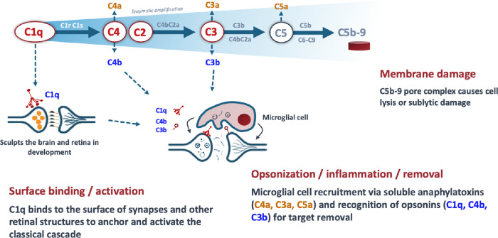
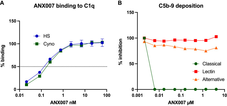
Purpose: C1q and the classical complement cascade are key regulators of synaptic pruning, and their aberrant activation has been implicated in neurodegenerative ophthalmic diseases including geographic atrophy and glaucoma. The antigen-binding fragment antibody ANX007 specifically recognizes globular head groups of C1q to block substrate binding and functionally inhibit classical complement cascade activation. ANX007 was assessed in nonclinical studies of biodistribution and C1q target engagement in the eye following intravitreal (IVT) administration in cynomolgus monkeys.

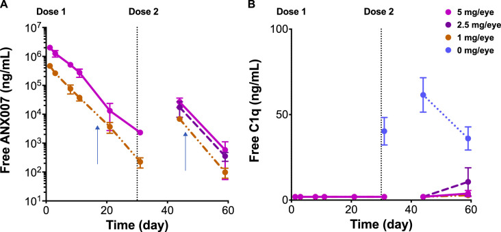
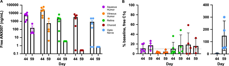
Methods: Female juvenile cynomolgus monkeys (n = 12) received a single bilateral dose of 1 or 5 mg ANX007/eye, with vitreous and non-perfused tissue samples collected approximately 4 weeks later. In a separate study, male (n = 6/5) and female (n = 6/5) animals received repeat bilateral dosing of 1, 2.5, or 5 mg ANX007/eye on days 1 and 29, with aqueous and vitreous collections on day 44 or day 59. Tissues from the 5 mg/eye repeat-dose group were perfused, and retina, choroid, and optic nerve samples were collected approximately 2 and 4 weeks post-last dose.

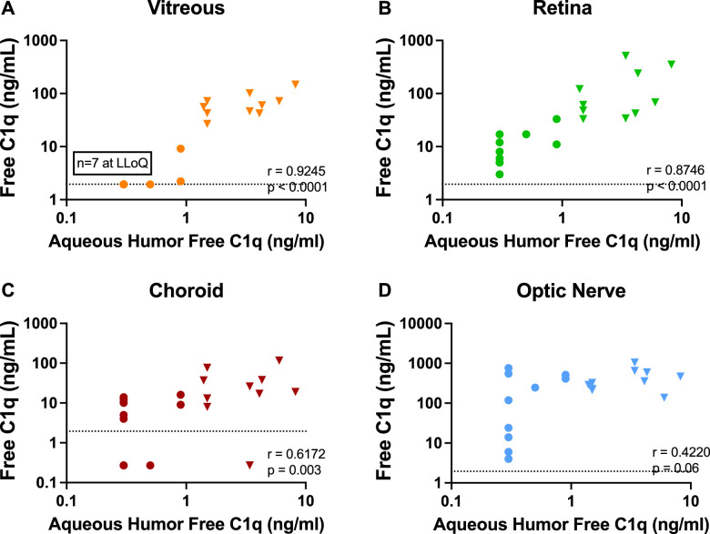
Results: Following a single dose of ANX007, vitreous levels of free drug were measurable through 4 weeks at both the 1 and 5 mg dose levels, with approximately 3-day half-life. With repeat dose of 5 mg/eye, free-ANX007 was measurable 4 weeks post-last dose in perfused retina and choroid and up to approximately 2 weeks post-last dose in optic nerve. There was a strong correlation between C1q target engagement and free drug levels in aqueous and vitreous humors and retinal tissue.

Conclusions: Following IVT administration, ANX007 distributes to sites within the retina that are relevant to neurodegenerative ophthalmic disease with clear evidence of C1q target engagement. Based on its mechanism of action inhibiting C1q and its downstream activity, ANX007 is predicted to mitigate tissue damage driven by classical complement activation in the retina. These data support further clinical evaluation of ANX007.


Conflict of interest statement

Disclosure: A. Grover, Annexon (E); S. Sankaranarayanan, Escape Bio (E), Annexon (E); V. Mathur, Acelot Inc. (E), Annexon (E); P. Suri, Annexon (E); H. Qiu, Annexon (E); Y. Andrews-Zwilling, Annexon (E); K. Mease, Annexon (C), ToxStrategies Inc. (E); L.K. Taylor, Annexon (E); E. Cahir-McFarland, Annexon (E); S. Keswani, Annexon (E); T. Yednock, Annexon (E)

Figures

Figure 1.
Figure 1.
Actions associated with C1q activation via the classical complement pathway cascade.
Figure 2.
Figure 2.
(A) EC50 of binding to human and cynomolgus monkey C1q is equivalent. ANX007 binding to C1q was determined in vitro using a one-sided enzyme-linked immunosorbent assay with C1q purified from human and cynomolgus serum (maximum luminescence counts to human was 1258074 and to cynomolgus monkey was 1051459). (B) ANX007 inhibits classical but not lectin or alternative complement pathway activation. ANX007 effects on complement activation assays were measured in Wieslab assays (EURO DIAGNOSTICA AB, Sweden) using specific activators of the classical, lectin, and alternative pathways. Cyno, cynomolgus monkey; EC50, half maximal effective concentration; HS, human serum.
Figure 3.
Figure 3.
Mean vitreous pharmacokinetics and pharmacodynamics at dose levels of 0, 1, 2.5, and 5 mg ANX007 per eye. Doses were administered at day 1 and day 29. Day 44 is approximately 2 weeks after the second dose, and day 59 is approximately 4 weeks post-last dose. N = 2 for 1 to 29 days, and N = 6 at 44 and 59 days. Error bars = standard error of the mean. Graphs represent combined data from single and multiple dose studies. (A) Free-ANX007 levels. (B) Free-C1q levels. Vertical arrows in (A) represent 14 days after the single and repeat-dose intravitreal injections of ANX007, respectively.
Figure 4.
Figure 4.
Mean ocular tissue and fluid pharmacokinetics and pharmacodynamics following two 5 mg/eye doses of ANX007 at 1-month intervals. Day 44 is approximately 2 weeks post-last dose, and day 59 is approximately 4 weeks post-last dose. (A) Free-ANX007 levels. (B) Free-C1q (% of baseline using the average of the control group). Baseline free-C1q levels (mean ± standard deviation) were: aqueous humor 3.5 ± 2.3 ng/mL, vitreous humor 67 ± 35 ng/mL, retina 151 ± 165 ng/mL, choroid 35 ± 35 ng/mL, and optic nerve 430 ± 273 ng/mL. Control animals are indicated as day 1/baseline. The 5 mg/eye dose was the only dose that was perfused. N = 3–6 eyes. Assay lower limit of quantitation is shown in (B) with the dotted horizontal line. Error bars = standard error of the mean.
Figure 5.
Figure 5.
Correlation of aqueous humor C1q suppression. (A) Vitreous humor. Ocular tissues (B) retina, (C) choroid, and (D) optic nerve. Presented data are a combination of control (triangles) and 5 mg/eye ANX007-treated (circles) animals and show levels of free-C1q in perfused tissues and aqueous humor following 2 ANX007 doses of 5 mg/eye at a 1-month interval. Spearman r = correlation. Assay lower limit of quantitations (LLOQs) are detailed in the Methods section and are shown with dotted horizontal lines.

References

    1. Stevens B, Allen NJ, Vazquez LE, et al. .. The classical complement cascade mediates CNS synapse elimination. Cell. 2007; 131: 1164–1178. - PubMed
    1. Schafer DP, Lehrman EK, Kautzman AG, et al. .. Microglia sculpt postnatal neural circuits in an activity and complement-dependent manner. Neuron . 2012; 74: 691–705. - PMC - PubMed
    1. Stephan AH, Barres BA, Stevens B.. The complement system: an unexpected role in synaptic pruning during development and disease. Annu Rev Neurosci. 2012; 35: 369–389. - PubMed
    1. Stasi K, Nagel D, Yang X, et al. .. Complement component 1Q (C1Q) upregulation in retina of murine, primate, and human glaucomatous eyes. Invest Ophthalmol Vis Sci . 2006; 47(3): 1024–1029. - PubMed
    1. Ziabska K, Ziemka-Nalecz M, Pawelec P, Sypecka J, Zalewska T.. Aberrant complement system activation in neurological disorders. Int J Mol Sci . 2021; 22: 4675. - PMC - PubMed

Publication types

Substances