. 2023 Feb 2;8(5):e161284.
doi: 10.1172/jci.insight.161284.

The different natural estrogens promote endothelial healing through distinct cell targets

Affiliations

The different natural estrogens promote endothelial healing through distinct cell targets

Morgane Davezac et al. JCI Insight. .

Abstract

The main estrogen, 17β-estradiol (E2), exerts several beneficial vascular actions through estrogen receptor α (ERα) in endothelial cells. However, the impact of other natural estrogens such as estriol (E3) and estetrol (E4) on arteries remains poorly described. In the present study, we report the effects of E3 and E4 on endothelial healing after carotid artery injuries in vivo. After endovascular injury, which preserves smooth muscle cells (SMCs), E2, E3, and E4 equally stimulated reendothelialization. By contrast, only E2 and E3 accelerated endothelial healing after perivascular injury that destroys both endothelial cells and SMCs, suggesting an important role of this latter cell type in E4's action, which was confirmed using Cre/lox mice inactivating ERα in SMCs. In addition, E4 mediated its effects independently of ERα membrane-initiated signaling, in contrast with E2. Consistently, RNA sequencing analysis revealed that transcriptomic and cellular signatures in response to E4 profoundly differed from those of E2. Thus, whereas acceleration of endothelial healing by estrogens had been viewed as entirely dependent on endothelial ERα, these results highlight the very specific pharmacological profile of the natural estrogen E4, revealing the importance of dialogue between SMCs and endothelial cells in its arterial protection.

Keywords: Cardiovascular disease; Endocrinology; Endothelial cells; Sex hormones; Vascular Biology.


Conflict of interest statement

Conflict of interest: JFA has received a research grant from Mithra. JMF is a consultant at Mithra, a company that develops estetrol-based women’s healthcare products.

Figures

Figure 1
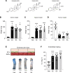
Figure 1. 17β-Estradiol (E2), estriol (E3), and estetrol (E4) accelerate endothelial healing following carotid artery endovascular injury.
Four-week-old female mice were ovariectomized and 2 weeks later implanted subcutaneously with vehicle (Veh), E2, E3, or E4 pellets for 2 weeks. Mice were then subjected to endovascular injury of the carotid artery. Carotid reendothelialization was analyzed 5 days after injury (n = 7–11 per group). (A) Chemical structures of E2, E3, and E4. (B) Uterine weight. (C) Vaginal weight. (D) Thymic weight. (E) Representative Evans blue staining of carotids with outlined deendothelialized areas (scale bar: 1 mm) and (F) quantitative analysis of reendothelialization, expressed as a percentage of reendothelialized area compared with day 0. ECs, endothelial cells. Results are expressed as mean ± SEM. To test the effect of the different treatments, Kruskal-Wallis test (B and D) or 1-way ANOVA (C and F) was performed. *P < 0.05, **P < 0.01, ****P < 0.0001 versus Veh-treated group.
Figure 2
Figure 2. In contrast to E2 and E3, E4 does not accelerate endothelial healing after carotid artery perivascular injury.
Four-week-old female mice were ovariectomized and 2 weeks later were implanted subcutaneously with vehicle (Veh), E2, E3, or E4 pellets or a combination of 2 of these estrogens for 2 weeks. Mice were subjected to perivascular injury of the carotid artery. Carotid reendothelialization was analyzed 3 days after injury (n = 5–9 per group). (A) Representative Evans blue staining of carotids with outlined deendothelialized areas (scale bar: 1 mm) and (B) quantitative analysis of reendothelialization, expressed as a percentage of reendothelialized area compared with day 0. ECs, endothelial cells. Results are expressed as mean ± SEM. To test the effect of the different treatments, 1-way ANOVA was performed. **P < 0.01, ***P < 0.001, ****P < 0.0001 versus Veh-treated group; ††P < 0.01 for difference between E2 and E2+E4; §§§P < 0.001 for difference between E3 and E3+E4.
Figure 3
Figure 3. ERα in smooth muscle cells is necessary for E4’s effect on endothelial healing but dispensable for E3’s effect.
(A) Four-week-old ovariectomized αSMACreERT2+ERαlox/lox female mice and their respective control littermates were implanted with vehicle (Veh), E4, or E3 pellets for 2 weeks and subjected to endovascular injury of the carotid artery. Quantitative analysis of reendothelialization 5 days after injury, relative to day 0, are depicted in response to (B) E4 (n = 5–6 per group) or (C) E3 (n = 5–7 per group). Results are expressed as mean ± SEM. To test the effect of E4 and E3 treatments in each genotype, 2-way ANOVA was performed. **P < 0.01 versus Veh-treated group.
Figure 4
Figure 4. E4 does not require membrane-initiated ERα signaling to accelerate endothelial healing and antagonizes this pathway in endothelial cells.
Four-week-old ovariectomized (A) C451A-ERα (n = 6–7 per group) and (B) R264A-ERα (n = 7–11 per group) female mice and their respective control WT littermates were implanted with vehicle (Veh) or E4 pellets for 2 weeks and subjected to endovascular injury of the carotid artery. Schematic representation of each mouse model and quantitative analysis of reendothelialization 5 days after injury relative to day 0 are depicted. Results are expressed as mean ± SEM. To test the effect of E4 treatments in each genotype, 2-way ANOVA was performed. (C) Estrogen-deprived ERα-TeloHAECs were incubated with DMSO, E2 (1 × 10⁻⁸ M), E4 (1 × 10⁻⁶ M), or a combination of E2 and E4 for 5 minutes. Proximity ligation assay for ERα-SRC interaction was performed. Interactions are represented by red dots. Nuclei were counterstained with DAPI (scale bars: 20 μm). (D) Quantification of the number of dots per ERα-positive cell from 1 representative experiment. The experiment was replicated 3 times. Results are expressed as mean ± SEM. To test the effect of the different treatments, 1-way ANOVA was performed. *P < 0.05, **P < 0.01, ****P < 0.0001 versus Veh-treated group. ††††P < 0.0001 for difference between E2 and E2+E4.
Figure 5
Figure 5. E4 still accelerates endothelial healing in the presence of exogenous and endogenous estrogens.
(A) Four-week-old C57BL/6 female mice were ovariectomized and 2 weeks later were implanted with vehicle (Veh), E2, E4, or a combination of E2 and E4 pellets. Two weeks later, mice were subjected to endovascular injury of the carotid artery. Carotid reendothelialization was analyzed 5 days after injury (n = 5–9 per group). (B) Uterine weight. (C) Quantitative analysis of reendothelialization, expressed as a percentage of reendothelialized area compared with day 0. (D) Six-week-old gonad-intact C57BL/6 female mice were implanted with Veh or E4 pellets. Two weeks later, mice were subjected to endovascular injury of the carotid artery. Carotid reendothelialization was analyzed 5 days after injury (n = 5–6 per group). (E) Representative estrous cycles before and after Veh and E4 treatment. (F) Quantitative analysis of reendothelialization, expressed as a percentage of reendothelialized area compared with day 0. Results are expressed as mean ± SEM. To test the effect of the different treatments, Kruskall-Wallis test (B and C) or 2-tailed Student’s t test (F) was performed. *P < 0.05, **P < 0.01, ***P < 0.001.
Figure 6
Figure 6. E4 displays a specific transcriptional program that differs from E2 in carotid arteries.
(A) Four-week-old C57BL/6 female mice were ovariectomized and treated with a vehicle (Veh) or E4 for 2 weeks. RNAs were isolated from uninjured carotid arteries and sequenced (n = 4–5 per group). (B) Heatmap illustrating the relative expression values of all genes significantly regulated following E4 treatment (fold change >2 or <0.5 versus control with Benjamini-Hochberg–corrected P < 0.05). Hierarchical clustering regroups each sample with its corresponding treatment group. (C) GSEA representing the different hallmark pathways regulated by E4. Calculated false discovery rate (FDR) q value is given for each term. (D) Venn diagram representing the overlap of genes regulated by E2 and E4. (E) t-SNE of single-cell RNA sequencing data from carotid arteries of WT mice, organized by cell cluster (23). SMC, smooth muscle cells; Fibro, fibroblasts; Macro, macrophages; EC, endothelial cells. (F) Feature plots of E4-regulated genes (left) and E2-regulated genes (right) identified by RNA sequencing.
Figure 7
Figure 7. E4 treatment decreases Cxcl10 mRNA levels in vivo in injured carotid arteries and in vitro in SMCs.
(A) Four-week-old C57BL/6 female mice were ovariectomized and after 2 weeks of recovery were implanted with vehicle (Veh) or E4 pellets. Two weeks later, mice were subjected to endovascular injury of the carotid artery. RNAs were isolated from injured and contralateral noninjured carotid arteries 24 hours later. (B) RT-qPCR analysis of Cxcl10 mRNA in noninjured and injured carotid arteries (n = 9–10 per group). (C) Stably transduced vascular SMCs expressing full-length ERα (ERα-VSMCs) were serum starved for 24 hours and then pretreated with DMSO or E4 (1 × 10⁻⁶ M) for 24 hours before IFN-γ stimulation. (D) RT-qPCR analysis of CXCL10 mRNA in ERα-VSMCs (n = 6 per group from 2 independent experiments). Results are expressed as mean ± SEM. Two-way ANOVA was performed to test the effect of the different treatments. *P < 0.05, **P < 0.01, ****P < 0.0001.

References

    1. Arnal J-F, et al. Estrogen receptors and endothelium. Arterioscler Thromb Vasc Biol. 2010;30(8):1506–1512. doi: 10.1161/ATVBAHA.109.191221. - DOI - PubMed
    1. Filipe C, et al. Estradiol accelerates endothelial healing through the retrograde commitment of uninjured endothelium. Am J Physiol Heart Circ Physiol. 2008;294(6):H2822–H2830. doi: 10.1152/ajpheart.00129.2008. - DOI - PubMed
    1. Gimbrone MA, García-Cardeña G. Endothelial cell dysfunction and the pathobiology of atherosclerosis. Circ Res. 2016;118(4):620–636. doi: 10.1161/CIRCRESAHA.115.306301. - DOI - PMC - PubMed
    1. Toutain CE, et al. Estrogen receptor alpha expression in both endothelium and hematopoietic cells is required for the accelerative effect of estradiol on reendothelialization. Arterioscler Thromb Vasc Biol. 2009;29(10):1543–1550. doi: 10.1161/ATVBAHA.109.192849. - DOI - PubMed
    1. Arnal J-F, et al. Membrane and nuclear estrogen receptor alpha actions: from tissue specificity to medical implications. Physiol Rev. 2017;97(3):1045–1087. doi: 10.1152/physrev.00024.2016. - DOI - PubMed

Publication types