. 2023 Feb;27(4):538-552.
doi: 10.1111/jcmm.17678. Epub 2023 Feb 2.

Vascular endothelial growth factor A is a potential prognostic biomarker and correlates with immune cell infiltration in hepatocellular carcinoma

Affiliations

Vascular endothelial growth factor A is a potential prognostic biomarker and correlates with immune cell infiltration in hepatocellular carcinoma

Yuchen Qi et al. J Cell Mol Med. 2023 Feb.

Abstract

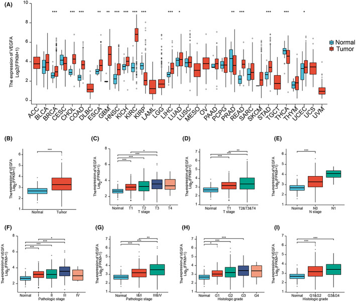
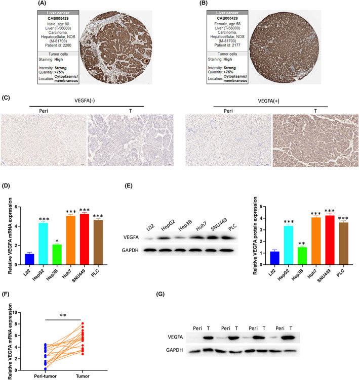
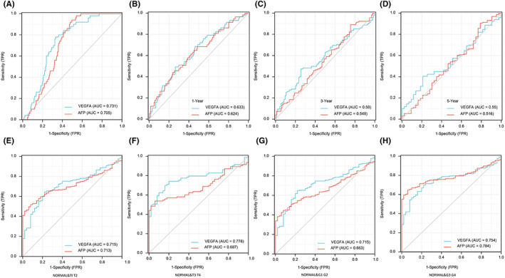
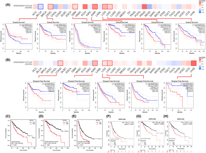
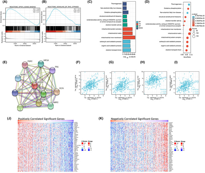
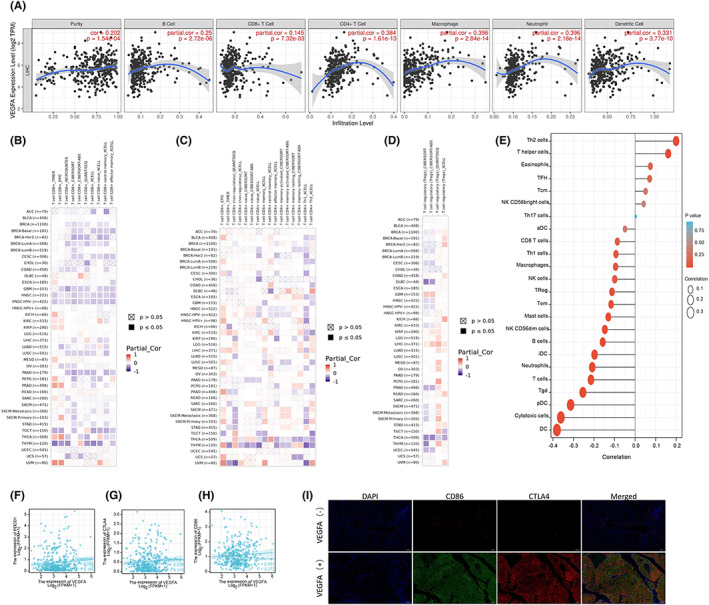
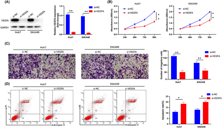
Hepatocellular carcinoma (HCC) is the fourth leading cause of cancer-related deaths among cancer patients. Vascular endothelial growth factor A (VEGFA) is involved in regulating biological processes, such as angiogenesis and vascular permeability, and is very closely related to the pathogenesis of various tumours, especially vascular-rich, solid tumours. Clinical data of patients with HCC and other tumours were analysed through public databases, such as the TCGA database, Gene Expression Omnibus database, Human Protein Atlas database, STRING, Tumour Immune Estimation Resource and Kaplan-Meier Plotter. The tumour tissues and adjacent normal tissues of patients with HCC from Hunan Provincial People's Hospital were collected to verify the expression of VEGFA by immunohistochemistry, immunofluorescence, Western blotting and qPCR. VEGFA expression is elevated in multiple tumour types and correlates with the prognosis of tumour patients. VEGFA is involved in regulating the tumour microenvironment and immune cell function in tumour development. Inhibition of VEGFA reduces proliferation, invasion, and migration and promotes apoptosis in HCC cells. VEGFA is a potential predictive biomarker for the diagnosis and prognosis of HCC.

Keywords: VEGFA; hepatocellular carcinoma; immune infiltrates; prognostic biomarker.


Conflict of interest statement

The authors confirm that there are no conflicts of interest.

Figures

FIGURE 1
FIGURE 1
Expression of VEGFA in tumour and normal tissues. (A) VEGFA expression levels in different types of tumour and normal tissues from TCGA database. (B) VEGFA expression levels in HCC from TCGA database. (C–I) VEGFA expression in different stages of HCC from TCGA database. *p < 0.05, **p < 0.01, ***p < 0.001. ACC, adrenocortical carcinoma; BLCA, bladder urothelial carcinoma; BRCA, breast invasive carcinoma; CHOL, cervical and endocervical cancers (CESC), cholangiocarcinoma; COAD, colon adenocarcinoma; DLBC, lymphoid neoplasm diffuse large B‐cell lymphoma; ESCA, oesophageal carcinoma; GBM, glioblastoma multiforme; HNSC, head and neck squamous cell carcinoma; KICH, kidney chromophobe; KIRC, kidney renal clear cell carcinoma; KIRP, kidney renal papillary cell carcinoma; LAML, acute myeloid leukaemia; LGG, brain lower grade glioma; HCC, hepatocellular carcinoma; LUAD, lung adenocarcinoma; LUSC, lung squamous cell carcinoma; MESO, mesothelioma; OV, ovarian serous cystadenocarcinoma; PAAD, pancreatic adenocarcinoma; PCPG, pheochromocytoma and paraganglioma; PRAD, prostate adenocarcinoma; READ, rectum adenocarcinoma; SKCM, skin cutaneous melanoma; STAD, stomach adenocarcinoma; TGCT, testicular germ cell tumours; THCA, thyroid carcinoma; UCEC, uterine corpus corpus endometrial carcinoma
FIGURE 2
FIGURE 2
Expression of VEGFA in HCC. (A, B) VEGFA was highly expressed in HCC tumours from the HPA. (C) VEGFA expression in peritumoral and HCC tissues by immunohistochemistry (IHC). (D) VEGFA mRNA expression in normal and HCC cells was detected by PCR. (E) VEGFA protein expression in normal hepatocytes and HCC cells was detected by Western blotting. (F) VEGFA mRNA expression in peritumoral and HCC tissues was detected by PCR. (G) VEGFA protein expression in peritumoral and HCC tissues was detected by Western blot. peri, peritumoral tissue; T, tumour tissue
FIGURE 3
FIGURE 3
ROC curve was established by TCGA Program database. (A) Diagnostic efficacy of VEGFA and AFP in HCC. (B–D) Diagnostic efficacy of VEGFA and AFP in different stages of HCC. (E–H) Differences in the diagnosis of VEGFA and AFP between normal patients and patients with HCC at different timepoints
FIGURE 4
FIGURE 4
Kaplan–Meier survival curve analysis of the prognostic significance of VEGFA expression in different types of human cancers. (A) The correlation between VEGFA expression levels and OS in different tumours was analysed using the GEPIA2 website, TGCA database and GTEx database. (B) The correlation between VEGFA expression levels and DFS in different tumours was analysed using the GEPIA2 website, TGCA database and GTEx database. (C–E) The expression level of VEGFA was negatively correlated with OS, DSS and PFI of HCC by TCGA database. (F–H) Kaplan–Meier Plotter was used to analyse the expression levels of VEGFA in the GEO, EGA and TCGA databases, and there was a negative correlation with OS, DSS and PFI of HCC
FIGURE 5
FIGURE 5
Enrichment analysis of VEGFA functional networks. (A, B) Enrichment plots by GSEA. (C, D) Enrichment of GO terms and KEGG for genes related to VEGFA. (E) PPI network of VEGFA. (F–I) Correlation between VEGFA expression levels and HIF1A, FLT1, FN1 and NRP1 expression levels. (J) The heatmap shows the top 50 genes positively related to VEGFA in the HCC cohort. (K) The heatmap shows the top 50 genes negatively related to VEGFA in the HCC cohort
FIGURE 6
FIGURE 6
Correlation analysis of VEGFA expression and infiltration levels of immune cells in tumour tissues. (A) VEGFA expression was positively correlated with tumour purity and infiltration levels of B cells, CD8+ T cells, CD4+ T cells, macrophages and DCs in HCC tissues based on the TIMER database. (B–D) The relationship between VEGFA mRNA expression and CD8+ T cells, CD4+ T cells and regulatory T cells in various tumours was evaluated using a variety of algorithms based on the TIMER database. (E) Correlation between VEGFA expression level and infiltration of various immune cells (Th2 Cell, T helper Cell, Eosinophils, TFH, Tcm, NK CD58bright Cell, Th17‐cell, sDC, CD8+ T‐cell, Th1‐Cell, Macrophages, NK‐Cell, Treg, Tem, Mast cell, NK CD58dim Cell, B‐cell, iDC, Neutrophils, T‐cell, Tgd, pDC, Cytotoxic Cell, DC) in HCC tissues. (F–H) Correlation between VEGFA expression level and PDCD1, CTLA4 and CD86 expression levels in HCC tissues based on TCGA database. (I) The correlation between VEGFA expression levels and PDCD1, CTLA4 and CD86 expression levels in HCC tissues was detected by immunofluorescence
FIGURE 7
FIGURE 7
Inhibition of VEGFA reduced the proliferation, invasion and migration of HCC cells and promoted apoptosis. (A) Transfection efficiency of si‐VEGFA in Huh7 and SUN449 cells. (B) Inhibition of VEGFA significantly inhibited the proliferation of HCC cells as shown by CCK8 assay. (C) Inhibition of VEGFA significantly inhibited HCC migration as shown by transwell assays. (D) Inhibition of VEGFA promoted apoptosis of HCC cells

References

    1. Yang JD, Hainaut P, Gores GJ, Amadou A, Plymoth A, Roberts LR. A global view of hepatocellular carcinoma: trends, risk, prevention and management. Nat Rev Gastroenterol Hepatol. 2019;16(10):589‐604. - PMC - PubMed
    1. Sung H, Ferlay J, Siegel RL, et al. Global cancer statistics 2020: GLOBOCAN estimates of incidence and mortality worldwide for 36 cancers in 185 countries. CA Cancer J Clin. 2021;71(3):209‐249. - PubMed
    1. Bray F, Ferlay J, Soerjomataram I, Siegel RL, Torre LA, Jemal A. Global cancer statistics 2018: GLOBOCAN estimates of incidence and mortality worldwide for 36 cancers in 185 countries. CA Cancer J Clin. 2018;68(6):394‐424. - PubMed
    1. McGlynn KA, Petrick JL, El‐Serag HB. Epidemiology of hepatocellular carcinoma. Hepatology. 2021;73(Suppl 1):4‐13. - PMC - PubMed
    1. Akinyemiju T, Abera S, Ahmed M, et al. The burden of primary liver cancer and underlying etiologies from 1990 to 2015 at the global, regional, and National Level: results from the global burden of disease study 2015. JAMA Oncol. 2017;3(12):1683‐1691. - PMC - PubMed

Publication types

MeSH terms

Substances