Review
. 2023 Feb 3;132(3):355-378.
doi: 10.1161/CIRCRESAHA.122.322332. Epub 2023 Feb 2.

Integrin-Dependent Cell-Matrix Adhesion in Endothelial Health and Disease

Affiliations
Review

Integrin-Dependent Cell-Matrix Adhesion in Endothelial Health and Disease

Jurjan Aman et al. Circ Res. .

Abstract

The endothelium is a dynamic, semipermeable layer lining all blood vessels, regulating blood vessel formation and barrier function. Proper composition and function of the endothelial barrier are required for fluid homeostasis, and clinical conditions characterized by barrier disruption are associated with severe morbidity and high mortality rates. Endothelial barrier properties are regulated by cell-cell junctions and intracellular signaling pathways governing the cytoskeleton, but recent insights indicate an increasingly important role for integrin-mediated cell-matrix adhesion and signaling in endothelial barrier regulation. Here, we discuss diseases characterized by endothelial barrier disruption, and provide an overview of the composition of endothelial cell-matrix adhesion complexes and associated signaling pathways, their crosstalk with cell-cell junctions, and with other receptors. We further present recent insights into the role of cell-matrix adhesions in the developing and mature/adult endothelium of various vascular beds, and discuss how the dynamic regulation and turnover of cell-matrix adhesions regulates endothelial barrier function in (patho)physiological conditions like angiogenesis, inflammation and in response to hemodynamic stress. Finally, as clinical conditions associated with vascular leak still lack direct treatment, we focus on how understanding of endothelial cell-matrix adhesion may provide novel targets for treatment, and discuss current translational challenges and future perspectives.

Keywords: angiogenesis; cell-matrix adhesions; edema; endothelium; inflammation; integrins.


Figures

Figure 1.
Figure 1.
Diseases associated with clinical vascular leak. Depicted is the vascular tree and associated cell types. Clinically relevant vascular leak predominantly takes place at the capillary and venular level. The clinical sequelae of vascular leak may vary according to etiological factors involved and to the affected vascular bed, ranging from general to organ-specific manifestations like in ARDS and angioedema. In addition, the vasoactive substances involved in vascular leak may vary according to clinical condition. Of note, many of these cytokines have been identified in cross-sectional studies. Ang-2 sensitizes the endothelium to vasoactive agents, among others via integrin binding. ARDS indicates acute respiratory distress syndrome; Ang-2, angiopoietin-2; DAMPs, disease-associated molecular patterns; IFN, interferon; Il, interleukin; Il1R, interleukin 1 receptor; MMP, matrix metalloproteinase; ROS, reactive oxygen species; sRAGE, soluble receptor for advanced glycation end products; TNF, tumor necrosis factor; VEGF, vascular endothelial growth factor; and vWF, von Willebrand Factor. Created with Biorender.com.
Figure 2.
Figure 2.
Basics of endothelial cell (EC)–cell and cell–extracellular matrix (ECM) adhesion. A, Properties of cell–cell junction proteins in ECs. Created with Biorender.com. B, Examples of vascular endothelial-cadherin (VE-cadherin) distribution in AJs in endothelial cells. The dotted box in the left image indicates linear junctions, arrows point to focal AJs (FAJs). In the middle image, arrows indicate cadherin fingers. C, Integrins are αβ heterodimeric cell-surface receptors that exist in a dynamic equilibrium between the bent conformation (with low affinity for ligand) and the extended conformation (with high affinity for ligand). The shift from bent-to-extended is called integrin activation and is promoted by binding of talin (talin-1 or talin-2) and kindlin (kindlin-1, -2, or -3) to the NPxY/NxxY motifs in the β-cytoplasmic tail, which can also recruit a variety of other proteins such as tensin. Ligand-bound integrins are connected to actin filaments and recruit many other factors with a structural, adapter, or signaling role, thus building adhesion complexes such as nascent adhesions. These can contain several different integrins, are not connected to actin stress fibers, and are promoted by Rac activity. In contrast, FAs are connected to stress fibers, and as many FA components are recruited in a tension-dependent manner, they grow and mature after increased RhoA activity. Whereas FAs are predominantly peripheral, specialized adhesions called FBs arise from the translocation of integrin α5β1 in a RhoA- and tensin-dependent manner from the cell periphery to the cell center, a process associated with cell contractility and FN fibrillogenesis. Created with Biorender.com. D, Examples of cell-ECM adhesions in ECs. Paxillin and P(Y) mark nascent adhesions (arrows)/FAs, elongated central adhesions positive for β1 are FBs. FAs indicates focal adhesions; FAK, focal adhesion kinase; FBs, fibrillar adhesions; FN, fibronectin; IPP, ILK-PINCH-parvin complex; kind2, kindlin-2; FN, fibronectin; p(Y), phosphorylated tyrosine; Src, proto-oncogene tyrosine-protein kinase; VASP, vasodilator-stimulated phosphoprotein; and vin, vinculin.
Figure 3.
Figure 3.
Integrin–extracellular matrix (ECM) interactions in the blood vessel wall. A, Main integrins and their ligands in the layers of blood vessels (see also Table 1). B, Schematic representation of some of the most common vascular ECM proteins and the main binding sites for integrins, growth factors, and other ECM molecules. Several more vascular ECM proteins are recognized by integrins, including decorin and versican. Integrins organize many matrix proteins into fibrils, such as collagen I (mainly integrins α11/α2β1) and fibronectin (FN; mainly integrin α5β1), and can also bind soluble forms of FN, vWF, and the γC fragment of fibrinogen. Many integrins recognize the RGD (Arginine-Glycine-Aspartate) sequence, for instance in fibrinogen, perlecan, vWF, fibrillins, and FN. In the endothelial basement membrane, laminins and collagen are abundant in quiescent endothelium. Upon injury, inflammation, or remodeling, the deposition of matrix and plasma proteins such as FN, vWF and fibrinogen is increased, as well as in provisional matrices associated with wound healing and angiogenesis. FN contains an RGD and PHSRN (also called the synergy site) motif. FN exists in several splice variants, and increased expression of the extra domain (ED)A and EDB isoforms of FN is associated with tissue remodeling. The variable region in FN, containing a binding site for α4 integrins, is also subject to alternative splicing and not depicted here. Note that the interaction between fibrillin and LTBP, which is responsible for regulated transforming growth factor (TGF)-β activation, is probably indirect. BMP indicates bone morphogenetic protein; Col, collagen; FGF, fibroblast growth factor; GFOGER, Glycine-Phenylanaline-Hydroxyproline-Glycine-Glutamic acid-Arginine; HGF, hepatocyte growth factor; LN, laminin; LTBP, latent transforming growth factor (TGF)-β binding protein; PDGF, platelet-derived growth factor; VE, versican; VEGF, vascular endothelial growth factor; and vWF, von Willebrand Factor. Created with Biorender.com.
Figure 4.
Figure 4.
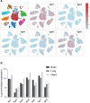
Expression profiles of integrin subunits in various endothelia. Data were obtained by in silico analysis of single-cell sequencing databases. Integrin subunit expression in various organs of adult mice, obtained from EC Atlas,; Carmeliet laboratory (https://endotheliomics.shinyapps.io/ec_atlas; A) and Rehman laboratory (http://rehmanlab.org/ribo; B).
Figure 5.
Figure 5.
Endosomal traffic of integrins and associated proteins in endothelial cells (ECs). A, Integrins can be internalized by caveolin- or clathrin-dependent endocytosis, mediated by several adaptor proteins (Dab2, Numb, AP2, GIPC1) that bind to motifs in integrin cytoplasmic tails (NPxY/NxxY in the β-tail, YxxΦ and SDA in some α-tails such as α5). Integrins can also be endocytosed in combination with other receptors; for example α5β1 co-internalizes with neuropilin-1 (Nrp-1), mediated by GIPC1 and myosin-VI (Myo VI). B, Internalized integrins and associated proteins are delivered to early endosomes (EEs) in a RIN2/Rab5 dependent manner. C, In EEs, sorting toward the recycling route takes place under control of several factors, including RhoJ, syntaxin-6 (STX6), sorting nexin-9 (SNX9), and Arf6, whose activation is stimulated by HGF and VEGF. Factors indicated in red have been identified as regulators of integrin trafficking in other cell types than ECs. D, Some recycling pathways combine with the biosynthetic route to deliver de novo synthesized FN from the Golgi together with recycled α5β1 to the cell-surface, where it is captured by liprin-α1. E, Internalized integrins can also be routed to lysosomes for degradation. F, VEGFR2 can exist on the cell-surface in complex with several other receptors, including integrins and Nrp-1, and follows similar routes as integrins. VEGFR2 internalization is promoted by Dab2 and Numb, as well as GIPC1 binding to Nrp-1, and myosin-VI mediates transport along actin filaments to EEs. Protection from degradation depends on RIN2/Rab5C, and recycling is promoted by Nrp-1 via Rab11+ vesicles, or via Rab4+ vesicles, where VEGFR2 co-traffics with integrin αvβ3 (G). Despite the identification of many individual components, a comprehensive view of how and where all of these components interact is largely lacking. In addition, while for many of these factors a role in protein trafficking during angiogenesis has been established, their role in barrier function remains less clear. AP2 indicates adaptor protein 2; Arf, ADP ribosylation factor; BRAG2, brefeldin A-resistant ArfGEF 2; Dab2, disabled-2; EE, early endosome; EV, endocytic vesicle; FN, fibronectin; GDP, guanine diphosphate; GIPC, GAIP interacting protein, C terminus; Grp1, general receptor for 3-phosphoinositides 1; GTP, guanine triphosphate; HGF, hepatocyte growth factor; Lys, lysosome; Myo VI, Myosin-VI; NPxY, Asparagine-Proline-x-Tyrosine; NxxY, Asparagine-x-x-Tyrosine; PGC, post-Golgi carrier; PTPRF, protein tyrosine phosphatase receptor type F; Rab4, Ras-like protein in brain 4; Rab5, Ras-like protein in brain 5; Rabank-5, rabankyrin-5; RE, recycling endosome; RhoJ, Ras homologous J; RIN2, Ras And Rab Interactor 2; SDA, Serine-Aspartic acid-Alanine; SE, sorting endosome; SNX27, Sorting Nexin 27; Ub, ubiquitin; VEGF, vascular endothelial growth factor; VEGFR, vascular endothelial growth factor receptor; and vps3, vacuolar protein sorting 3. Created with Biorender.com.
Figure 6.
Figure 6.
Crosstalk between cell–extracellular matrix (ECM) and cell–cell complexes in endothelial barrier function. FAs and AJs share many common components, including GTPases and their GEFs (guanine nucleotide exchange factors), adaptor proteins, actin-associated proteins, and several kinases (see also Table 2). Integrin αvβ3 and several β1 integrins can strengthen cell–cell junctions such as AJs (red arrow), although the mechanisms remain poorly understood. Integrins maintain overall cellular architecture and increase global Rac activation, leading to cortical actin assembly and strengthening of AJs. Rac activation is also locally increased at AJs in response to flow, by a complex including vascular endothelial cadherin (VE-cadherin), PTPRF, Notch, and the Rac GEF Trio, which also promotes Rac activation from FAs. In the absence of β1 integrins there is increased presence of VE-cadherin in vesicles but it is unclear if cell–ECM complexes actively repress VE-cadherin internalization or whether this is merely an effect of altered barrier properties. During barrier-disruptive conditions, integrin α5β1 increases RhoA activation from FAs or FBs, which increases their assembly and in addition promotes pulling on AJs (indicated by membrane deformation) by actin stress fiber formation and the recruitment of mechanosensitive proteins including vinculin, VASP, and zyxin. Integrin α5β1 also binds soluble ligands involved in barrier disruption, such as Ang-2, and elicits pro-inflammatory signaling through NF-κB, which increases FN deposition and further stimulates α5β1 ligation and signaling. This positive feedforward loop is further stimulated by a mechanosensitive complex consisting of PECAM-1, VEGFR2, and VE-cadherin and the mechanosensitive channel Piezo-1, which increase integrin activation in response to disturbed flow. α-act indicates α-actinin; AJ, adherens junctions; Ang-2, angiopoietin-2; β-pix, p21-activated protein kinase exchange factor beta; FAs, focal adhesions; FAJs, focal adherens junctions; FBs, fibrillar adhesions; IPP, ILK-PINCH-parvin complex; kind2, kindlin-2; NTM, Notch transmembrane domain; FN, fibronectin; LN, laminin; NF-κB, nuclear factor-κB; PECAM-1, platelet endothelial cell adhesion molecule-1; PTPRF, protein tyrosine phosphatase receptor type F; VASP, vasodilator-stimulated phosphoprotein; VEGFR2, vascular endothelial growth factor receptor-2; and vin, vinculin. Created with Biorender.com.
Figure 7.
Figure 7.
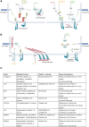
Pharmacological intervention at cell–extracellular matrix (ECM) adhesions. A, Targeting kinases and GTPases involved in turnover and spatial distribution of FAs. MAP4K4 is inhibited by several kinase inhibitors and phosphorylates moesin, which competes with talin for binding to the β1 integrin tail, thus favoring β1 integrin inactivation. MAP4K4 also activates Arf6 (inhibited by SecinH3), which drives endocytic recycling of β1 integrins, regulating their surface expression. c-Src, FAK, and the Abelson kinases are also inhibited by several tyrosine kinase inhibitors. c-Src and FAK regulate among others the phosphorylation of paxillin and FA turnover, involved in both barrier disruption and restoration. The Abl tyrosine kinase Arg increases FA disassembly and central redistribution of FAs. B, Integrin αvβ3 and peripheral, talin-bound β1 integrins enhance the integrity of VE-cadherin containing cell-cell junctions, protecting the endothelial barrier against barrier-disruptive agents. During barrier disruption, a talin-tensin switch parallels the formation of α5β1-containing FBs, which are located more centrally. While several compounds exist that target αvβ3 or α5β1, their potential role in barrier function remains to be established. The GTPase Rap1 can be activated by 007-acetoxymethyl and promotes integrin inside-out signaling, resulting in both integrin activation and cell spreading. Color coding for compounds and arrows: green indicates a barrier-protective effect, red indicates a barrier-disruptive effect, orange indicates compounds with unknown effect on endothelial barrier integrity. Created with Biorender.com. C, Summary of regulators of endothelial cell-matrix adhesion as targets for therapeutic intervention. Act indicates activator; AJ, adherens junction; Arg, Abl-related gene; cAMP, cyclic adenosine monophosphate; Epac, exchange protein activated by cAMP; FA, focal adhesion; FAK, focal adhesion kinase; FB, fibrillar adhesions; FN, fibronectin; inh, inhibitor; MAP4K4, Mitogen-Activated Protein Kinase Kinase Kinase Kinase; pax, paxillin; and VE-cadherin, vascular endothelial cadherin.

Similar articles

Cited by

References

    1. Claesson-Welsh L, Dejana E, McDonald DM. Permeability of the endothelial barrier: identifying and reconciling controversies. Trends Mol Med. 2021;27:314–331. doi: 10.1016/j.molmed.2020.11.006 - PMC - PubMed
    1. Shyy JYJ, Chien S. Role of integrins in endothelial mechanosensing of shear stress. Circ Res. 2002;91:769–775. doi: 10.1161/01.res.0000038487.19924.18 - PubMed
    1. Komarova YA, Kruse K, Mehta D, Malik AB. Protein interactions at endothelial junctions and signaling mechanisms regulating endothelial permeability. Circ Res. 2017;120:179–206. doi: 10.1161/circresaha.116.306534 - PMC - PubMed
    1. Yano K, Liaw PC, Mullington JM, Shih SC, Okada H, Bodyak N, Kang PM, Toltl L, Belikoff B, Buras J, et al. . Vascular endothelial growth factor is an important determinant of sepsis morbidity and mortality. J Exp Med. 2006;203:1447–1458. doi: 10.1084/jem.20060375 - PMC - PubMed
    1. London NR, Zhu W, Bozza FA, Smith MCP, Greif DM, Sorensen LK, Chen L, Kaminoh Y, Chan AC, Passi SF, et al. . Targeting Robo4-dependent Slit signaling to survive the cytokine storm in sepsis and influenza. Sci Transl Med. 2010;2:23ra–2319. - PMC - PubMed

Publication types

MeSH terms