

Bilateral Calcaneus Transfers for the Treatment of Congenital Tibial Deficiencies: A Novel Surgical Technique and Case Report
- PMID: 36732306
- PMCID: PMC9726303
- DOI: 10.5435/JAAOSGlobal-D-22-00070
Bilateral Calcaneus Transfers for the Treatment of Congenital Tibial Deficiencies: A Novel Surgical Technique and Case Report
Abstract
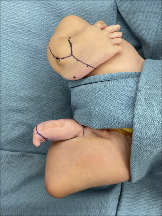
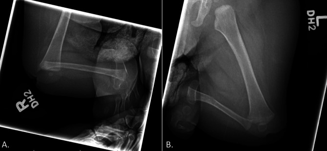
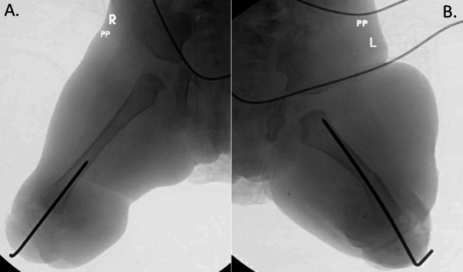
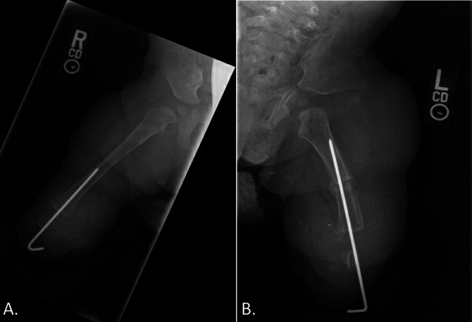
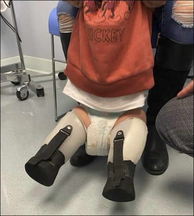
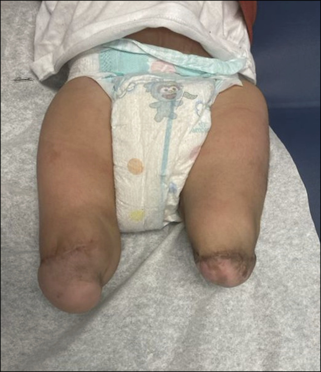
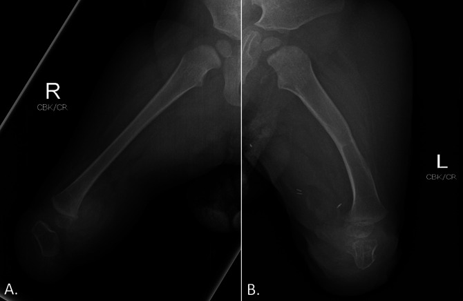
Tibial deficiency (also known as tibial hemimelia) is a rare condition with variable presentation. A 2-month-old patient presented with absent bilateral tibias. When the patient was 1 year, a novel reconstructive surgery was done. A bilateral fibular resection with pedicled calcaneus transfer was done, allowing for transfer of the calcaneus along with the overlying glabrous skin and soft tissues to the end of the femur. The patient was permitted to weight-bear after the 4-week postoperative follow-up. At the six-month follow-up, the patient was able to pull to stand and walk with assistance without any reports of pain.
Copyright © 2022 The Authors. Published by Wolters Kluwer Health, Inc. on behalf of the American Academy of Orthopaedic Surgeons.
Figures

Similar articles
-
One-stage reconstruction of composite bone and soft-tissue defects in traumatic lower extremities.Plast Reconstr Surg. 2004 Nov;114(6):1457-66. doi: 10.1097/01.prs.0000138811.88807.65. Plast Reconstr Surg. 2004. PMID: 15509933 Review.
-
Asymmetric limb lengthening in the treatment of tibial hemimelia caused by osteomyelitis: A case report.Medicine (Baltimore). 2019 Jan;98(3):e14031. doi: 10.1097/MD.0000000000014031. Medicine (Baltimore). 2019. PMID: 30653110 Free PMC article.
-
Residual malformations and leg length discrepancy after treatment of fibular hemimelia.J Orthop Surg Res. 2011 Sep 27;6:51. doi: 10.1186/1749-799X-6-51. J Orthop Surg Res. 2011. PMID: 21951397 Free PMC article.
-
Limb salvage treatment for Gollop-Wolfgang complex (femoral bifurcation, complete tibial hemimelia, and hand ectrodactyly).J Pediatr Orthop B. 2013 Sep;22(5):457-63. doi: 10.1097/BPB.0b013e3283620640. J Pediatr Orthop B. 2013. PMID: 23660549
-
Recent advances in surgery of lower limb deficiencies.Clin Orthop Relat Res. 1980 May;(148):97-105. Clin Orthop Relat Res. 1980. PMID: 6991192 Review.
Cited by
-
New Reconstruction Technique in Combined Tibial and Fibular Hemimelia.J Am Acad Orthop Surg Glob Res Rev. 2025 Jul 21;9(7):e25.00040. doi: 10.5435/JAAOSGlobal-D-25-00040. eCollection 2025 Jul 1. J Am Acad Orthop Surg Glob Res Rev. 2025. PMID: 40689695 Free PMC article.
References
-
- Orimolade E, Ikem I, Oginni L, Odunsi A: Femoral bifurcation with ipsilateral tibia hemimelia: Early outcome of ablation and prosthetic fitting. Niger J Clin Pract 2011;14:492-494. - PubMed
-
- Sahoo PK, Sahu MM, Das SP: Clinical spectrum of congenital tibial hemimelia in 35 limbs of 24 patients: A single center observational study from India. Eur J Med Genet 2019;62:103666. - PubMed
-
- Paley D, Chong DY: Tibial hemimelia. Pediatr Lower Limb Deformities, Newark, NJ, Springer; 2016:455-481.
Publication types
MeSH terms
LinkOut - more resources
Full Text Sources
