. 2023 Feb 2;14(1):572.
doi: 10.1038/s41467-023-36250-4.

SARS-CoV-2 humoral and cellular immunity following different combinations of vaccination and breakthrough infection

Affiliations

SARS-CoV-2 humoral and cellular immunity following different combinations of vaccination and breakthrough infection

Jernej Pušnik et al. Nat Commun. .

Abstract

The elicited anti-SARS-CoV-2 immunity is becoming increasingly complex with individuals receiving a different number of vaccine doses paired with or without recovery from breakthrough infections with different variants. Here we analyze the immunity of individuals that initially received two doses of mRNA vaccine and either received a booster vaccination, recovered from a breakthrough infection, or both. Our data suggest that two vaccine doses and delta breakthrough infection or three vaccine doses and optionally omicron or delta infection provide better B cell immunity than the initial two doses of mRNA vaccine with or without alpha breakthrough infection. A particularly potent B cell response against the currently circulating omicron variant (B. 1.1.529) was observed for thrice vaccinated individuals with omicron breakthrough infection; a 46-fold increase in plasma neutralization compared to two vaccine doses (p < 0.0001). The T cell response after two vaccine doses is not significantly influenced by additional antigen exposures. Of note, individuals with hybrid immunity show better correlated adaptive immune responses compared to those only vaccinated. Taken together, our data provide a detailed insight into SARS-CoV-2 immunity following different antigen exposure scenarios.


Conflict of interest statement

The authors declare no competing interests. The idea, the plan, the concept, the protocol, the conduct, the data analysis, and the writing of the manuscript of this study were independent of any third parties, including the funding agency.

Figures
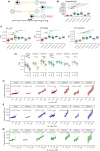
Fig. 1
Fig. 1. Plasma antibody response after different combinations of vaccination and breakthrough infection.
a Schematic chronological representation of antigen exposures defining the seven groups compared in this study. Detailed information about the antigen exposure and sampling time points is provided as supplemental material. b S1-specific IgG levels in international units measured for the seven groups with different antigen exposure histories. Exact p values in sequential order from the upmost bracket: *p = 0.028, ***p = 0.00021, *p = 0.032, **p < 0.0038, ****p < 0.0001, ****p < 0.0001, *p = 0.011, ****p < 0.0001. c Plasma neutralization capacity against wild-type, delta, and omicron variants. Exact p values in sequential order from the upmost bracket of the left graph: ***p = 0.00071, **p = 0.0027, *p = 0.021, **p = 0.0023, ****p < 0.0001, *p = 0.04, **p = 0.0034, *p = 0.04, ****p < 0.0001, **p = 0.0035, **p = 0.0018, **p = 0.0081, **p = 0.0011, ****p < 0.0001. d Comparison of neutralization susceptibility of SARS-CoV-2 variants for each of the analyzed groups. The IC50 medians are given numerically for each boxplot. Exact p values in sequential order from the upmost bracket of the leftmost group: ****p < 0.0001, ****p < 0.0001, ****p < 0.0001, *p = 0.047, *p = 0.047, ***p = 0.00073, ***p = 0.00073, ****p < 0.0001, *p = 0.023, ****p < 0.0001, *p = 0.047, *p = 0.047, *p = 0.047, *p = 0.047. In panels bd the data is displayed as boxplots, indicating the first quartile, median, and third quartile, with individual data points. Correlation between plasma levels of S1-specific IgG and plasma neutralization capacity for e wild-type, f delta, g omicron variants. The 95% confidence intervals around the line of best fit are displayed as shading. The r and p values are given for each line. The following numbers of biologically independent samples were included in each group for all the graphs in this figure: 2xVacc, n = 54; 2xVacc+α, n = 7; 2xVacc+δ, n = 13; 3xVacc, n = 23; 3xVacc+ο, n = 10; 3xVacc+α, n = 7; 3xVacc+δ, n = 7. Differences between the groups were assessed using the Mann–Whitney test or Wilcoxon test for matched data. The strength of correlations was assessed by Spearman’s correlation test. Correction for multiple testing was performed using Holm’s method, all statistical tests were two-sided. Source data are provided as a Source data file.
Fig. 2
Fig. 2. SARS-CoV-2-specific antibodies in saliva.
S1-specific a IgG (exact p values in sequential order from the upmost bracket: **p = 0.0091, ***p = 0.0003, *p = 0.015, ***p < 0.00012, ****p < 0.0001, **p = 0.0032, **p = 0.0091), b IgA (exact p values in sequential order from the upmost bracket: **p = 0.0015, *p = 0.019, ****p < 0.0001, ***p < 0.00027, ****p < 0.0001, **p = 0.0027, **p = 0.0044, ****p < 0.0001), and c IgG+IgA (exact p values in sequential order from the upmost bracket: **p = 0.0098, ***p = 0.00041, **p < 0.0015, **p < 0.0023, ****p < 0.0001, ****p < 0.0001, *p < 0.017, *p < 0.016, ***p < 0.00034) levels in saliva for the seven groups with different antigen exposure histories. Data is displayed as boxplots (a, b), indicating the first quartile, median, and third quartile, or bars showing the median (c) with individual data points. d Relative proportions of IgA and IgG isotypes among the S1-specific antibodies in the saliva of individuals with different antigen exposure histories. Data are shown as a stacked bar plot for each individual. Statistically significant differences between the groups are indicated in the table next to the graph (*p = 0.011, ****p < 0.0001). e Correlations between the plasma and salivary S1-specific IgG for the seven antigen exposure groups. The 95% confidence intervals around the line of best fit are displayed as shading. The r and p values are given for each line. The following numbers of biologically independent samples were included in each group for all the graphs in this figure: 2xVacc, n = 51; 2xVacc+α, n = 7; 2xVacc+δ, n = 10; 3xVacc, n = 17; 3xVacc+ο, n = 10; 3xVacc+α, n = 7; 3xVacc+δ, n = 7. Differences between the groups were assessed using the two-sided Mann–Whitney test with Holm’s correction for multiple testing. The strength of correlations was assessed by the two-sided Spearman’s correlation test. Source data are provided as a Source data file.
Fig. 3
Fig. 3. SARS-CoV-2-specific memory B cell response after different combinations of vaccination and breakthrough infection.
a Representative flow cytometry pseudocolor plots for detection of S1-specific memory B cells with different BCRs. For the detailed gating strategy see Supplemental Fig. 2. Frequencies of b IgG+ (exact p values in sequential order from the upmost bracket: *p = 0.014, **p = 0.005, *p = 0.015, *p = 0.014, **p < 0.004, *p = 0.047), c IgA+, d IgM+, and e total S1-specific memory B cells (exact p values in sequential order from the upmost bracket: *p = 0.014, **p = 0.005, *p = 0.015, *p = 0.014, **p < 0.004, *p = 0.047) in peripheral blood of individuals belonging to the seven antigen exposure groups expressed as a percentage of B cells. The data is displayed as boxplots (bd), indicating the first quartile, median, and third quartile, or bars showing the median (e) with individual data points. f Relative proportions of S1-specific memory B cells bearing BCRs of a different isotype (color-coded) presented as stacked bar plots. Each section of the bar represents the median proportion of an isotype. The following numbers of biologically independent samples were included in each group for all the graphs in this figure: 2xVacc, n = 27; 2xVacc+α, n = 7; 2xVacc+δ, n = 13; 3xVacc, n = 23; 3xVacc+ο, n = 10; 3xVacc+α, n = 7; 3xVacc+δ, n = 7. Differences between the groups were assessed using the two-sided Mann–Whitney test with Holm’s correction for multiple testing. Source data are provided as a Source data file.
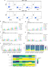
Fig. 4
Fig. 4. SARS-CoV-2-specific T cell response to different antigen exposure scenarios.
Frequencies of S-specific a representative flow cytometry pseudocolor plots demonstrating the detection of SARS-CoV-2-specific CD4+ and CD8+ T cells with different effector functions; cytotoxicity (CD107a and IFNγ expression), IFNγ-expression, IL-2 expression, and TNFα expression. For the detailed gating strategy see Supplemental Fig. 3. b CD4+ T cells and c CD8+ T cells with different effector functions as a percentage of bulk T cells. Frequencies of N-specific d CD4+ T cells and e CD8+ T cells with different effector functions as a percentage of bulk T cells. Different antigen exposure groups are color-coded. For panels be, the data is displayed as box-whisker-plots, indicating minimum, first quartile, median, third quartile, and maximum, with individual data points. f Frequencies of S or N-specific T cells with different functions, not discriminating the CD4+ and CD8+ T cells. The exact p values in sequential order from the leftmost bracket: **p = 0.01, *p = 0.012. Data is displayed as bars showing the median with individual data points. g Relative proportions of S-specific CD4+, N-specific CD4+, S-specific CD8+, and N-specific CD8+ T cells performing different functions for each antigen exposure group. Data are presented as stacked bar plots. CD4+/CD8+ T cells and their specificities for N or S proteins are color-coded. h Percentage of individuals with detectable SARS-CoV-2-specific CD4+ and CD8+ T cells within each group. The percentage of responders (individuals where the frequency of stimulation-responding T cells was higher than in the negative control) is color-coded. The following numbers of biologically independent samples were included in each group for all the graphs in this figure: 2xVacc, n = 27; 2xVacc+α, n = 7; 2xVacc+δ, n = 13; 3xVacc, n = 23; 3xVacc+ο, n = 10; 3xVacc+α, n = 7; 3xVacc+δ, n = 7. Differences between the groups were assessed using the two-sided Mann–Whitney test with Holm’s correction for multiple testing. Source data are provided as a Source data file.
Fig. 5
Fig. 5. Correlation between the parameters of the adaptive immune response to different antigen exposure scenarios.
Correlation matrices demonstrate the strength of correlations between the measured immune parameters for each of the antigen exposure groups. The strength of a correlation (Spearman’s correlation coefficient) is depicted by the size and color of the circle, significance is indicated by asterisks. The exact p values are given in the Source data file. Green lines separate correlations between different branches of the measured immune parameters; antibodies, memory B cells, and T cells. The following numbers of biologically independent samples were included in each group for all correlated parameters: 2xVacc, n = 54 (for plasma antibodies), 2xVacc, n = 51 (for plasma antibodies), 2xVacc, n = 27 (for B and T cells); 2xVacc+α, n = 7; 2xVacc+δ, n = 13 (for plasma antibodies, B cells, and T cells), 2xVacc+δ, n = 10 (for saliva antibodies); 3xVacc, n = 23 (for plasma antibodies, B cells, and T cells), 3xVacc, n = 17 (for saliva antibodies); 3xVacc+ο, n = 10; 3xVacc+α, n = 7; 3xVacc+δ, n = 7. The strength of correlations was assessed by the two-sided Spearman’s correlation test. Source data are provided as a Source data file.

References

    1. Eyre DW, et al. Effect of Covid-19 vaccination on transmission of alpha and delta variants. N. Engl. J. Med. 2022;386:744–756. doi: 10.1056/NEJMoa2116597. - DOI - PMC - PubMed
    1. Prunas O, et al. Vaccination with BNT162b2 reduces transmission of SARS-CoV-2 to household contacts in Israel. Science. 2022;375:1151–1154. doi: 10.1126/science.abl4292. - DOI - PMC - PubMed
    1. Macchia A, et al. Evaluation of a COVID-19 vaccine campaign and SARS-CoV-2 infection and mortality among adults aged 60 years and older in a middle-income country. JAMA Netw. Open. 2021;4:e2130800–e2130800. doi: 10.1001/jamanetworkopen.2021.30800. - DOI - PMC - PubMed
    1. Turner JS, et al. SARS-CoV-2 mRNA vaccines induce persistent human germinal centre responses. Nature. 2021;596:109–113. doi: 10.1038/s41586-021-03738-2. - DOI - PMC - PubMed
    1. Walsh EE, et al. Safety and immunogenicity of two RNA-based Covid-19 vaccine candidates. N. Engl. J. Med. 2020;383:2439–2450. doi: 10.1056/NEJMoa2027906. - DOI - PMC - PubMed

Publication types

Supplementary concepts