. 2023 Jan-Dec:19:17448069231158290.
doi: 10.1177/17448069231158290.

Toll-like receptor 4 signaling pathway in sensory neurons mediates remifentanil-induced postoperative hyperalgesia via transient receptor potential ankyrin 1

Affiliations

Toll-like receptor 4 signaling pathway in sensory neurons mediates remifentanil-induced postoperative hyperalgesia via transient receptor potential ankyrin 1

Xiaowen Liu et al. Mol Pain. 2023 Jan-Dec.

Abstract

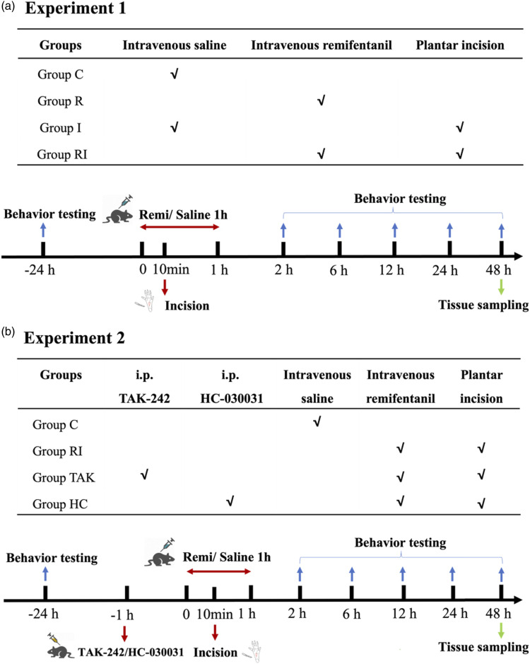
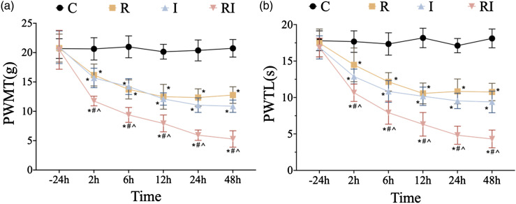
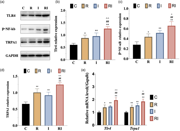
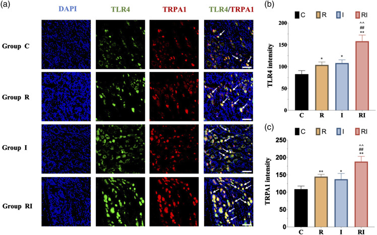
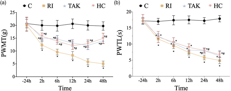
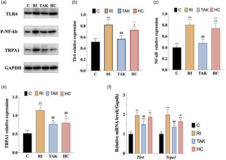
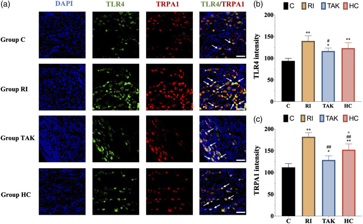
Background: Remifentanil-induced postoperative hyperalgesia (RIH) refers to a state of hyperalgesia or aggravated pre-existing pain after remifentanil exposure. There has been considerable interest in understanding and preventing RIH. However, the mechanisms responsible for RIH are still not completely understood. Toll-like receptor 4 (TLR4), a classic innate immune receptor, has been detected in sensory neurons and participates in various nociceptive conditions, whereas its role in RIH remains unclear. Transient receptor potential ankyrin 1 (TRPA1) always serves as a nociceptive channel, whereas its role in RIH has not yet been investigated. This study aimed to determine whether the TLR4 signaling pathway in sensory neurons engaged in the development of RIH and the possible involvement of TRPA1 during this process. Methods: A rat model of remifentanil-induced postoperative hyperalgesia (RIH) was established, which presented decreased paw withdrawal mechanical threshold (PWMT) and paw withdrawal thermal latency (PWTL). The mRNA and protein expression levels of TLR4, phosphorylated NF-κB, and TRPA1 in the dorsal root ganglion (DRG) from RIH model were analyzed by real-time PCR, western blot, and immunofluorescence. The TLR4 antagonist TAK-242 and the TRPA1 antagonist HC-030031 were applied to determine the role of sensory neuron TLR4 signaling and TRPA1 in RIH. Results: Compared with control, PWMT and PWTL were significantly decreased in RIH model. Moreover, the mRNA and protein expression of TLR4 and TRPA1 in DRG were upregulated after remifentanil exposure together with increased NF-κB phosphorylation. TLR4 antagonist TAK-242 mitigated mechanical pain in RIH together with downregulated expression of TLR4, phosphorylated NF-κB, and TRPA1 in DRG neurons. In addition, TRPA1 antagonist HC-030031 also alleviated mechanical pain and decreased TRPA1 expression in RIH without affecting TLR4 signaling in DRG. Conclusions: Taken together, these results suggested that activation of TLR4 signaling pathway engaged in the development of RIH by regulating TRPA1 in DRG neurons. Blocking TLR4 and TRPA1 might serve as a promising therapeutic strategy for RIH.

Keywords: Remifentanil-induced postoperative hyperalgesia; dorsal root ganglion; opioids; toll-like receptor 4 signaling; transient receptor potential ankyrin 1.


Conflict of interest statement

The author(s) declared no potential conflicts of interest with respect to the research, authorship, and/or publication of this article.

Figures

Figure
1.
Figure 1.
Schematic diagram of the experiment protocol. a. Experiment 1: To confirm the changes of pain behavior and protein expression after remifentanil exposure in RIH rats. b. Experiment 2: To explore the role of TLR4 signaling and TRPA1 after remifentanil exposure in RIH rats.
Figure 2.
Figure 2.
Time course of PWMT and PWTL after remifentanil and incision. PWMT, paw withdrawal mechanical threshold; PWTL, paw withdrawal thermal latency. a-b. Compared with control group, mechanical allodynia (a) and thermal hyperalgesia (b) were induced at 2 h, 6 h, 12 h, 24 h, and 48 h time points in group R (rats with remifentanil infusion), group I (rats with plantar incision), and group RI (rats with remifentanil infusion and incision). There was no statistically significant difference in PWMT and PWTL between group R and group I. Compared with group R and group I, mechanical allodynia (a) and thermal hyperalgesia (b) were aggravated in group RI, indicating a rat model of remifentanil-induced postoperative hyperalgesia was successfully established. All the data are means ± SD (n = 6) and analyzed by two-way ANOVA with Tukey’s post hoc comparisons. *p < .05 versus group C at the same time point; #p < .05 versus group R at the same time point; ^p < .05 versus group I at the same time point.
Figure 3.
Figure 3.
Expression of TLR4 signaling and TRPA1 in DRG neurons in rats with RIH. a. Representative bands of Western blot for the expression of TLR4, p-NF-κB, and TPRA1 in DRG at 48 h after remifentanil exposure. b–d. Compared with the control group, the expression of TLR4, p-NF-κB, and TPRA1 were significantly upregulated in group R and group I. Moreover, the expression of TLR4, p-NF-κB, and TPRA1 were further increased in group RI. e. The mRNA expression of Tlr4 and Trpa1 were elevated significantly in rats with RIH model, when compared to the other three groups. All the data are expressed as means ± SD (n = 4) and analyzed by one-way ANOVA with Tukey’s post hoc comparisons. *p < .05, **p < .01 versus group C; #p < .05, ##p < .01 versus group R; ^p < .05, ^^p < .01 versus group I.
Figure
4.
Figure 4.
Immunofluorescence of TLR4 and TRPA1 expression in DRG neurons. a. Representative immunofluorescence micrographs, with the scale bar equaling to 50 μm. b. The statistical results of TLR4 expression in DRG neurons. The expression of TLR4 in group RI was upregulated compared with any other groups. c. The expression of TRPA1 in group RI was upregulated compared with any other groups. All the data are expressed as means ± SD (n = 4) and analyzed by one-way ANOVA with Tukey’s post hoc comparisons. *p < 0.05, **p < 0.01 versus group C; ##p < 0.01 versus group R; ^^p < 0.01 versus group I.
Figure 5.
Figure 5.
Effects of inhibiting TLR4 or TRPA1 on mechanical and thermal sensitivity. PWMT, paw withdrawal mechanical threshold; PWTL, paw withdrawal thermal latency. a-b. For rats in group TAK, TLR4 was inhibited by TAK-242. For rats in group HC, TRPA1 was inhibited by HC-030031. Compared with group RI, mechanical allodynia (a) was alleviated at 2 h, 6 h, 12 h, 24 h, and 48 h time points in group TAK and HC. However, thermal hyperalgesia (b) in group TAK and group HC showed no significant differences at most time points compared to that in group RI. All the data are means ± SD (n = 6) and analyzed by two-way ANOVA with Tukey’s post hoc comparisons. *p < .05 versus group C at the same time point; #p < .05 versus group RI at the same time point.
Figure 6.
Figure 6.
Expression of TLR4 signaling and TRPA1 in DRG neurons after inhibiting TLR4 or TRPA1 in RIH model. a. Representative bands of Western blot for the expression of TLR4, p-NF-κB, and TPRA1 in DRG at 48 h after remifentanil exposure. b-d. When compared with group RI, inhibition of TLR4 decreased the expression of TLR4 (b), p-NF-κB (c), and TRPA1 (d). However, inhibition of TRPA1 did not affect the expression of TLR4 (b) or p-NF-κB (c). e. Consistent with the protein expression trends of TLR4 and TPRA1 after inhibiting TLR4, the mRNA expression levels of Tlr4 and Trpa1 were significantly downregulated in group TAK compared with group RI. All the data are expressed as means ± SD (n = 4) and analyzed by one-way ANOVA with Tukey’s post hoc comparisons. *p < .05, **p < .01 versus group C; #p < .05, ##p < 0.01 versus group RI; ^p < .05, ^^p < .01 versus group TAK.
Figure
7.
Figure 7.
Immunofluorescence of TLR4 and TRPA1 expression after inhibiting TLR4 or TRPA1 in DRG neurons. a. Representative immunofluorescence micrographs, with the scale bar equaling to 50 μm. b. The statistical results of TLR4 expression in DRG neurons. The upregulation of TLR4 in rats with RIH was inhibited after blocking TLR4. c. The statistical results of TRPA1 expression in DRG neurons. The expression of TRPA1 in group TAK and group HC were downregulated after blocking TLR4 or TRPA1 when compared with group RI. All the data are expressed as means ± SD (n = 4) and analyzed by one-way ANOVA with Tukey’s post hoc comparisons. *p < .05, **p < .01 versus group C; #p < .05, ##p < .01 versus group RI; ^p < 0.05 versus group TAK.

Similar articles

Cited by

References

    1. Colvin LA, Bull F, Hales TG. Perioperative opioid analgesia-when is enough too much? A review of opioid-induced tolerance and hyperalgesia. Lancet (London, England) 2019; 393(10180): 1558–1568. - PubMed
    1. Lee M, Silverman SM, Hansen H, Patel VB, Manchikanti L. A comprehensive review of opioid-induced hyperalgesia. Pain Physician 2011; 14(2): 145–161. - PubMed
    1. Higgins C, Smith BH, Matthews K. Evidence of opioid-induced hyperalgesia in clinical populations after chronic opioid exposure: a systematic review and meta-analysis. Br J Anaesth 2019; 122(6): e114–e126. - PubMed
    1. Neuman MD, Bateman BT, Wunsch H. Inappropriate opioid prescription after surgery. Lancet (London, England) 2019; 393(10180): 1547–1557. - PMC - PubMed
    1. Kim Y, Bae H, Yoo S, Park SK, Lim YJ, Sakura S, Kim JT. Effect of remifentanil on postoperative analgesic consumption in patients undergoing shoulder arthroplasty after interscalene brachial plexus block: a randomized controlled trial. J Anesth 2022; 36(4): 506–513. - PubMed

Publication types

Substances

LinkOut - more resources