. 2023 Jan 23:25:73-85.
doi: 10.1016/j.bioactmat.2023.01.008. eCollection 2023 Jul.

The combination of eddy thermal effect of biodegradable magnesium with immune checkpoint blockade shows enhanced efficacy against osteosarcoma

Affiliations

The combination of eddy thermal effect of biodegradable magnesium with immune checkpoint blockade shows enhanced efficacy against osteosarcoma

Jun Ge et al. Bioact Mater. .

Abstract

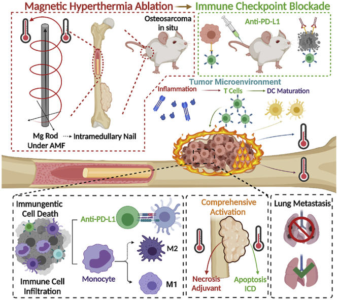
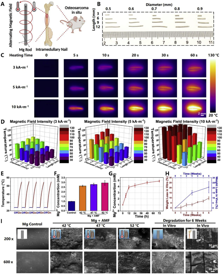
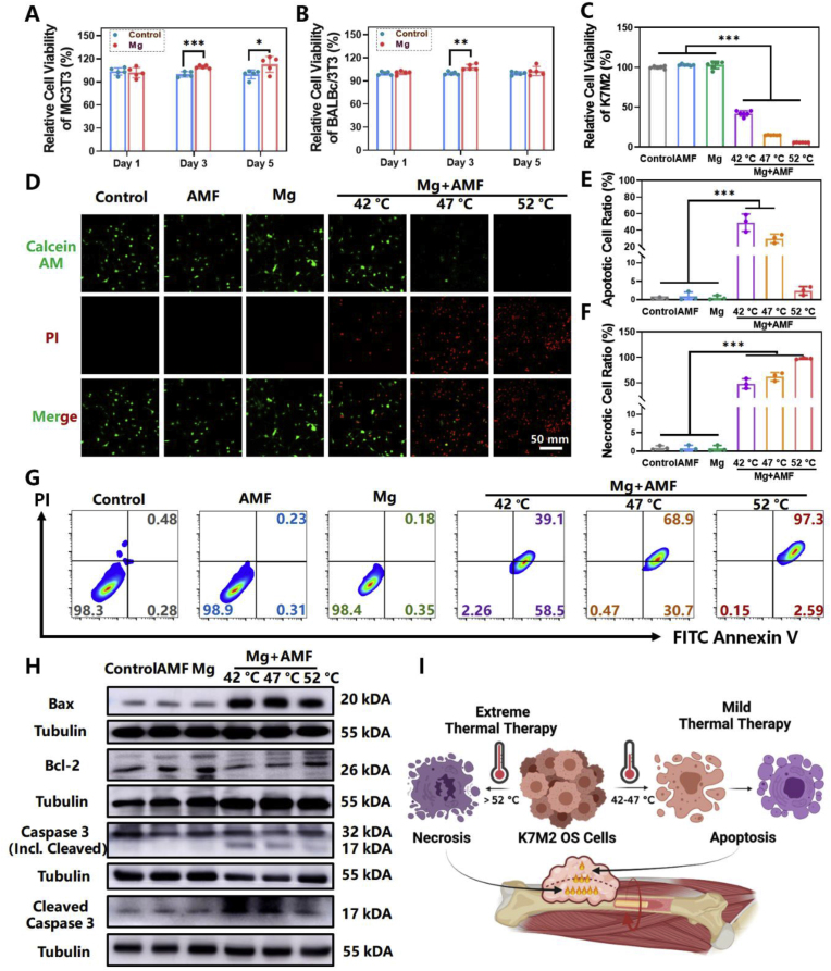
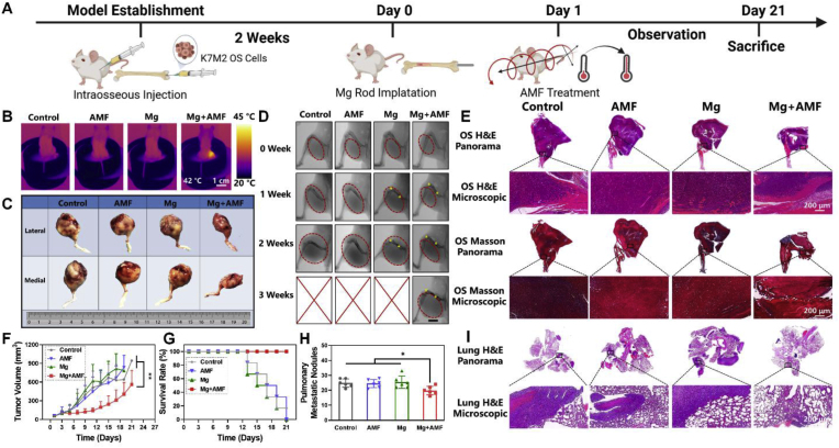
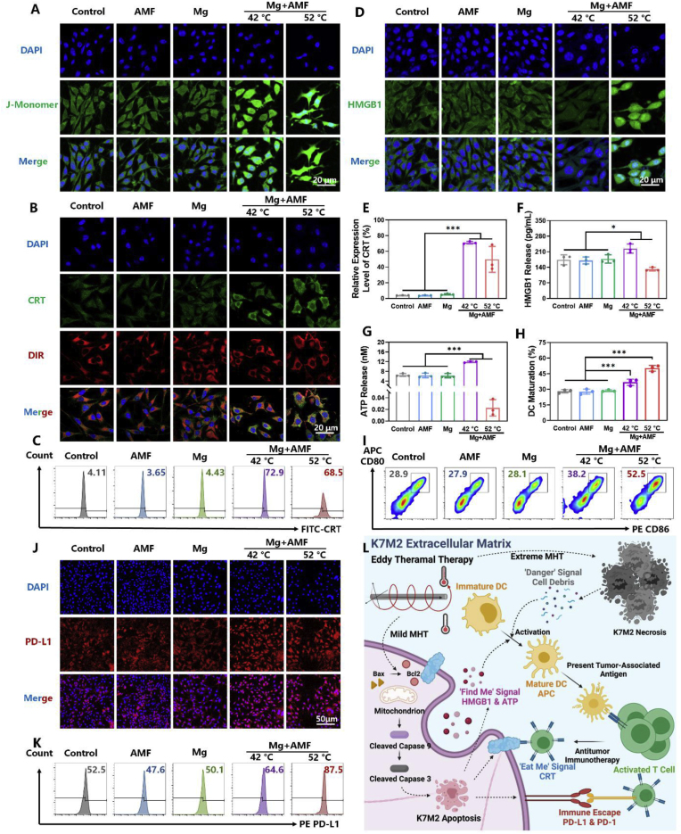
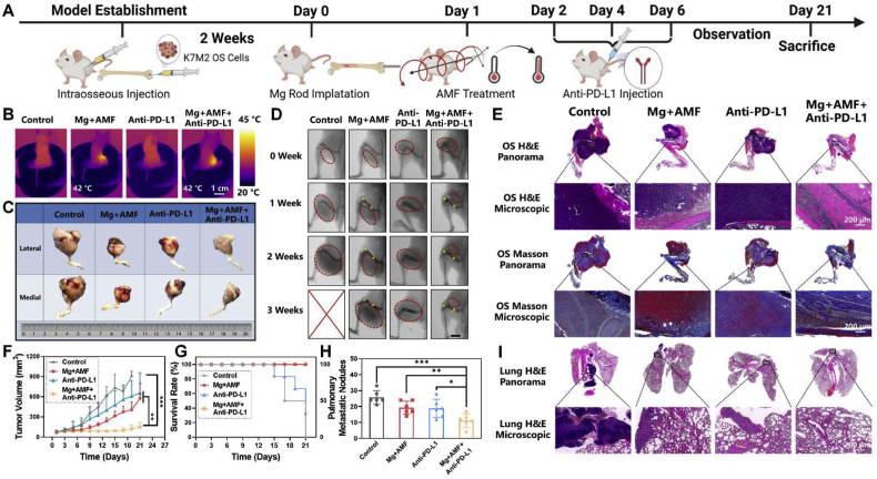
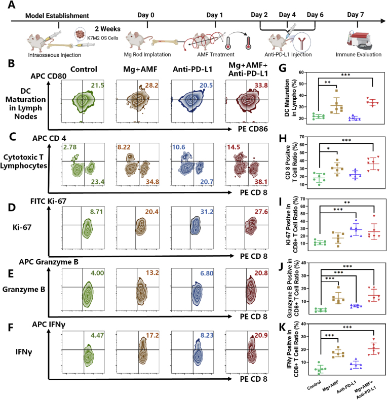
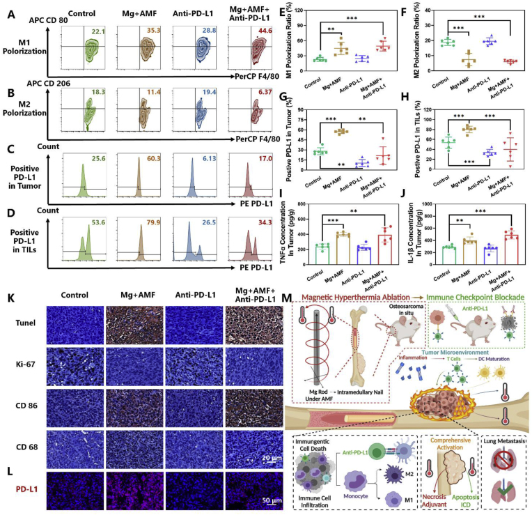
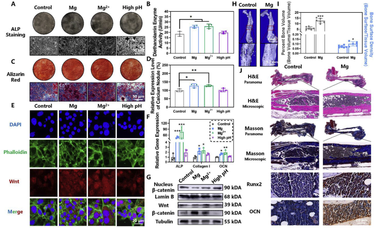
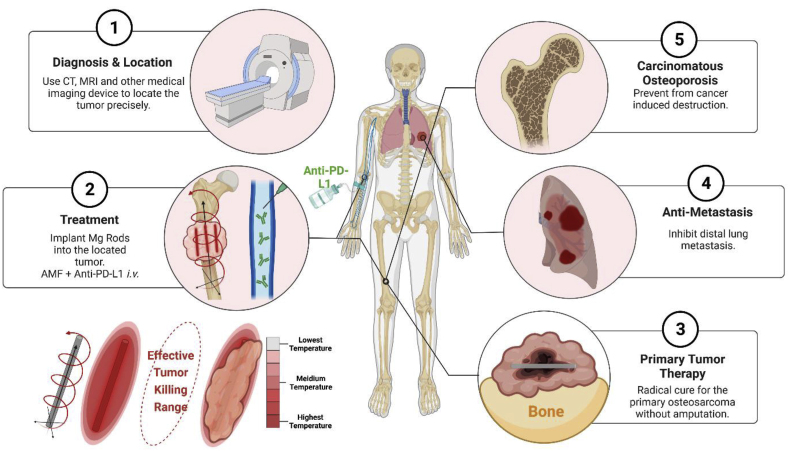
Osteosarcoma (OS) patients have a poor prognosis due to its high degree of heterogeneity and high rate of metastasis. Magnetic hyperthermia therapy (MHT) combined with immunotherapy is an effective strategy to treat solid and metastatic tumors. Here, we combined biodegradable magnesium (Mg) macroscale rods, which acted as an eddy thermo-magnetic agent under a low external alternating magnetic field, and immunotherapy to achieve a radical cure for OS. The eddy thermal effect (ETE) of the Mg rods (MgR) showed outstanding cytotoxic effects and enhanced the maturation of dendritic cells (DCs), and the mild MHT induced the immunogenic cell death (ICD) in the OS cells. Combined with immune checkpoint blockade (ICB) therapy, we obtained an excellent curative effect against OS, and a further evaluation demonstrated that the local MHT induced by the MgR increased T cells infiltration and the polarization of M1 macrophages. Interestingly, the biodegradable MgR also promoted bone osteogenesis. Our work highlighted the uneven ETE mediated by the biodegradable MgR induced a comprehensive immunologic activation in the OS tumor microenvironment (TME), which would inspire the application of MHT for the effective treatment of OS.

Keywords: Eddy thermal effect; Magnesium rods (MgR); Osteosarcoma; Osteosarcoma therapy; Tumor microenvironment (TME) immunotherapy.


Conflict of interest statement

The authors declare that they have no known competing financial interests or personal relationships that could have appeared to influence the work reported in this paper.

Figures

Image 1
Graphical abstract
Fig. 1
Fig. 1
Eddy thermal effects of magnesium rods. A. Schematic illustration for the eddy thermal therapy of MgR on OS. B. The general photograph of MgR. C. The eddy thermal images of the MgR under AMF. D. The temperature of the ETE depends on the magnetic field intensity, MgR diameter and MgR length. E. Heating cycle of the MgR under 5 kA · m−1 AMF. Heating time: 20 s. F. Mg ions released from MgR under AMF. G. Mg ions released from MgR in high glucose-medium (N = 3). H. Weight loss of MgR in vitro and in vivo for 6 weeks (N = 6). I. SEM images of MgR. The diameter and the length of MgR is 0.7 mm and 9 mm.
Fig. 2
Fig. 2
In vitro magnetic hyperthermia ablation of magnesium rod on OS. A. Cell viability of MC3T3-E1 after exposure to MgR for long term (N = 5). B. Cell viability of BALBc/3T3 after exposure to MgR for long term (N = 5). C. Cell viability of K7M2 8 h after the ETE of MgR (N = 6). D. Live/Dead staining 8 h after the ETE of MgR. Quantity analysis of the flow cytometer on the K7M2 OS cells after the ETE of MgR (N = 3). E. Apoptotic; F. Necrotic. G. Representative flow cytometer data of Annexin V/PI. H. Western blotting of K7M2 OS cells 1 h after the ETE of MgR. I. Schematic illustration for the magnetic hyperthermia ablation of OS. The diameter and the length of MgR is 0.7 mm and 9 mm.
Fig. 3
Fig. 3
In vivo inhibitory effect of the eddy thermal effect of Mg on OS. A. Schematic illustration for in vivo evaluation. B. Thermal pictures of mice implanted with MgR (D = 0.7 mm, L = 9 mm) under AMF. C. The general photographs of osteosarcoma harvested from mice. D. The X-ray images of mice after various treatments. Red dashed lines indicate the tumors. Yellow dots indicate the MgR implants.E. Representative H&E and Masson staining of osteosarcoma after the various treatments. F. The growth curve of osteosarcoma after the various treatments (N = 6). G. Survival rate of mice after the various treatments (N = 6). H. Pulmonary metastatic nodules of mice (N = 6). I. Representative H&E staining of lungs after the various treatments.
Fig. 4
Fig. 4
The eddy thermal effect of Mg rods leads to immunogenic cell death and dendritic cells maturation of osteosarcoma cells. CLSM images of A. JC-1, B. CRT and D. HMGB1 of K7M2 after MHT. C. Representative flow cytometer data of CRT after the various treatments (N = 3). E. Quantity analysis of the flow cytometer on CRT after the various treatments (N = 3). F. ELISA kit for the detecting HMGB1 release of K7M2 after MHT (N = 3). G. ATP release of K7M2 after MHT (N = 3). H. Quantity analysis of the flow cytometer on DC maturation (N = 3, in the gate of CD11c+). I. Representative flow cytometer data of DCs maturation after the various treatments. J. CLSM images of PD-L1 after MHT. K. Representative flow cytometer data of PD-L1 after the various treatments. L. Schematic illustration for the magnetic hyperthermia ablation leads to immunogenic cell death and dendritic cells maturation of osteosarcoma cells.
Fig. 5
Fig. 5
In vivo radical treatment of osteosarcoma by the eddy thermal effect of Mg rod combined with immune checkpoint blocking. A. Schematic illustration for the in vivo evaluation. B. Thermal pictures of mice implanted with MgR (D = 0.7 mm, L = 9 mm) under AMF. C. The general photographs of osteosarcoma harvested from mice. D. The X-ray images of mice after the various treatments. Red dashed lines indicate the tumors. Yellow dots indicate the MgR implants. E. Representative H&E and Masson staining of osteosarcoma after the various treatments. F. The growth curve of osteosarcoma after the various treatments (N = 6). G. Survival rate of mice after the various treatments (N = 6). H. Pulmonary metastatic nodules of mice (N = 6). I. Representative H&E staining of lungs after the various treatments.
Fig. 6
Fig. 6
The eddy thermal therapy of Mg rods improves DCsmaturation and T cellsfunction. A. Schematic illustration for in vivo evaluation. Representative flow cytometer data of B. DCs maturation in lymph nodes (in the gate of CD11c+), C. T-cell (in the gate of CD3+ and CD45+), D. Ki-67, E. Granzyme B and F. IFNγ in CD8+ T cells (in the gate of CD3+, CD45+ and CD8+). Quantity analysis of the flow cytometer (N = 6) on G. DCs maturation in lymph nodes, H. CD8+ T-cell in tumors, I. Ki-67, J. Granzyme B and K. IFNγ in CD8+ T cells.
Fig. 7
Fig. 7
The eddy thermal therapy of Mg rods modulates TME and enhanced immunotherapy. Representative flow cytometer data of A. M1 Polorization, B. M2 Polorization in tumors (in the gate of CD11b+ and CD45+) and C. PD-L1 positive in tumor (in the gate of CD45). D. PD-L1 positive in tumor infiltrating lymphocytes (in the gate of CD45+). Quantity analysis of the flow cytometer (N = 6) on E. M1 Polorization, F. M2 Polorization in tumors, G. PD-L1 ratio on tumor, H. PD-L1 ratio on TILs, and ELISA kit for detecting I. TNF-α and J. IL-1β in tumor after the various treatments (N = 6). K. Histological analysis of tumors after the various treatments. L. Histological immunofluorescence of PD-L1 of tumors after the various treatments. M. Schematic illustration of the comprehensive radical therapy of the ETE of MgR.
Fig. 8
Fig. 8
Magnesium rods enhance osteogenic activity. A. ALP staining and B. activity of MC3T3-E1 after 7 days of osteogenesis induction (N = 3). C. Alizarin red staining of MC3T3-E1 after 14 days of osteogenesis differentiation. D. Quantity analysis of alizarin red staining (N = 3). E. Confocal fluorescence images of WNT in MC3T3-E1 after 7 days of osteogenesis induction. F. Gene expression of ALP, collagen I, and OCN after various treatments (N = 3). G. Western blotting analysis of WNT/β-catenin pathway in MC3T3-E1 after 7 days of osteogenesis differentiation. H. Representative Micro-CT reconstruction images. I. Quantity analysis of Micro-CT (N = 6). J. Histological analysis of the fractured femur after the different treatments.
Scheme 1
Scheme 1
Schematic illustration of the clinical application of the MHT and immunotherapy combinatory treatment for OS. By implanting the Mg rods with the corresponding size into the tumor under imaging navigation, MHT combined with immune checkpoint blockade can effectively destroy the tumor from deep inside and prevent from further metastasis. The excellent osteogenic property of Mg rods may further prevent cancer-induced OS.

References

    1. Durfee R.A., Mohammed M., Luu H.H. Review of osteosarcoma and current management. Rheumatology and therapy. 2016;3(2):221–243. - PMC - PubMed
    1. Meazza C., Scanagatta P. Metastatic osteosarcoma: a challenging multidisciplinary treatment. Expet Rev. Anticancer Ther. 2016;16(5):543–556. - PubMed
    1. Bielack S.S., Hecker-Nolting S., Blattmann C., Kager L. Advances in the management of osteosarcoma. 2016;F1000Research:5. - PMC - PubMed
    1. Lindsey B.A., Markel J.E., Kleinerman E.S. Osteosarcoma overview. Rheumatology and therapy. 2017;4(1):25–43. - PMC - PubMed
    1. Yang B., Yin J., Chen Y., Pan S., Yao H., Gao Y., Shi J. 2D‐black‐phosphorus‐reinforced 3D‐printed scaffolds: a stepwise countermeasure for osteosarcoma. Adv. Mater. 2018;30(10) - PubMed

LinkOut - more resources