. 2023 Apr 1;44(13):1170-1185.
doi: 10.1093/eurheartj/ehad028.

Mitochondrial dysfunction in human hypertrophic cardiomyopathy is linked to cardiomyocyte architecture disruption and corrected by improving NADH-driven mitochondrial respiration

Affiliations

Mitochondrial dysfunction in human hypertrophic cardiomyopathy is linked to cardiomyocyte architecture disruption and corrected by improving NADH-driven mitochondrial respiration

Edgar E Nollet et al. Eur Heart J. .

Abstract

Aims: Genetic hypertrophic cardiomyopathy (HCM) is caused by mutations in sarcomere protein-encoding genes (i.e. genotype-positive HCM). In an increasing number of patients, HCM occurs in the absence of a mutation (i.e. genotype-negative HCM). Mitochondrial dysfunction is thought to be a key driver of pathological remodelling in HCM. Reports of mitochondrial respiratory function and specific disease-modifying treatment options in patients with HCM are scarce.

Methods and results: Respirometry was performed on septal myectomy tissue from patients with HCM (n = 59) to evaluate oxidative phosphorylation and fatty acid oxidation. Mitochondrial dysfunction was most notably reflected by impaired NADH-linked respiration. In genotype-negative patients, but not genotype-positive patients, NADH-linked respiration was markedly depressed in patients with an indexed septal thickness ≥10 compared with <10. Mitochondrial dysfunction was not explained by reduced abundance or fragmentation of mitochondria, as evaluated by transmission electron microscopy. Rather, improper organization of mitochondria relative to myofibrils (expressed as a percentage of disorganized mitochondria) was strongly associated with mitochondrial dysfunction. Pre-incubation with the cardiolipin-stabilizing drug elamipretide and raising mitochondrial NAD+ levels both boosted NADH-linked respiration.

Conclusion: Mitochondrial dysfunction is explained by cardiomyocyte architecture disruption and is linked to septal hypertrophy in genotype-negative HCM. Despite severe myocardial remodelling mitochondria were responsive to treatments aimed at restoring respiratory function, eliciting the mitochondria as a drug target to prevent and ameliorate cardiac disease in HCM. Mitochondria-targeting therapy may particularly benefit genotype-negative patients with HCM, given the tight link between mitochondrial impairment and septal thickening in this subpopulation.

Keywords: Cardiomyocyte architecture; Hypertrophic cardiomyopathy; Metabolism; Mitochondrial dysfunction; Mitochondrial therapy.


Conflict of interest statement

K.B.M. is a consultant for Bristol Myers Squibb and receives research support from Amgen.

Figures

Structured Graphical Abstract
Our analyses reveal substantial variation in mitochondrial dysfunction in patients with hypertrophic cardiomyopathy. At the cellular level, this was strongly linked to mitochondrial disorganization. In patients with no known causative sarcomere mutation, mitochondrial dysfunction was associated with septal thickening. Mitochondria were responsive to treatments aimed at boosting NADH-driven respiration, indicating therapeutic potential of the mitochondria despite severe cardiac remodelling.
Figure 1
Figure 1
Overview of samples and methods. In total, 59 myectomy tissue samples from patients with hypertrophic cardiomyopathy and 14 non-failing donor left ventricle heart tissue samples were used. Forty-seven patients with hypertrophic cardiomyopathy underwent genetic testing via next-generation sequencing of all known hypertrophic cardiomyopathy-linked genes. A causative mutation was found in 23 patients (genotype-positive; Gpositive); the other 24 patients were termed genotype-negative (Gnegative). Clinical information and echocardiographic features were available for all subjects included in this study. Respirometry was performed on hypertrophic cardiomyopathy samples to evaluate oxidative phosphorylation (N = 59) and fatty acid oxidation (N = 37) capacity. The effects of elamipretide treatment and raising NAD+ levels were studied in 13 samples. Transmission electron microscopy was done on 19 hypertrophic cardiomyopathy samples and 5 non-failing donor heart samples to assess cardiomyocyte ultrastructure. Abundance of OXPHOS protein subunits, proteins involved in mitochondria dynamics, and tubulin profile was determined via western blot on 12–19 hypertrophic cardiomyopathy samples and 5–8 non-failing donor heart samples.
Figure 2
Figure 2
Oxygen flux through oxidative phosphorylation (OXPHOS) pathways in myectomy tissue samples from patients with hypertrophic cardiomyopathy. The combination of substrates (green) and uncouplers/inhibitors (purple) that were used to evaluate respiration is shown in (A). Average trace throughout the experimental protocol and respiratory state of each combination of substrates, uncouplers, and inhibitors is shown in (B). Parameters in (C–F) are absolute values. Normalized NADH-linked respiration indicates NADH-linked respiration expressed as a fraction of OXPHOS capacity (G). Normalized succinate-linked respiration indicates succinate-linked respiration expressed as a fraction of uncoupled respiration (H). Electron transfer system excess capacity indicates the percentage difference between OXPHOS capacity and uncoupled respiration (I). Normalized leak indicates leak respiration expressed as a fraction of OXPHOS capacity (J). In all parameters that were acquired, no differences were observed between genotype-positive (Gpositive) patients (N = 23, 14 males, 9 females), genotype-negative (Gnegative) patients (N = 24, 16 males, 8 females), and patients with unknown genotype status (N = 12; 6 males, 6 females) (C–J). Data are expressed as mean ± standard error of the mean. Statistical tests: one-way analysis of variance with Tukey’s multiple comparisons test in (C, D, F, and J); Brown–Forsythe analysis of variance with Dunnett’s T3 multiple comparisons test in (E); the Kruskal–Wallis test with Dunn’s multiple comparisons test in (G–I). Blue and pink symbols indicate male and female patients, respectively. CytC, cytochrome-c; FCCP, carbonyl cyanide p-trifluoro-methoxyphenyl hydrazine; ROX, residual oxygen consumption.
Figure 3
Figure 3
Oxygen flux stimulated by oxidation of octanoylcarnitine in myectomy tissue samples from patients with hypertrophic cardiomyopathy. Octanoylcarnitine freely passes the mitochondrial membranes and is activated in the mitochondrial matrix via conversion to octanoyl-CoA, yielding NADH, FADH2, and acetyl-CoA via β-oxidation; acetyl-CoA generates NADH via stepwise conversion to citrate, α-ketoglutarate, and succinate, which is subsequently transported out of the mitochondria (A). Average trace throughout the experimental protocol is shown in (B). No differences were observed between genotype-positive (Gpositive; N = 15; 8 males, 7 females), genotype-negative (Gnegative; N = 14, 7 males, 7 females) patients, and patients with unknown genotype status (N = 8, 4 males, 4 females) (Kruskal–Wallis test with Dunn’s multiple comparisons test) (C). Data are expressed as mean ± standard error of the mean. Blue and pink symbols indicate male and female patients respectively. CytC, cytochrome-c.
Figure 4
Figure 4
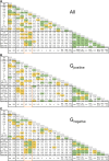
Correlation matrices showing correlations between clinical, echocardiographic, and mitochondrial function parameters in patients with hypertrophic cardiomyopathy. Matrix A shows correlations of all patients combined; matrix B shows genotype-positive (Gpositive) patients; matrix C shows genotype-negative (Gnegative) patients. Values indicate Pearson’s r. Correlations significant at P < 0.05 are marked in yellow; correlations significant at P < 0.01 are marked in green. Correlations between mitochondrial functional parameters and echocardiographic parameters are highlighted by an orange frame. BMI, body mass index; LAD, left atrial diameter; LADi, left atrial diameter indexed to body surface area; IVS, interventricular septum thickness; IVSi, interventricular septum thickness indexed to body surface area; LVD, left ventricular diameter at end-diastole; LVDi, left ventricular diameter at end-diastole indexed to body surface area; Dec. time, deceleration time; Rest LV pg, resting left ventricular outflow tract pressure gradient; Prov. LV pg, left ventricular outflow tract pressure gradient after Valsalva manoeuvre; OXPHOS, total oxidative phosphorylation capacity; Unc., uncoupled respiration; Succ.-linked, succinate-linked respiration; FAO, fatty acid oxidation; Norm. NADH, NADH-linked respiration normalized to OXPHOS; Norm. succ., succinate-linked respiration normalized to uncoupled respiration; Norm. leak, leak respiration normalized to OXPHOS.
Figure 5
Figure 5
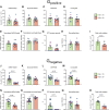
Mitochondrial respiration in myectomy tissue samples from genotype-negative (Gnegative) and genotype-positive (Gpositive) patients with hypertrophic cardiomyopathy with an indexed interventricular septum thickness (IVSi) <10 and ≥10. Parameters in (A–D, I, J–M, and R) represent absolute values. Normalized NADH-linked respiration indicates NADH-linked respiration expressed as a fraction of OXPHOS capacity (E and H). Normalized succinate-linked respiration indicates succinate-linked respiration expressed as a fraction of uncoupled respiration (F and O). Electron transfer system excess capacity indicates the percentage difference between OXPHOS capacity and uncoupled respiration (G and P). Normalized leak indicates leak respiration expressed as a fraction of OXPHOS capacity (H and Q). Data are expressed as mean ± standard error of the mean. Sample sizes: Gpositive patients in (A–H) N = 23 (14 males, 9 females) and (I) N = 15 (8 males, 7 females); Gnegative patients in (JQ) N = 24 (16 males, 8 females) and (R) N = 14 (7 males, 7 females). Statistical tests: unpaired Student’s t-test in (BF, H, I, KO, Q, and R); Welch’s t-test in (J); Mann–Whitney test in (A, G, and P). A Bonferroni correction was applied in (A–H and J–Q). Blue and pink symbols indicate male and female patients, respectively.
Figure 6
Figure 6
Overview of transmission electron microscopy analyses in tissue samples from non-failing donor hearts and myectomy samples from patients with hypertrophic cardiomyopathy. (A and B) Examples of how images were annotated to quantify myofibrillar area, mitochondrial area, and individual mitochondrial morphology, the results of which are displayed in (C–E). (F–H) Depict correlations between oxidative phosphorylation (OXPHOS) capacity and myofibrillar area, mitochondrial area, and mitochondrial size in myectomy samples from patients with hypertrophic cardiomyopathy. Examples of TEM images of non-failing donor heart samples and hypertrophic cardiomyopathy myectomy tissue samples in which all interfibrillar mitochondria are annotated; varying degrees of mitochondrial disorganization are visible in hypertrophic cardiomyopathy (I). Percentage disorganized mitochondria in non-failing donor heart tissue and hypertrophic cardiomyopathy myectomy tissue (J). Correlations between percentage disorganized mitochondria and mitochondrial function parameters in myectomy samples from patients with hypertrophic cardiomyopathy (K–P). (K–M and P) Absolute respiration values. Normalized NADH-linked respiration indicates NADH-linked respiration expressed as a fraction of OXPHOS capacity (N). Normalized succinate-linked respiration indicates succinate-linked respiration expressed as a fraction of uncoupled respiration (O). Scale bars indicate 2 µm (A, B, and I). Data are expressed as mean ± standard error of the mean. Correlations are displayed as linear regression; dotted lines indicate a 95% confidence interval. R2 indicates the coefficient of determination. Sample sizes: non-failing donors N = 5 (2 males, 3 females); genotype-positive (Gpositive) patients N = 10 (7 males, 3 females); genotype-negative (Gnegative) patients N = 8 (5 males, 3 females) in (C–H). Non-failing donors N = 5 (2 males, 3 females); Gpositive patients N = 10 (7 males, 3 females); Gnegative patients N = 9 (6 males, 3 females) in J. Gpositive patients N = 10 (7 males, 3 females); Gnegative patients N = 9 (6 males, 3 females) in (K–P). Statistical tests: Welch’s t-test in (C); unpaired Student’s t-test in (D, E, and J). Bonferroni corrections were applied to all group comparisons (C–E and J) and to all correlations (F–H and K–P). Blue and pink symbols indicate male and female patients, respectively.
Figure 7
Figure 7
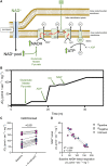
Effect of NAD+ supplementation on mitochondrial function ex vivo in myectomy samples from patients with hypertrophic cardiomyopathy. (A) The proposed mechanism via which NAD+ supplementation boosts the capacity of NADH-linked respiration. NAD+ enters the mitochondrial matrix via SLC25A51, raising the mitochondrial NAD+ pool and increasing NAD+ availability for NADH production and subsequent respiration via complex I. Average trace during the experimental protocol is shown in (B). The absolute rate of NADH-linked respiration before and after adding NAD+ is seen in (C) (paired two-tailed t-test). (D) The association between NADH-linked respiration before adding NAD+ and the absolute respiration increase that followed upon adding NAD+. Correlation is displayed as linear regression; dotted lines indicate a 95% confidence interval. R2 indicates the coefficient of determination. Sample sizes: N = 13 (4 males, 9 females) in (C and D). Blue and pink symbols indicate male and female patients, respectively. Gpositive, genotype-positive; Gnegative, genotype-negative.
Figure 8
Figure 8
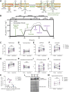
Effect of elamipretide incubation on mitochondrial function ex vivo in myectomy samples from patients with hypertrophic cardiomyopathy. (A) The proposed mechanism via which elamipretide improves mitochondrial respiratory function. Elamipretide associates with cardiolipin, stabilizing respiratory supercomplexes which ameliorates electron transfer through complexes. Average traces for both treatment conditions during the experimental protocol are shown in (B). (C–F) Absolute respiration values. Normalized NADH-linked respiration indicates NADH-linked respiration expressed as a fraction of OXPHOS capacity (G). Normalized succinate-linked respiration indicates succinate-linked respiration expressed as a fraction of uncoupled respiration (H). Electron transfer system (ETS) excess capacity indicates the percentage difference between OXPHOS capacity and uncoupled respiration (I). Normalized leak indicates leak respiration expressed as a fraction of OXPHOS capacity (J). (K) The association between NADH-linked respiration in vehicle-treated samples and the absolute difference in NADH-linked respiration relative to the matched elamipretide-treated samples. Representative Blue Native gel electrophoresis lanes showing supercomplex-bound complex I in myectomy samples from patients with hypertrophic cardiomyopathy that were treated with vehicle or 100 µM elamipretide (L); quantification in (M). Treatment effects were analysed using the paired two-tailed t-test (C–J and M). A Bonferroni correction was applied in (C–J). Correlation (K) is displayed as linear regression; dotted lines indicate a 95% confidence interval. R2 indicates the coefficient of determination. Sample sizes: N = 13 (4 males, 9 females) in (C–K); N = 5 (1 male, 4 females) in (L and M). Blue and pink symbols indicate male and female patients, respectively. CytC, cytochrome-c; FCCP, carbonyl cyanide p-trifluoro-methoxyphenyl hydrazine; ROX, residual oxygen consumption; Gpositive, genotype-positive; Gnegative, genotype-negative; TPS, total protein stain.

Comment in

References

    1. Semsarian C, Ingles J, Maron MS, Maron BJ. New perspectives on the prevalence of hypertrophic cardiomyopathy. J Am Coll Cardiol 2015;65:1249–1254. 10.1016/j.jacc.2015.01.019 - DOI - PubMed
    1. Ommen SR, Mital S, Burke MA, Day SM, Deswal A, Elliott P, et al. . 2020 AHA/ACC guideline for the diagnosis and treatment of patients with hypertrophic cardiomyopathy: a report of the American College of Cardiology/American Heart Association Joint Committee on Clinical Practice Guidelines. J Am Coll Cardiol 2020;76:e159–e240. 10.1016/j.jacc.2020.08.045 - DOI - PubMed
    1. Maron BJ. Clinical course and management of hypertrophic cardiomyopathy. N Engl J Med 2018;379:1977. 10.1056/NEJMra1710575 - DOI - PubMed
    1. Ho CY, Charron P, Richard P, Girolami F, Van Spaendonck-Zwarts KY, Pinto Y. Genetic advances in sarcomeric cardiomyopathies: state of the art. Cardiovasc Res 2015;105:397–408. 10.1093/cvr/cvv025 - DOI - PMC - PubMed
    1. Neubauer S, Kolm P, Ho CY, Kwong RY, Desai MY, Dolman SF, et al. . Distinct subgroups in hypertrophic cardiomyopathy in the NHLBI HCM registry. J Am Coll Cardiol 2019;74:2333–2345. 10.1016/j.jacc.2019.08.1057 - DOI - PMC - PubMed

Publication types