

RCAS1 increases cell morphological changes in murine fibroblasts by reducing p38 phosphorylation
- PMID: 36734265
- PMCID: PMC9926866
- DOI: 10.3892/mmr.2023.12949
RCAS1 increases cell morphological changes in murine fibroblasts by reducing p38 phosphorylation
Abstract
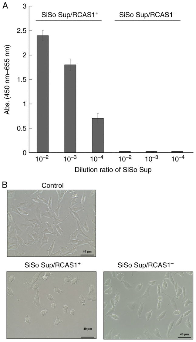
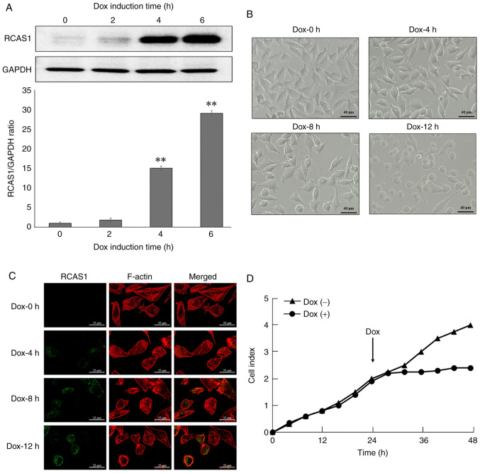
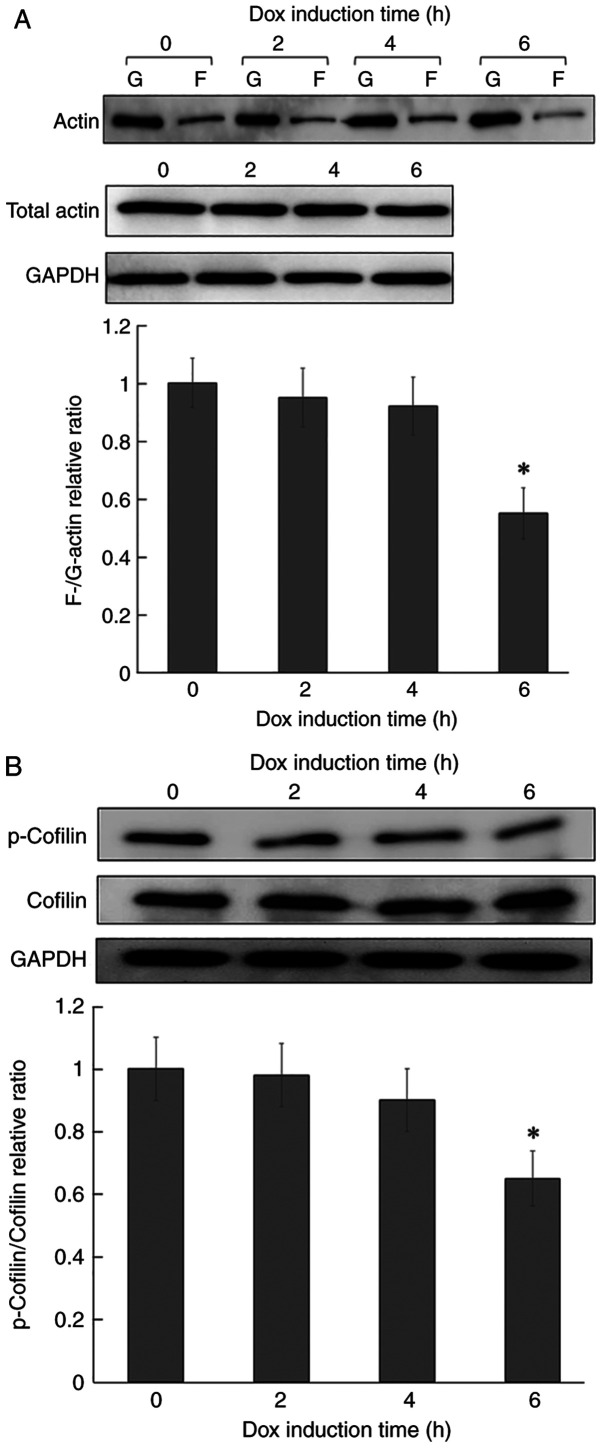
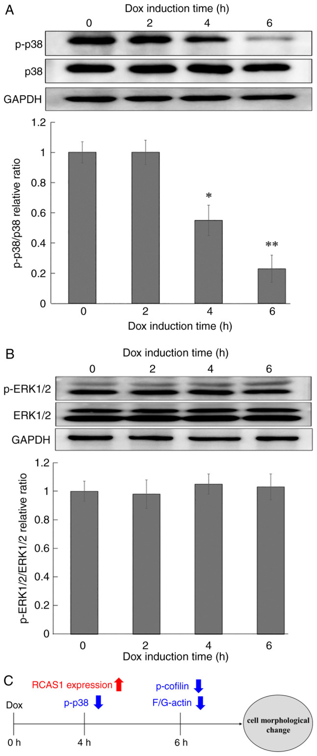
Receptor‑binding cancer antigen expressed on SiSo cells (RCAS1) is a tumor‑associated antigen that is expressed in a number of human malignancies. RCAS1 acts as a ligand for a putative RCAS1 receptor that is present on various human cells including T and B lymphocytes and natural killer cells, in which it induces cell growth inhibition and apoptosis. It has been suggested that RCAS1 might serve an important role in tumor cell evasion from the host immune system. In fact, RCAS1 expression is related to malignant characteristics including tumor size, invasion depth, clinical stage and poor overall survival. The authors previously established doxycycline‑induced RCAS1 overexpression murine fibroblast L cells to analyze the biological functions of RCAS1 and reported that its expression inhibited cell cycle progression via the downregulation of cyclin D3, which subsequently induced apoptosis. Additionally, it was found that RCAS1 expression induced cell morphological changes prior to caspase‑mediated apoptosis. Thus, the present study examined signaling pathways associated with changes in cell morphology that were induced by RCAS1 expression. The data showed that increased RCAS1 expression caused a reduction in actin stress fibers and decreased cofilin phosphorylation. Recent studies have shown that p38 signaling regulates actin polymerization. The data the present study showed that increased RCAS1 expression significantly decreased p38 phosphorylation.
Keywords: actin dynamics; cell morphological change; p38 MAP kinase; receptor‑binding cancer antigen expressed on SiSo cells.
Conflict of interest statement
The authors declare that they have no competing interests.
Figures
Similar articles
-
Analysis of cell cycle arrest and apoptosis induced by RCAS1.Int J Mol Med. 2010 May;25(5):717-22. doi: 10.3892/ijmm_00000396. Int J Mol Med. 2010. PMID: 20372814
-
Receptor-binding cancer antigen expressed on SiSo cells induces apoptosis via ectodomain shedding.Exp Cell Res. 2010 Jul 1;316(11):1795-803. doi: 10.1016/j.yexcr.2010.01.011. Epub 2010 Jan 15. Exp Cell Res. 2010. PMID: 20079734
-
The membrane molecule RCAS1 induces immune cell apoptosis via the RCAS1-RCAS1R pathway.Int J Mol Med. 2013 Jun;31(6):1319-26. doi: 10.3892/ijmm.2013.1326. Epub 2013 Apr 3. Int J Mol Med. 2013. PMID: 23563217
-
Novel therapeutic strategies to target RCAS1, which induces apoptosis via ectodomain shedding.Histol Histopathol. 2011 Nov;26(11):1475-86. doi: 10.14670/HH-26.1475. Histol Histopathol. 2011. PMID: 21938684 Review.
-
The biological role of the unique molecule RCAS1: a bioactive marker that induces connective tissue remodeling and lymphocyte apoptosis.Front Biosci. 2008 Jan 1;13:1106-16. doi: 10.2741/2748. Front Biosci. 2008. PMID: 17981616 Review.
References
-
- Pisa P, Halapi E, Pisa EK, Gerdin E, Hising C, Bucht A, Gerdin B, Kiessling R. Selective expression of interleukin 10, interferon gamma, and granulocyte-macrophage colony-stimulating factor in ovarian cancer biopsies. Proc Natl Acad Sci USA. 1992;89:7708–7712. doi: 10.1073/pnas.89.16.7708. - DOI - PMC - PubMed
MeSH terms
Substances
LinkOut - more resources
Full Text Sources
Medical
Molecular Biology Databases
Research Materials
