Review
. 2023 Feb 7;12(3):e028121.
doi: 10.1161/JAHA.122.028121. Epub 2023 Feb 3.

Biomechanical and Mechanobiological Drivers of the Transition From PostCapillary Pulmonary Hypertension to Combined Pre-/PostCapillary Pulmonary Hypertension

Affiliations
Review

Biomechanical and Mechanobiological Drivers of the Transition From PostCapillary Pulmonary Hypertension to Combined Pre-/PostCapillary Pulmonary Hypertension

Betty J Allen et al. J Am Heart Assoc. .

Abstract

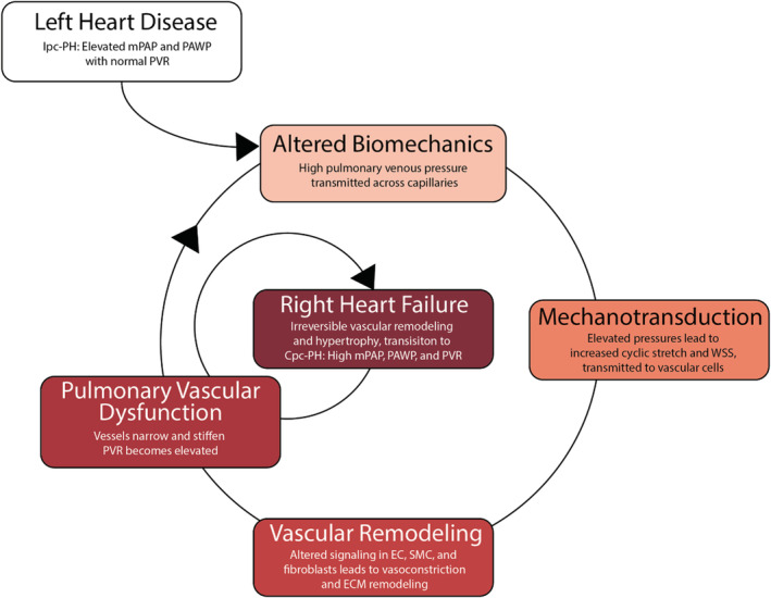
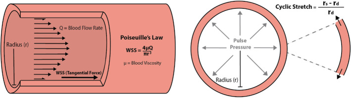
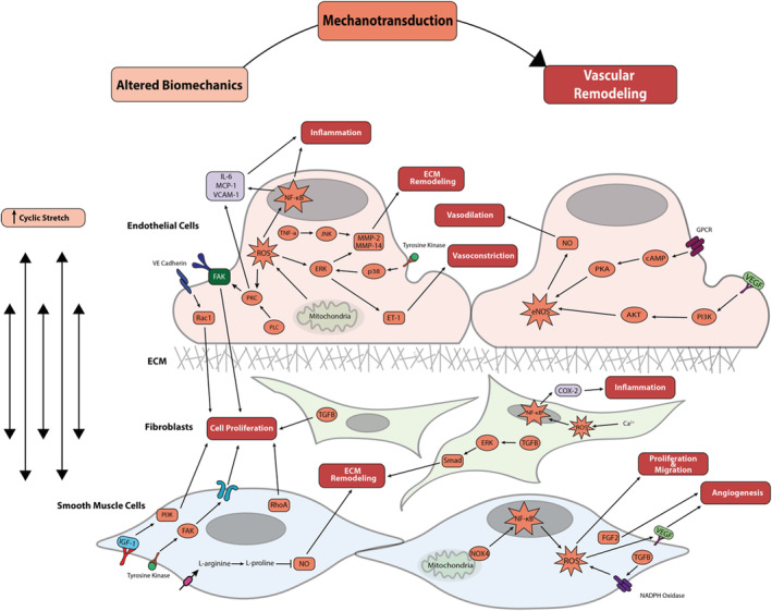
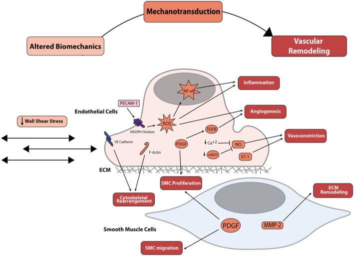
Combined pre-/postcapillary pulmonary hypertension (Cpc-PH), a complication of left heart failure, is associated with higher mortality rates than isolated postcapillary pulmonary hypertension alone. Currently, knowledge gaps persist on the mechanisms responsible for the progression of isolated postcapillary pulmonary hypertension (Ipc-PH) to Cpc-PH. Here, we review the biomechanical and mechanobiological impact of left heart failure on pulmonary circulation, including mechanotransduction of these pathological forces, which lead to altered biological signaling and detrimental remodeling, driving the progression to Cpc-PH. We focus on pathologically increased cyclic stretch and decreased wall shear stress; mechanotransduction by endothelial cells, smooth muscle cells, and pulmonary arterial fibroblasts; and signaling-stimulated remodeling of the pulmonary veins, capillaries, and arteries that propel the transition from Ipc-PH to Cpc-PH. Identifying biomechanical and mechanobiological mechanisms of Cpc-PH progression may highlight potential pharmacologic avenues to prevent right heart failure and subsequent mortality.

Keywords: biomechanics; mechanotransduction; pulmonary hypertension due to left heart failure; pulmonary vascular remodeling.


Figures

Figure 1
Figure 1. Mechanotransduction is the key step in the pathophysiologic progression of pulmonary vascular disease in the setting of left heart failure.
The transition from isolated postcapillary pulmonary hypertension to combined pre−/postcapillary pulmonary hypertension is characterized by pulmonary vascular remodeling and results in right heart failure. Cpc‐PH, combined pre−/postcapillary pulmonary hypertension; EC, endothelial cell; Ipc‐PH, isolated postcapillary pulmonary hypertension; mPAP, mean pulmonary artery pressure; PAWP, pulmonary artery wedge pressure; PVR, pulmonary vascular resistance; SMC, smooth muscle cell; and WSS, wall shear stress.
Figure 2
Figure 2. Schematic representation of wall shear stress (WSS) and cyclic stretch in a vessel.
WSS is directly proportional to blood flow rate (Q) and blood viscosity (μ) and is inversely proportional to the radius of the vessel lumen (r). Cyclic stretch is the difference between lumen radius at systole (rs) and the lumen radius at diastole (rd) normalized by the radius at diastole.
Figure 3
Figure 3. Mechanotransduction of increased cyclic stretch because of left heart failure in endothelial cells, fibroblasts, and smooth muscle cells triggers a cascade of pathologic vascular remodeling.
Boxes within the cell signaling cascade are colored to match their corresponding box at the top of the figure (altered biomechanics, mechanotransduction, or vascular remodeling). AKT indicates protein kinase B; cAMP indicates cyclic adenosine monophosphate; Cox‐2 indicates cyclooxygenase‐2; ECM, extracellular matrix; eNOS, endothelial nitric oxide synthase; ERK, extracellular signal‐regulated kinase; FAK, focal adhesion kinase; FGF‐2, fibroblast growth factor‐2; GPCR, G protein‐coupled receptor; IGF‐1, insulin‐like growth factor‐1; IL‐6, interleukin‐6; JNK, c‐Jun N‐terminal kinase; MCP‐1, myocyte chemoattractant protein‐1; MMP, matrix metalloproteinase; NF‐κB, nuclear factor‐κB; NOX‐4, NADPH oxidase‐4; P13K, p13 kinase; PKA, protein kinase A; PKC, protein kinase C; PLC, phospholipase C; Rac1, Ras‐related C3 botulinum toxin substrate 1; RhoA, ras homolog family member A; ROS, reactive oxygen species; TGFβ, transforming growth factor beta; TNF‐α, tumor necrosis factor‐α; VCAM‐1, vascular cell adhesion protein 1; VE cadherin, vascular endothelial cadherin; and VEGF, vascular endothelial growth factor.
Figure 4
Figure 4. Mechanotransduction of low wall shear stress because of left heart failure in endothelial cells and smooth muscle cells results in pathologic vascular remodeling throughout the vessel wall.
Boxes within the cell signaling cascade are colored to match their corresponding box at the top of the figure (altered biomechanics, mechanotransduction, or vascular remodeling). ECM indicates extracellular matrix; eNOS, endothelial nitric oxide synthase; ET‐1, endothelin‐1; F‐Actin, anti‐filamentous actin; MMP, matrix metalloproteinase; NF‐κB, nuclear factor‐κB; PDGF, platelet‐derived growth factor; PECAM‐1, platelet endothelial cell adhesion molecule‐1; ROS, reactive oxygen species; SMC, smooth muscle cell; TGFβ, transforming growth factor beta; and VE cadherin, vascular endothelial cadherin.

References

    1. Mozaffarian D, Benjamin EJ, Go AS, Arnett DK, Blaha MJ, Cushman M, Das SR, de Ferranti S, Després JP, Fullerton HJ, et al. Heart disease and stroke statistics‐2016 update: a report from the American Heart Association. Circulation. 2016;133:e38–e360. doi: 10.1161/CIR.0000000000000350 - DOI - PubMed
    1. Lam CSP, Roger VL, Rodeheffer RJ, Borlaug BA, Enders FT, Redfield MM. Pulmonary hypertension in heart failure with preserved ejection fraction. J Am Coll Cardiol. 2009;53:1119–1126. doi: 10.1016/j.jacc.2008.11.051 - DOI - PMC - PubMed
    1. Guazzi M. Alveolar‐capillary membrane dysfunction in heart failure: evidence of a pathophysiologic role. Chest. 2003;124:1090–1102. doi: 10.1378/chest.124.3.1090 - DOI - PubMed
    1. Miller WL, Grill DE, Borlaug BA. Clinical features, hemodynamics, and outcomes of pulmonary hypertension due to chronic heart failure with reduced ejection fraction: pulmonary hypertension and heart failure. JACC Heart Fail. 2013;1:290–299. doi: 10.1016/j.jchf.2013.05.001 - DOI - PubMed
    1. Vanderpool RR, Saul M, Nouraie M, Gladwin MT, Simon MA. Association between hemodynamic markers of pulmonary hypertension and outcomes in heart failure with preserved ejection fraction. JAMA Cardiol. 2018;3:298–306. doi: 10.1001/jamacardio.2018.0128 - DOI - PMC - PubMed

Publication types