. 2023 Mar;37(3):e22777.
doi: 10.1096/fj.202201614R.

Targeted deletion of Fgf9 in tendon disrupts mineralization of the developing enthesis

Affiliations

Targeted deletion of Fgf9 in tendon disrupts mineralization of the developing enthesis

Elahe Ganji et al. FASEB J. 2023 Mar.

Erratum in

Abstract

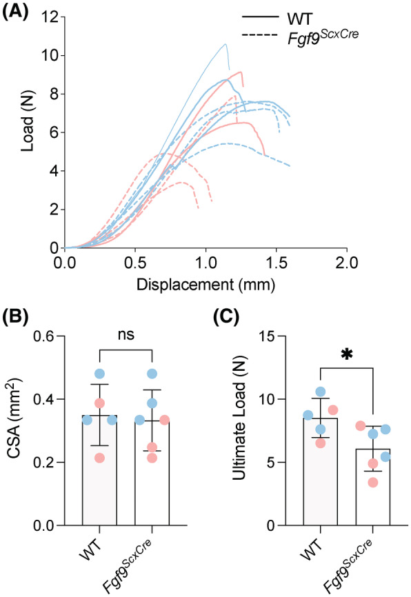
The enthesis is a transitional tissue between tendon and bone that matures postnatally. The development and maturation of the enthesis involve cellular processes likened to an arrested growth plate. In this study, we explored the role of fibroblast growth factor 9 (Fgf9), a known regulator of chondrogenesis and vascularization during bone development, on the structure and function of the postnatal enthesis. First, we confirmed spatial expression of Fgf9 in the tendon and enthesis using in situ hybridization. We then used Cre-lox recombinase to conditionally knockout Fgf9 in mouse tendon and enthesis (Scx-Cre) and characterized enthesis morphology as well as mechanical properties in Fgf9ScxCre and wild-type (WT) entheses. Fgf9ScxCre mice had smaller calcaneal and humeral apophyses, thinner cortical bone at the attachment, increased cellularity, and reduced failure load in mature entheses compared to WT littermates. During postnatal development, we found reduced chondrocyte hypertrophy and disrupted type X collagen (Col X) in Fgf9ScxCre entheses. These findings support that tendon-derived Fgf9 is important for functional development of the enthesis, including its postnatal mineralization. Our findings suggest the potential role of FGF signaling during enthesis development.

Keywords: attachment; enthesis; fibroblast growth factor; musculoskeletal; postnatal; tendon.


Figures

FIGURE 1
FIGURE 1
Interfaces analyzed included the supraspinatus enthesis of the mouse shoulder and the Achilles enthesis of the mouse foot (left panel). The middle and right panels, respectively, show the morphology of the developing enthesis (stained with Toluidine Blue) and expression of Fgf9 (shown in magenta) of the neonatal Achilles' enthesis and tendon. Fluorescent in situ hybridization for Mm‐Fgf9. Scale bar = 100 μm. Figure made using Biorender.
FIGURE 2
FIGURE 2
At 8 weeks of age, Fgf9 ScxCre mice had smaller humeral epiphyses and calcaneal apophyses compared to age‐matched WT littermates. (A and B) Schematics showing segmentation of bone, with blue pseudo‐color indicating trabecular bone and white pseudo‐color indicating cortical bone, of (A) the humeral epiphysis and (B) calcaneal apophyses. (C and D) Humeral epiphyses and calcaneal apophyses were smaller in Fgf9 ScxCre mice compared to age‐matched controlled but epi/apophyseal bone volume ratio (BV/TV did not differ. Blue and pink dots denote male and female mice, respectively. Data presented as mean ± 95% CI; *p < .05; **p < .01.
FIGURE 3
FIGURE 3
Insertional thickness in mature 8 week‐old Fgf9 ScxCre entheses was significantly thinner compared to age‐matched WT mice. Respective grayscale microCT images of (A and A') supraspinatus and (C and C') Achilles entheses and regions of repeated thickness measurement (blue lines) are shown. (B and D) Measured thickness was significantly thinner at the (B) supraspinatus and (D) Achilles attachments. Blue and pink dots denote male and female mice, respectively. Data presented as mean ± 95% CI and *p < .05; **p < .01. Scale bar = 600 μm.
FIGURE 4
FIGURE 4
The structurally mature supraspinatus and Achilles enthesis of 8‐week‐old Fgf9 ScxCre mice had thinner cortical bone compared to age‐matched WT mice (orange arrows), as well as smaller apophyses. Fgf9 ScxCre mice also had acellular metachromatic regions (orange dashed outline in Achilles entheses), shown in Toluidine blue stained sections of Achilles, compared to WT mice; scale bar = 100 μm scale for all panels.
FIGURE 5
FIGURE 5
During post‐natal development, the cellular density of the enthesis decreased. (A) Total cell count and (A') cell count per area (cellularity) were measured using ImageJ using the outlined regions of the enthesis (shown as white dashed areas in C). Histological representation of the Achilles enthesis stained using (B) H&E and (C) Toluidine Blue during developing stages, as well as for the (B' and C') young‐adult enthesis and (B'' and C'') adult enthesis. Scale bar = 200 μm. Data presented as mean ± 95% CI; *p < .05; ***p < .0007; ****p < .0001.
FIGURE 6
FIGURE 6
Cellular density was higher in mature supraspinatus entheses of 8‐week‐old Fgf9 ScxCre mice compared to WT. (A) Representative fluorescent images of supraspinatus and Achilles' regions of interest (scale bar 20 μm). (B) Nuclear density and (C) number of nuclei presented for both Fgf9 ScxCre and WT mice at 8 weeks of age; Pink dots/lines = female mice; Blue dots/lines = male mice. Scale bar = 50 μm. Data presented as mean ± 95% CI and p < .05.
FIGURE 7
FIGURE 7
Targeted Fgf9 deletion disrupts tensile mechanics of mature Achilles enthesis. Achilles tendon entheses of 8‐week‐old Fgf9 ScxCre mice had reduced ultimate load compared to age‐matched WT mice. (A) Overlaid load–displacement curves, (B) CSA, and (C) maximum load for all samples tested. Pink dots/lines = female mice; Blue dots/lines = male mice. Error bars denote mean ± 95% CI, *p < .05; ns = not significantly different.
FIGURE 8
FIGURE 8
Loss of Fgf9 in tendon and enthesis leads to reduced area of active mineralization at the SOC of the young mice at 3 weeks of age. (A and B) 3‐week‐old Fgf9 ScxCre mice had smaller hypertrophic chondrocytes, however (C) the secondary ossification center (SOC) was not significantly smaller compared to age‐matched WT mice. Additionally, (D–F) the size of the ColX area in the SOC was smaller in Fgf9 ScxCre mice compared to age‐matched WT littermates. *p < .05; ns = not significantly different (p > .05). Scale bars = 100 μm.

References

    1. Blitz E, Viukov S, Sharir A, et al. Bone ridge patterning during musculoskeletal assembly is mediated through SCX regulation of Bmp4 at the tendon‐skeleton junction. Dev Cell. 2009;17(6):861‐873. - PMC - PubMed
    1. Zelzer E, Blitz E, Killian ML, Thomopoulos S. Tendon‐to‐bone attachment: from development to maturity. Birth Defects Research Part C Embryo Today. 2014;102(1):101‐112. - PMC - PubMed
    1. Galatz L, Rothermich S, VanderPloeg K, Petersen B, Sandell L, Thomopoulos S. Development of the supraspinatus tendon‐to‐bone insertion: localized expression of extracellular matrix and growth factor genes. J Orthop Res. 2007;25(12):1621‐1628. - PubMed
    1. Thomopoulos S, Genin GM, Galatz LM. The development and morphogenesis of the tendon‐to‐bone insertion—what development can teach us about healing. J Musculoskelet Neuronal Interact. 2010;10(1):35‐45. - PMC - PubMed
    1. Blitz E, Sharir A, Akiyama H, Zelzer E. Tendon‐bone attachment unit is formed modularly by a distinct pool of Scx ‐ and Sox9 ‐positive progenitors. Development. 2013;140(13):2680‐2690. - PubMed

Publication types

Substances

LinkOut - more resources