. 2023 Feb 3;9(5):eade4361.
doi: 10.1126/sciadv.ade4361. Epub 2023 Feb 3.

Cytidine deaminases catalyze the conversion of N(S, O)4-substituted pyrimidine nucleosides

Affiliations

Cytidine deaminases catalyze the conversion of N(S, O)4-substituted pyrimidine nucleosides

Nina Urbelienė et al. Sci Adv. .

Abstract

Cytidine deaminases (CDAs) catalyze the hydrolytic deamination of cytidine and 2'-deoxycytidine to uridine and 2'-deoxyuridine. Here, we report that prokaryotic homo-tetrameric CDAs catalyze the nucleophilic substitution at the fourth position of N4-acyl-cytidines, N4-alkyl-cytidines, and N4-alkyloxycarbonyl-cytidines, and S4-alkylthio-uridines and O4-alkyl-uridines, converting them to uridine and corresponding amide, amine, carbamate, thiol, or alcohol as leaving groups. The x-ray structure of a metagenomic CDA_F14 and the molecular modeling of the CDAs used in this study show a relationship between the bulkiness of a leaving group and the volume of the binding pocket, which is partly determined by the flexible β3α3 loop of CDAs. We propose that CDAs that are active toward a wide range of substrates participate in salvage and/or catabolism of variously modified pyrimidine nucleosides. This identified promiscuity of CDAs expands the knowledge about the cellular turnover of cytidine derivatives, including the pharmacokinetics of pyrimidine-based prodrugs.


Figures

Fig. 1.
Fig. 1.. A diversity of reactions catalyzed by CDAs.
The conversion of N4-benzoyl-2′-deoxycytidine (25) (A), S4-benzylthiouridine (42) (D), 4-benzyloxy-5-fluoro-uridine (50) (G), and capecitabine (61) (5′-deoxy-5-fluoro-N4-pentyloxycarbonylcytidine) (J) are shown. High-performance liquid chromatography–mass spectrometry chromatography spectra (254 nm) of reaction products: 2′-deoxyuridine (2′dU) (B), uridine (U) (E), 5-fluoro-uridine (5FU) (H), and 5-fluoro-5′deoxyuridine (545′dU) (K), and typical GC-MS chromatography spectra outtakes of extracted reaction products: benzamide (C), benzyl mercaptan (F), benzyl alcohol (I), and penthyl carbamate (L) are shown after the reaction schemes, respectively. m/z, mass/charge ratio.
Fig. 2.
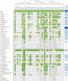
Fig. 2.. The clustering of the substrate ranges of the CDAs.
The list of the tested substrates is shown on the left side (fig. S7). The ID code representing each CDA is given on the top. The phylogenetic tree was added above the ID code at the top of the figure. The phylogenetic analysis of CDAs was conducted using the neighbor-joining tree routine of MEGA X software. The alignment was performed using ClustalW. The activity of enzymes is defined as follows: 2, activity is observed after 3 hours of the incubation at RT; 1, activity is observed after overnight incubation (weakly active); 0, inactive toward the substrate; N/A, not analyzed. The specific activity of CDA_F14 (nanomole per min per milligram) is shown on the right side. Blue color intensity reflects activity. The SD of measurement is shown in the datafile S1.
Fig. 3.
Fig. 3.. t-SNE representation of the CDA close homolog sequence space.
Coordinates of t-SNE embedding of 1708 cluster representatives were used for plotting. The size of a given dot was visualized on the basis of the cluster size it represents and coral orange color with label if the cluster contained a tested CDA variant.
Fig. 4.
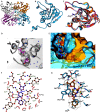
Fig. 4.. The crystal structure of CDA_F14.
(A) Homotetramer of CDA_F14 with subunits colored in gray, gold, blue, and violet. (B) Subunit of CDA_F14 consisted of a core of five β strands (β1 to β5) sandwiched by five α helices (α1 to α5), and η1 and η2 symbols indicate 310-helix; zinc atom (blue sphere) is coordinated by three cysteine residues located in α helices α2 (Cys53) and α3 (Cys91 and Cys88). (C) Comparison of CDA domain structures from different organisms: CDA_F14 is colored in blue, CDA_Mmu (PDB 2FR6) in gold, CDA_Bsu (PDB 1JTK) in coral, and CDA_Hsa (PDB 1MQ0) in violet. (D) Active site of CDA_F14 occupied by two molecules of crystallization agent 2-methyl-2,4-pentanediol (in purple); water molecules are shown as blue spheres. (E) Comparison of substrate binding pocket in CDA_F14 (blue) and in CDA_Mmu (gold); N4-benzoyl-2′-deoxycytidine and 2′-deoxycytidine are fitted in the active center, and the β3α3 loop is shown in front. (F and G) 2D and 3D schematic view of the active site of crystalized CDA_F14 with fitted N4-benzoyl-2′-deoxycytidine. The bond lengths are given in angstroms. The bond length between zinc and 4-hydroxyl group was 1.94 Å. Hydrogen and coordination bonds are shown as dotted lines. 2D view generated using LigPlot+ program (62), other views with Chimera1.16 (53).
Fig. 5.
Fig. 5.. The flexibility of amino acids residues in CDA_F14: N4-benzoyl-2′-deoxycytidine complex.
Root mean square fluctuation value (RMSF) measurements represent individual residue flexibility during a 100-ns simulation. The loop 80-to-86 region is highlighted in gray, and the several conformations for the loop are shown. The molecular dynamics trajectories were generated by using CPPTRAJ program from the AmberTools package.
Fig. 6.
Fig. 6.. 2DRMSD plots for the protein structure during a 100 ns simulation of the CDA_F14: N4-benzoyl-2′-deoxycytidine complex.
(A) 2D root mean squared deviation (2DRMSD) plot for the whole protein structure and (B) 2DRMSD plot only for the loop 80-to-86 atoms. Two conformations during the simulation can be observed for the whole protein, especially for the 80-to-86 loop region.
Fig. 7.
Fig. 7.. A putative role of CDAs in a catabolism of the modified pyrimidine nucleosides.
The noncanonical nucleobases originate due to various epigenetic events, stress responses, or during action of mutagens. This research shows that various CDAs can catalyze the nucleophilic substitution at the fourth position of the heterocyclic ring of N4-acyl-cytidines, N4-alkyl-cytidines, S4-alkylthio-uridine, and O4-alkyl-uridine derivatives subsequently, leading to the formation of uridine and respective amide, amine, carbamate, thiol, or alcohol.

References

    1. P. Boccaletto, F. Stefaniak, A. Ray, A. Cappannini, S. Mukherjee, E. Purta, M. Kurkowska, N. Shirvanizadeh, E. Destefanis, P. Groza, G. Avşar, A. Romitelli, P. Pir, E. Dassi, S. G. Conticello, F. Aguilo, J. M. Bujnicki, MODOMICS: A database of RNA modification pathways. 2021 update. Nucleic Acids Res. 50, D231–D235 (2022). - PMC - PubMed
    1. A. J. Sood, C. Viner, M. M. Hoffman, DNAmod: The DNA modification database. J. Cheminform. 11, 30 (2019). - PMC - PubMed
    1. P. J. McCown, A. Ruszkowska, C. N. Kunkler, K. Breger, J. P. Hulewicz, M. C. Wang, N. A. Springer, J. A. Brown, Naturally occurring modified ribonucleosides. Wiley Interdiscip. Rev. RNA 11, e1595 (2020). - PMC - PubMed
    1. S. Hong, X. Cheng, DNA base flipping: A general mechanism for writing, reading, and erasing DNA modifications. Adv. Exp. Med. Biol. 945, 321–341 (2016). - PMC - PubMed
    1. W. L. Nyhan, Nucleotide Synthesis Via Salvage Pathway, in eLS (John Wiley & Sons Ltd, 2014).