

Adenomyosis: single-cell transcriptomic analysis reveals a paracrine mesenchymal-epithelial interaction involving the WNT/SFRP pathway
- PMID: 36736810
- PMCID: PMC11257082
- DOI: 10.1016/j.fertnstert.2023.01.041
Adenomyosis: single-cell transcriptomic analysis reveals a paracrine mesenchymal-epithelial interaction involving the WNT/SFRP pathway
Abstract
Objective: To assess the cellular and molecular landscape of adenomyosis.
Design: Single-cell analysis of genome-wide messenger RNA (mRNA) expression (single-cell RNA sequencing) of matched tissues of endometrium, adenomyosis, and myometrium using relatively large numbers of viable cells.
Setting: Not applicable.
Patient(s): Patients (n = 3, age range 40-44 years) undergoing hysterectomy for diffuse adenomyosis.
Main outcome measure(s): Definition of the molecular landscape of matched adenomyotic, endometrial and myometrial tissues from the same uterus using single-cell RNA sequencing and comparison of distinct cell types in these tissues to identify disease-specific cell populations, abnormal gene expression and pathway activation, and mesenchymal-epithelial interactions.
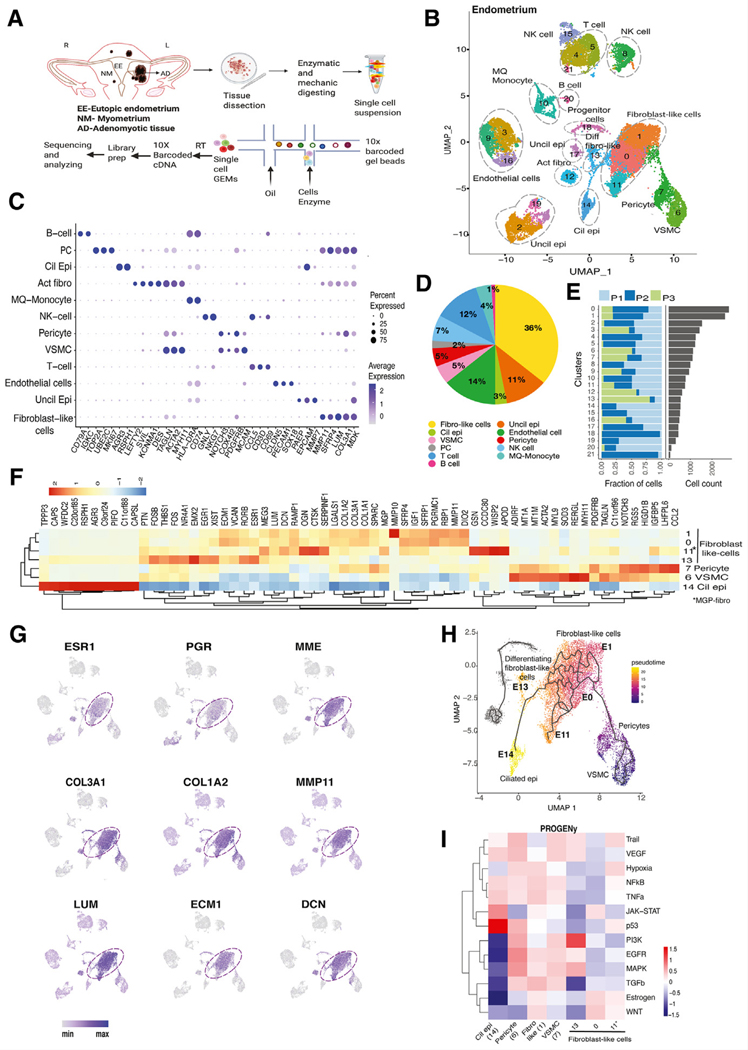
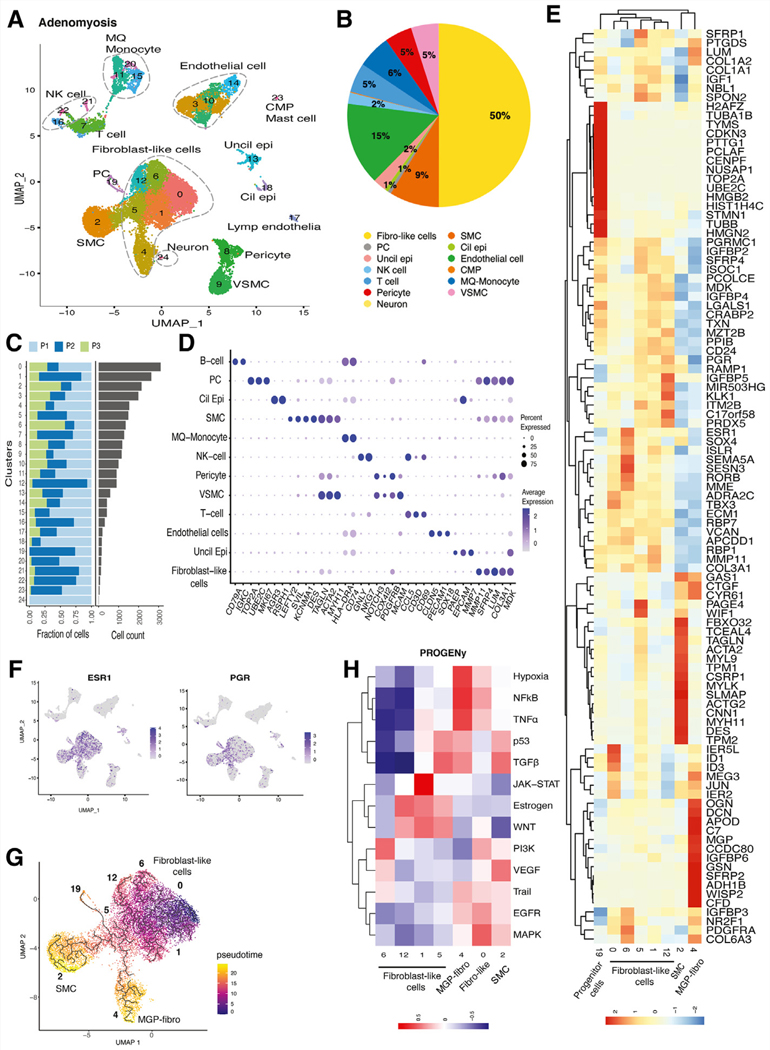
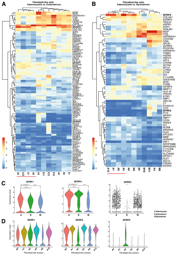
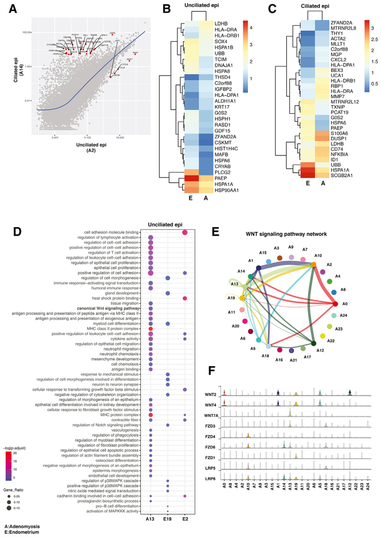
Result(s): The largest cell population in the endometrium was composed of closely clustered fibroblast groups, which comprise 36% of all cells and seem to originate from pericyte progenitors differentiating to estrogen/progesterone receptor-expressing endometrial stromal- cells. In contrast, the entire fibroblast population in adenomyosis comprised a larger (50%) portion of all cells and was not linked to any pericyte progenitors. Adenomyotic fibroblasts eventually differentiate into extracellular matrix protein-expressing fibroblasts and smooth muscle cells. Hierarchical clustering of mRNA expression revealed a unique adenomyotic fibroblast population that clustered transcriptomically with endometrial fibroblasts, suggestive of an endometrial stromal cell population serving as progenitors of adenomyosis. Four other adenomyotic fibroblast clusters with disease-specific transcriptomes were distinct from those of endometrial or myometrial fibroblasts. The mRNA levels of the natural WNT inhibitors, named, secreted frizzled-related proteins 1, 2, and 4, were higher in these 4 adenomyotic fibroblast clusters than in endometrial fibroblast clusters. Moreover, we found that multiple WNTs, which originate from fibroblasts and target ciliated and unciliated epithelial cells and endothelial cells, constitute a critical paracrine signaling network in adenomyotic tissue. Compared with endometrial tissue, unciliated and ciliated epithelial cells in adenomyosis comprised a significantly smaller portion of this tissue and exhibited molecular evidence of progesterone resistance and diminished regulation of estrogen signaling.
Conclusion(s): We found a high degree of heterogeneity in fibroblast-like cells in the adenomyotic uterus. The WNT signaling involving differential expression of secreted frizzled-related proteins, which act as decoy receptors for WNTs, in adenomyotic fibroblasts may have a key role in the pathophysiology of this disease.
Objetivo:: evaluar el panorama celular y molecular de la adenomiosis.
Diseño:: Análisis unicelular de la expresión de ARN mensajero (ARNm) de todo el genoma (secuenciación de ARN unicelular) de tejidos emparejados de endometrio, adenomiosis y miometrio utilizando cantidades relativamente grandes de ćelulas viables.
Entorno:: No aplicable.
Paciente(s):: Pacientes (n = 3, rango de edad de 40 a 44 años) sometidas a histerectomía por adenomiosis difusa.
Principales medidas de resultado:: Definición del panorama molecular de tejidos adenomióticos, endometriales y miometriales emparejados del mismo útero utilizando secuenciación de ARN de una sola ćelula y comparación de distintos tipos de ćelulas en estos tejidos para identificar poblaciones de ćelulas específicas de la enfermedad, expresión génica anormal y la activación de la vía, y las interacciones mesenquimales-epiteliales.
Resultado(s):: La población celular más grande en el endometrio estaba compuesta por grupos de fibroblastos estrechamente agrupados, que comprenden el 36 % de todas las ćelulas y parecen originarse a partir de los progenitores de pericitos que se diferencian a ćelulas del estroma endometrial que expresan receptores de estrógeno/progesterona. Por el contrario, toda la población de fibroblastos en la adenomiosis comprendía una porción más grande (50%) de todas las ćelulas y no estaba vinculada a ningún progenitor de pericitos. Los fibroblastos adenomióticos finalmente se diferencian en fibroblastos que expresan proteínas de la matriz extracelular y ćelulas de músculo liso. El agrupamiento jerárquico de la expresión de ARNm reveló una población de fibroblastos adenomióticos única que se agrupaba transcriptómicamente con fibroblastos endometriales, lo que sugiere una población de ćelulas del estroma endometrial que actuá como progenitores de la adenomiosis. Otros cuatro grupos de fibroblastos adenomióticos con transcriptomas específicos de la enfermedad eran distintos de los de los fibroblastos endometriales o miometriales. Los niveles de ARNm de los inhibidores naturales de WNT, denominados proteínas secretadas relacionadas con frizzled 1, 2 y 4, fueron más altos en estos 4 grupos de fibroblastos adenomióticos que en los grupos de fibroblastos endometriales. Además, encontramos que múltiples WNT, que se originan a partir de fibroblastos y se dirigen a ćelulas epiteliales ciliadas y no ciliadas y ćelulas endoteliales, constituyen una red de señalización paracrina crítica en el tejido adenomiótico. En comparación con el tejido endometrial, las ćelulas epiteliales ciliadas y no ciliadas en la adenomiosis comprendían una porción significativamente menor de este tejido y exhibieron evidencia molecular de resistencia a la progesterona y disminución de la regulación de la señalización de estrógenos.
Conclusión(es):: Encontramos un alto grado de heterogeneidad en las ćelulas similares a fibroblastos en el útero adenomiótico. La señalización de WNT que implica la expresión diferencial de proteínas secretadas relacionadas con frizzled, que actuán como receptores señuelo para WNT, en fibroblastos adenomióticos puede tener un papel clave en la fisiopatología de esta enfermedad.
Keywords: Adenomyosis; SFRP; endometriosis; endometrium; fibroblast; scRNA-seq.
Copyright © 2023 The Authors. Published by Elsevier Inc. All rights reserved.
Figures
References
-
- Garcia-Solares J, Donnez J, Donnez O, Dolmans MM. Pathogenesis of uterine adenomyosis: invagination or metaplasia? Fertil Steril 2018;109:371–9. - PubMed
-
- Bird CC, McElin TW, Manalo-Estrella P. The elusive adenomyosis of the uterus–revisited. Am J Obstet Gynecol 1972;112:583–93. - PubMed
-
- Struble J, Reid S, Bedaiwy MA. Adenomyosis: a clinical review of a challenging gynecologic condition. J Minim Invasive Gynecol 2016;23:164–85. - PubMed
Publication types
MeSH terms
Substances
Grants and funding
LinkOut - more resources
Full Text Sources
Medical
Research Materials
