. 2023 Jul:257:43-53.
doi: 10.1016/j.trsl.2023.01.005. Epub 2023 Feb 1.

Topical captopril: a promising treatment for secondary lymphedema

Affiliations

Topical captopril: a promising treatment for secondary lymphedema

Stav Brown et al. Transl Res. 2023 Jul.

Abstract

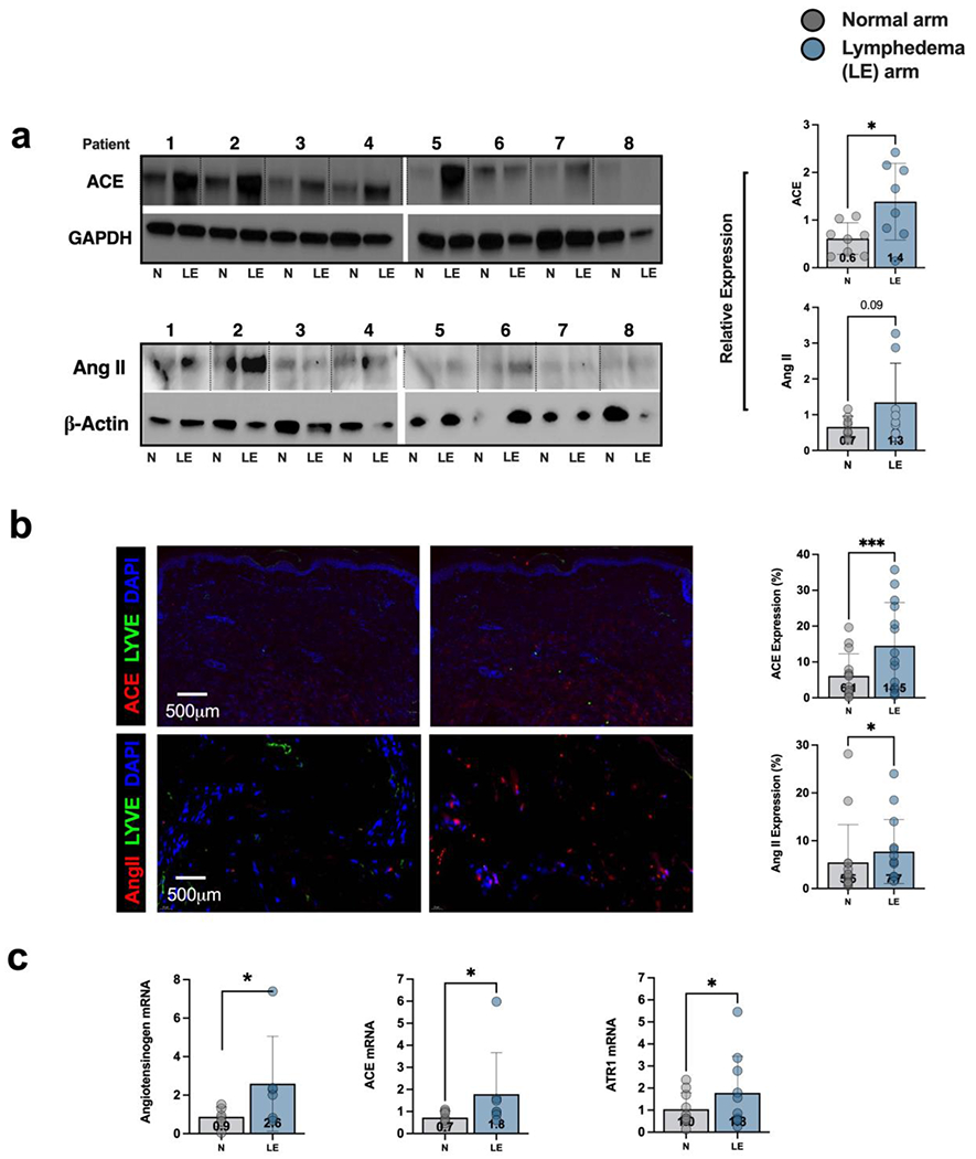
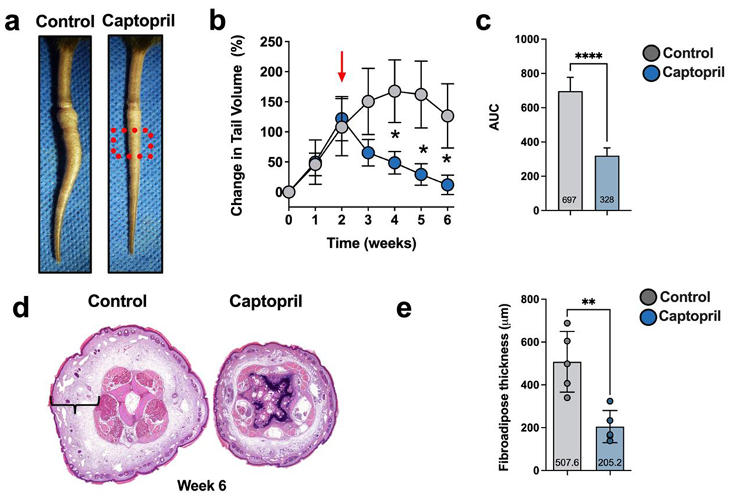
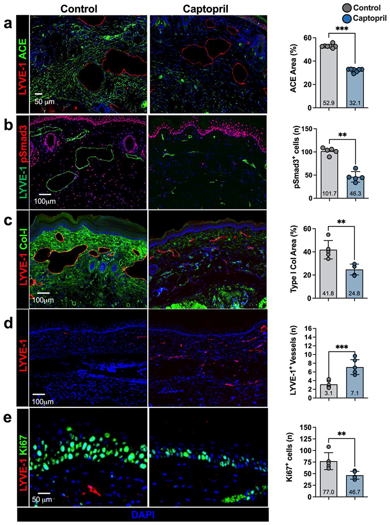
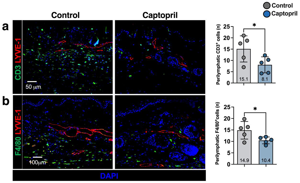
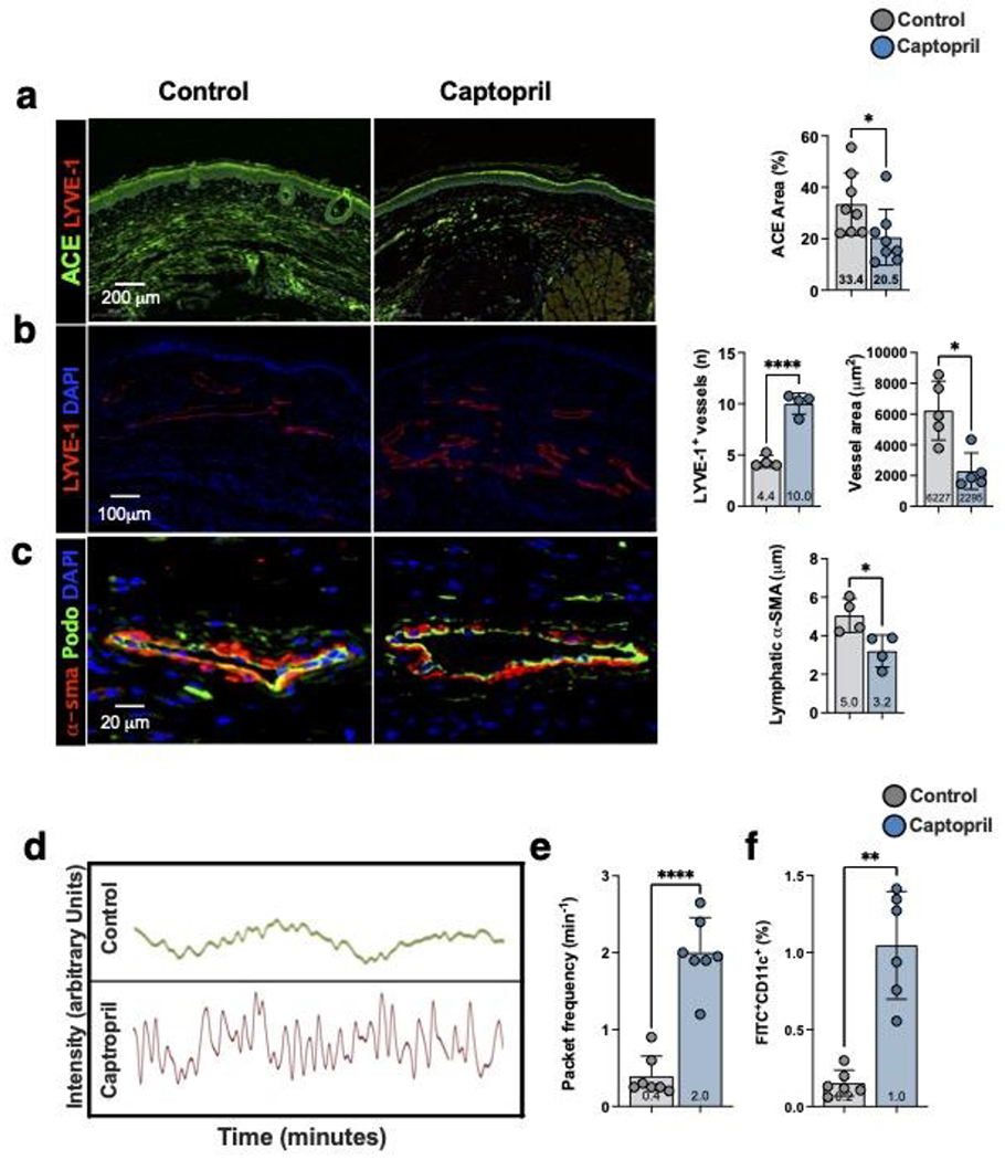
Transforming growth factor-beta 1 (TGF-β1)-mediated tissue fibrosis is an important regulator of lymphatic dysfunction in secondary lymphedema. However, TGF-β1 targeting can cause toxicity and autoimmune complications, limiting clinical utility. Angiotensin II (Ang II) modulates intracellular TGF-β1 signaling, and inhibition of Ang II production using angiotensin-converting enzyme (ACE) inhibitors, such as captopril, has antifibrotic efficacy in some pathological settings. Therefore, we analyzed the expression of ACE and Ang II in clinical lymphedema biopsy specimens from patients with unilateral breast cancer-related lymphedema (BCRL) and mouse models, and found that cutaneous ACE expression is increased in lymphedematous tissues. Furthermore, topical captopril decreases fibrosis, activation of intracellular TGF-β1 signaling pathways, inflammation, and swelling in mouse models of lymphedema. Captopril treatment also improves lymphatic function and immune cell trafficking by increasing collecting lymphatic pumping. Our results show that the renin-angiotensin system in the skin plays an important role in the regulation of fibrosis in lymphedema, and inhibition of this signaling pathway may hold merit for treating lymphedema.


Figures

Figure 1.
Figure 1.. Expression of ACE is increased in clinical lymphedema skin biopsy samples.
a) Western blot analysis and quantification of ACE and Ang II expression in matched tissue obtained from the lymphedematous (LE) and contralateral normal (N) upper extremities of patients with breast cancer-related lymphedema (BCRL). Patient number is shown above each matched pair and separated by dotted lines. Each dot in the plots represents the average of two separate western blots. Data are mean ± standard deviation. Statistical analysis was performed using a matched t-test (*p<0.05). b) Representative immunofluorescent images (left) and quantification (right) of ACE and Ang II in matched biopsies obtained from the lymphedematous (LE) and normal (N) upper extremities of patients with BRCL. Each dot in the graph represents the average of 2 high powered views (HPF; 40x) for each patient. Data are mean ± standard deviations. Statistical analysis was performed using a matched t-test (* p<0.05). c) Gene expression of angiotensinogen, ACE, and angiotensin receptor I (AT1R) in matched biopsies obtained from the lymphedematous (LE) normal (N) upper extremities of patients with BRCL. Each dot in the graph represents the average of qPCR performed in triplicate for each patient and quantified using the delta delta method. Data are mean ± standard deviations. Statistical analysis was performed using a matched t-test on log normalized values (* p<0.05).
Figure 2.
Figure 2.. Topical captopril decreases edema and fibrosis in the mouse tail model of lymphedema 6 weeks after surgery.
a) Representative images of tail skin after treatment with control or topical captopril beginning 2 weeks after surgery. The red dotted box is the area analyzed using histology in H&E section shown in Figure 2d and Figures 3 and 4. b) Quantification of tail volume changes using the truncated cone formula in mice treated with control or topical captopril. The red arrow indicates the time point when treatment was initiated. Statistical analysis was performed using a two-way ANOVA (* p<0.05). c) Comparison of the area under the curve (AUC) in tail volume graph comparing captopril and control treated mice. Statistical analysis was performed using a t-test(**** p<0.0001). d) Representative H&E staining of distal tail cross-sections in captopril and control treated mice. Brackets indicate fibroadipose thickness. e) Quantification of tissue fibroadipose thickness in captopril and control treated mice. Each dot represents the average of 4 measurements for each mouse (n=7) used in the experiment. Data are mean ± standard deviation. Statistical analysis was performed using the Student’s t test (**** p<0.0001).
Figure 3.
Figure 3.. Topical captopril decreases ACE expression, SMAD phosphorylation, fibroadipose deposition, and hyperkeratosis in the mouse tail model of lymphedema.
a-e) Representative immunofluorescent images (left) and quantification (right) of ACE (a), pSmad3 (b), collagen I (Col-I; c), LYVE-1 (d), and Ki67 (e) in captopril and control treated mice 6 weeks after tail surgery. Each dot represents the average of 3 high powered views for each animal used in the analysis (n=7). Data are mean ± standard deviation. Statistical analysis was performed using the Student’s t-test (*p<0.05, **p<0.01, ***p<0.001).
Figure 4.
Figure 4.. Topical captopril decreases inflammation in a mouse tail model of lymphedema.
a-b) Representative immunofluorescent images (left) and quantification (right) of LYVE-1 and CD3+ (a) and F4/80+ (b) cells in captopril and control treated mice 6 weeks after tail surgery. Each dot represents the average of 3 high powered fields for each animal (n=5) used in the analysis. Statistical analysis was performed using the Student’s t-test (*p<0.05).
Figure 5.
Figure 5.. Topical captopril increases lymphangiogenesis and lymphatic function following PLND.
a) Representative immunofluorescent images (left) and quantification (right) of ACE in the skin of captopril or control treated mice 4 weeks after PLND. Each dot represents the average of 3 high power fields per animal (n=8) used in the study. Statistical analysis was performed using the Student’s t-test (*p<0.05). b) Representative immunofluorescent images (left) and quantification (right) localizing LYVE-1+ lymphatic vessels in the skin of captopril or control treated mice 4-weeks after PLND. Quantification of LYVE-1+ vessel diameter is also shown in the bar graphs on the right. For the lymphatic vessel number, each dot represents the average number of lymphatic vessels in 3 low power fields (10x) per animal (n=4) used in the study. For the lymphatic vessel area, each dot represents the average of all vessels included in the images of 3 low power field per animal (n=5) used in the study. Statistical analysis was performed using the Student’s t-test (*p<0.05, ****p<0.0001). c) Representative immunofluorescent images localizing podoplanin (Podo) and α-SMA in collecting lymphatic vessels of captopril or control treated mice 4-weeks after PLND (left). Quantification of perilymphatic α-SMA thickness is shown to the right. Each dot represents the average thickness of α-SMA area in 4 quadrants of a collecting lymphatic for each animal (n=4) in the study. Statistical analysis was performed using the Student’s t-test (*p<0.05). d) Representative graphs depicting changes in near infrared fluorescence (NIR) in a collecting lymphatic vessel over time in captopril or control mice. Each peak represents a packet frequency. e) Quantification of NIR collecting lymphatic vessel packet frequency in captopril and control treated mice. Each dot represents the average of 3 recordings in each mouse (n=7). Statistical analysis was performed using the Student’s t-test (*p<0.0001). f) Quantification of dendritic cell trafficking in captopril and control treated mice. FITC-acetone was painted on the distal hindlimb skin and the percentage of FITC+CD11c+ DCs in ipsilateral inguinal lymph nodes was quantified. Each dot represents the average of two flow-cytometry experiments for each mouse (n=6). Statistical analysis was performed using the Student’s t-test (**p<0.01).

References

    1. Rockson SG and Rivera KK, Estimating the population burden of lymphedema. Ann N Y Acad Sci, 2008. 1131: p. 147–54. - PubMed
    1. Cormier JN, et al., Lymphedema beyond breast cancer: a systematic review and meta-analysis of cancer-related secondary lymphedema. Cancer, 2010. 116(22): p. 5138–49. - PubMed
    1. Dayan JH, et al., Lymphedema: Pathogenesis and Novel Therapies. Annu Rev Med, 2018. 69: p. 263–276. - PubMed
    1. Kataru RP, et al., Fibrosis and secondary lymphedema: chicken or egg? Transl Res, 2019. 209: p. 68–76. - PMC - PubMed
    1. Mihara M, et al., Pathological steps of cancer-related lymphedema: histological changes in the collecting lymphatic vessels after lymphadenectomy. PLoS One, 2012. 7(7): p. e41126. - PMC - PubMed

Publication types

MeSH terms

Substances