Review
. 2023 Feb 3;8(1):56.
doi: 10.1038/s41392-023-01330-w.

Osteoarthritis: pathogenic signaling pathways and therapeutic targets

Affiliations
Review

Osteoarthritis: pathogenic signaling pathways and therapeutic targets

Qing Yao et al. Signal Transduct Target Ther. .

Abstract

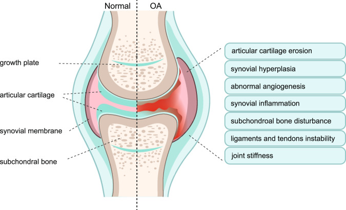
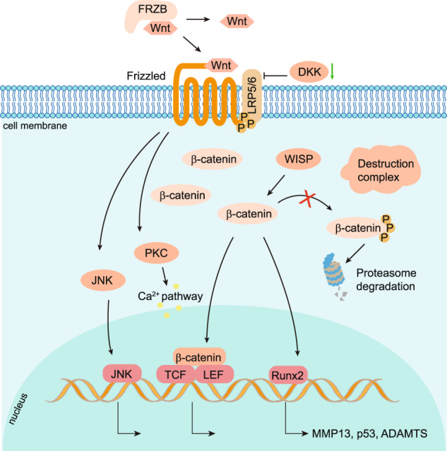
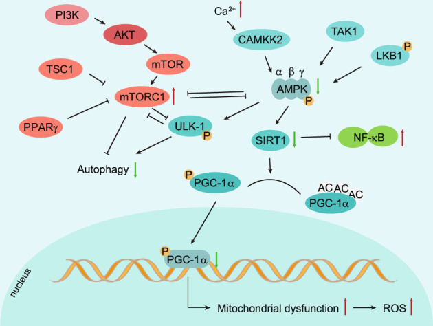
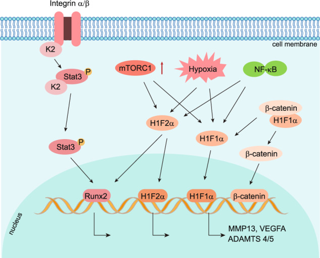
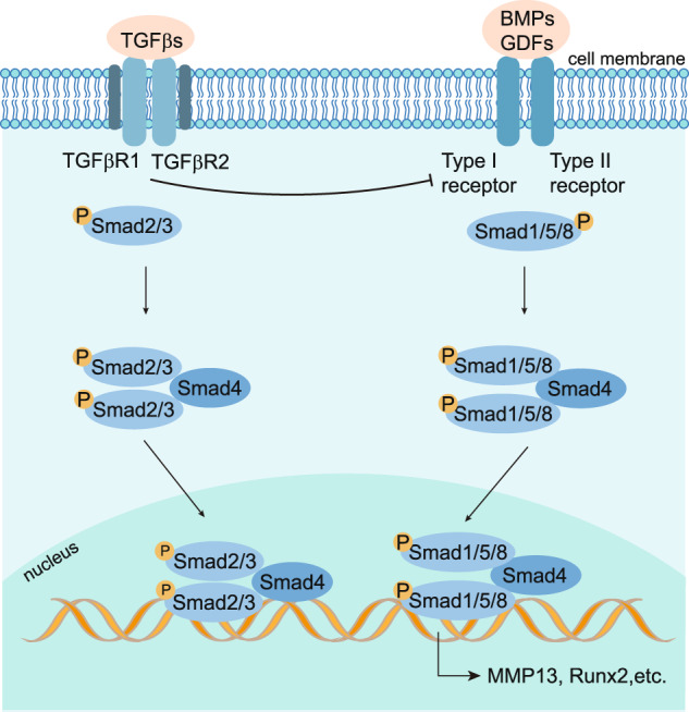
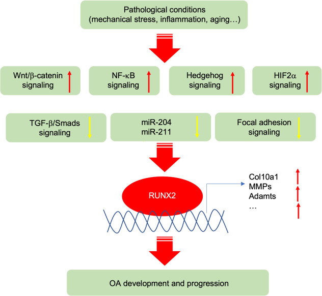
Osteoarthritis (OA) is a chronic degenerative joint disorder that leads to disability and affects more than 500 million population worldwide. OA was believed to be caused by the wearing and tearing of articular cartilage, but it is now more commonly referred to as a chronic whole-joint disorder that is initiated with biochemical and cellular alterations in the synovial joint tissues, which leads to the histological and structural changes of the joint and ends up with the whole tissue dysfunction. Currently, there is no cure for OA, partly due to a lack of comprehensive understanding of the pathological mechanism of the initiation and progression of the disease. Therefore, a better understanding of pathological signaling pathways and key molecules involved in OA pathogenesis is crucial for therapeutic target design and drug development. In this review, we first summarize the epidemiology of OA, including its prevalence, incidence and burdens, and OA risk factors. We then focus on the roles and regulation of the pathological signaling pathways, such as Wnt/β-catenin, NF-κB, focal adhesion, HIFs, TGFβ/ΒΜP and FGF signaling pathways, and key regulators AMPK, mTOR, and RUNX2 in the onset and development of OA. In addition, the roles of factors associated with OA, including MMPs, ADAMTS/ADAMs, and PRG4, are discussed in detail. Finally, we provide updates on the current clinical therapies and clinical trials of biological treatments and drugs for OA. Research advances in basic knowledge of articular cartilage biology and OA pathogenesis will have a significant impact and translational value in developing OA therapeutic strategies.


Conflict of interest statement

The authors declare no competing interests.

Figures

Fig. 1
Fig. 1
Phenotypes of Osteoarthritis (OA). Clinic evidence shows that the majority of OA patients have a diversity of OA phenotypes, including articular cartilage erosion, synovial hyperplasia, abnormal angiogenesis, synovial inflammation, subchondral bone disturbance, ligaments and tendons instability, and joint stiffness. Left-half side shows the structure of the normal synovial joint. Right-half side showed the possible alterations of synovial joint structure and symptoms in osteoarthritis
Fig. 2
Fig. 2
Histological characteristics of OA articular cartilage. Articular cartilage is composed of a dense extracellular matrix (ECM) and sparse distribution of chondrocytes. A cross-sectional diagram of articular cartilage can be divided into the superficial zone, middle zone, and deep zone (the lower part is calcified). Chondrocytes and ECM in the OA cartilage are extensively changed. Cell apoptosis and necrosis are increased in OA chondrocytes with inhibited anabolism and boosted catabolism leading to suppressed ECM synthesis and enhanced ECM degradation. Inflammatory factors, such as TNFα secreted by chondrocytes and synovial membrane fibroblasts, stimulate inflammation in the whole joint. Synovial hyperplasia and fibrosis, and abnormal blood vessel invasion from subchondral bone is observed in OA synovial joint. Increased chondrocyte hypertrophy and oxidative stress with excessive ROS production are reported
Fig. 3
Fig. 3
Activated Wnt canonical and non-canonical signaling pathways in OA. In healthy articular cartilage, the Wnt signaling pathway in articular cartilage and synovium is profoundly inhibited by the absence of Wnt-Wnt receptor interactions and β-catenin degradation through the proteasome pathway. In OA chondrocytes and synovial membrane cells, Wnt ligands bind to Frizzled/LRP5/6 receptors to activate the canonical pathway, in which the destruction complex moves to the cell membrane and releases β-catenin to translocate to the nucleus and binding with TCF/LEF family to control target gene transcription. For the non-canonical Wnt pathway, the protein kinase C (PKC) and c-Jun N-terminal Kinase (JNK) are activated to upregulate downstream target genes and pathways, including the calcium pathway
Fig. 4
Fig. 4
The activity of NF-κB signaling in OA chondrocytes. The NF-κB signaling is activated by a variety of ligands, including TNFα, IL-1β, and LPS, leading to phosphorylation (P) and proteasome degradation of IκB. Receptors of NF-κB signaling include TNFR, TLR, LTβR and etc. Activated IKKs lead to IκB degradation by regulating its phosphorylation. Thus, the translocation of NF-κB to the nucleus results in the transcription of the downstream target genes encoding RUNX2, MMPs, ADAMTS, and HIF2α
Fig. 5
Fig. 5
Regulation of chondrocyte autophagy and metabolism by AMPK and mTOR signaling pathways. Adenosine monophosphate (AMP)-activated protein kinase (AMPK) is a heterotrimeric complex that contains subunits α, β, and γ. LKB1 (liver kinase B1), CAMKK2 (calcium-sensitive kinase) and TAK1 (transforming growth factor-β-activated protein kinase-1) are three major enzymes that can introduce AMPK phosphorylation. In OA chondrocytes, inhibited AMPK phosphorylation decreases ULK1 expression, which is an essential initiator of autophagy. Decreased phosphorylation of AMPK also downregulates the expression of PGC-1α through SIRT-1 in articular chondrocytes, leading to mitochondrial dysfunction
Fig. 6
Fig. 6
Role of focal adhesion and HIFs pathway in OA chondrocytes. Focal adhesion protein Kindlin-2(K2), which is co-localized with integrin α on the cell membrane, is highly expressed in adult articular chondrocytes. In OA chondrocytes, loss of Kindlin-2 promotes chondrocyte hypertrophy and catabolism by activating the phosphorylation of Stat3 and upregulating transcription factor Runx2. HIF-1α and HIF-2α are both activated by hypoxia. HIF-1α inhibits Wnt signaling by blocking β-catenin translocation to the nucleus and downregulates MMP13 expression in healthy articular cartilage in mice. HIF-1α λ is upregulated in OA chondrocytes and induces abnormal blood vessel invasion by upregulating VEGFA. HIF-2α directly induces the expression of OA risk factor, Runx2, and catabolic genes encoding MMP1, MMP3, MMP13, ADAMTS4, and NOS2, to promote cartilage destruction in OA chondrocytes. Besides hypoxia, the activation of the NF-κB pathway and the upregulation of mTORC1 induce the overexpression and excessive accumulation of HIFs protein in articular chondrocytes
Fig. 7
Fig. 7
Schematic representation of TGFβ/BMP signaling pathway in OA chondrocytes. TGF-β/BMP family ligands bind to type I and type II receptors on the cell surface. TGF-βs bind TGFβRI and TGFβRII, and BMPs bind BMPRI and BMPRII. TGFβRIII is a coreceptor that facilitates interaction with TβRI and TβRII. TGF-βs induce the phosphorylation of Smad2/3, and BMPs mediate Smad1/5/8 phosphorylation. By forming complexes with Smad4, phosphorylated Smad2/3 and Smad1/5/8 translocate into the nucleus to regulate target gene expression
Fig. 8
Fig. 8
Runx2 in OA pathogenesis. A diagram showing the upstream and downstream of Runx2 in OA pathogenesis. Runt-related transcription factor 2 (Runx2) is upregulated by activation of Wnt/β-catenin, NF-κB, Hedgehog, HIF-2α signaling, and by suppression of TGF-β/Smads and focal adhesion signaling in OA chondrocytes. miR-204 and miR-211 inhibit the expression of Runx2 protein in articular chondrocytes in vivo. Red arrow: upregulated expression. Yellow arrow: downregulated expression

References

    1. Hunter DJ, Bierma-Zeinstra S. Osteoarthritis. Lancet. 2019;393:1745–1759. - PubMed
    1. Quicke JG, Conaghan PG, Corp N, Peat G. Osteoarthritis year in review 2021: epidemiology & therapy. Osteoarthr. Cartil. 2022;30:196–206. - PubMed
    1. Little CB, Hunter DJ. Post-traumatic osteoarthritis: from mouse models to clinical trials. Nat. Rev. Rheumatol. 2013;9:485–497. - PubMed
    1. Leung YY, Thumboo J. An overview of OA research in two urban APLAR populations. Int. J. Rheum. Dis. 2011;14:130–135. - PubMed
    1. Lu K, et al. Molecular signaling in temporomandibular joint osteoarthritis. J. Orthop. Transl. 2022;32:21–27. - PMC - PubMed

Publication types

MeSH terms