. 2023 Feb 3;10(1):70.
doi: 10.1038/s41597-023-01943-4.

Chákṣu: A glaucoma specific fundus image database

Affiliations

Chákṣu: A glaucoma specific fundus image database

J R Harish Kumar et al. Sci Data. .

Erratum in

Abstract

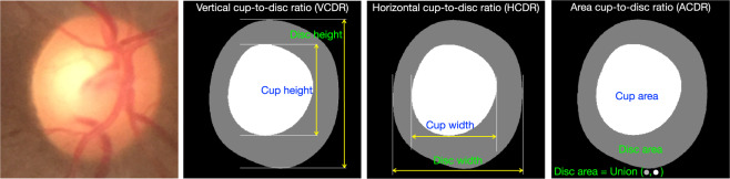
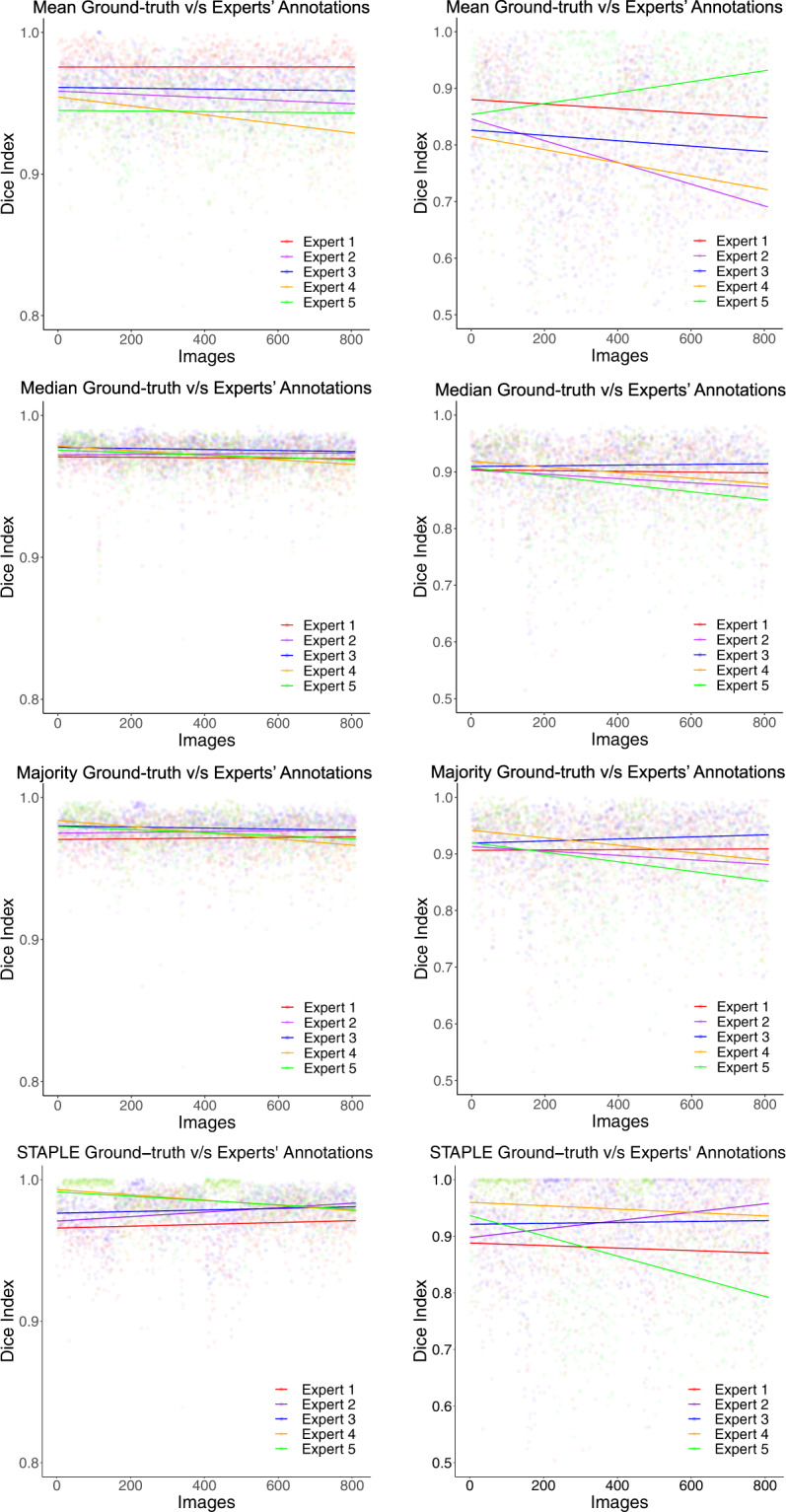
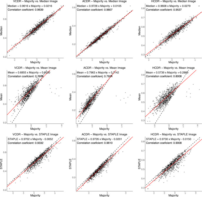
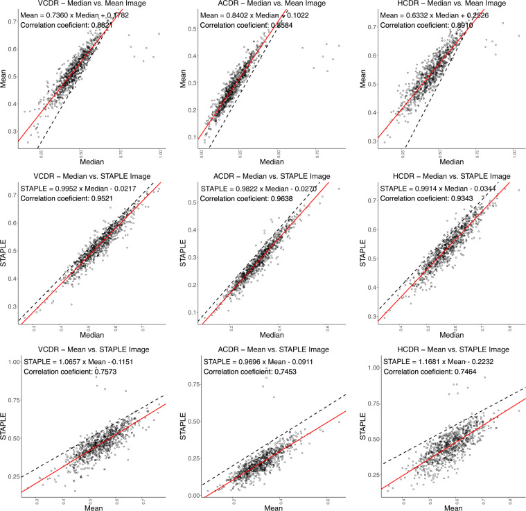
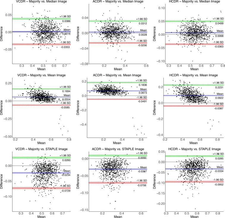
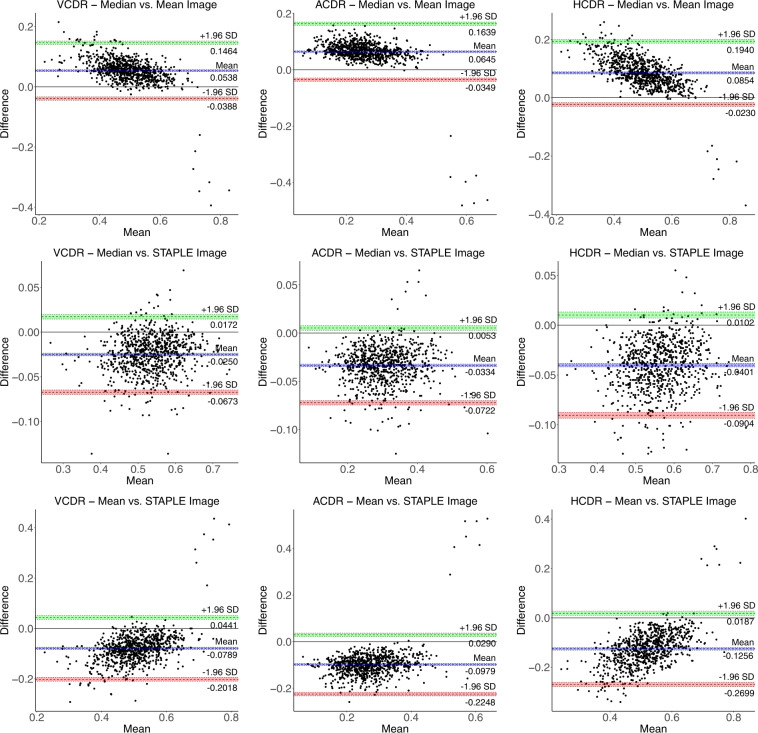
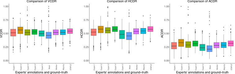
We introduce Chákṣu-a retinal fundus image database for the evaluation of computer-assisted glaucoma prescreening techniques. The database contains 1345 color fundus images acquired using three brands of commercially available fundus cameras. Each image is provided with the outlines for the optic disc (OD) and optic cup (OC) using smooth closed contours and a decision of normal versus glaucomatous by five expert ophthalmologists. In addition, segmentation ground-truths of the OD and OC are provided by fusing the expert annotations using the mean, median, majority, and Simultaneous Truth and Performance Level Estimation (STAPLE) algorithm. The performance indices show that the ground-truth agreement with the experts is the best with STAPLE algorithm, followed by majority, median, and mean. The vertical, horizontal, and area cup-to-disc ratios are provided based on the expert annotations. Image-wise glaucoma decisions are also provided based on majority voting among the experts. Chákṣu is the largest Indian-ethnicity-specific fundus image database with expert annotations and would aid in the development of artificial intelligence based glaucoma diagnostics.


Conflict of interest statement

The authors declare no competing interests.

Figures

Fig. 1
Fig. 1
Vertical CDR (VCDR), horizontal CDR (HCDR), and area CDR (ACDR) from the segmented OD and OC.
Fig. 2
Fig. 2
Examples of optic disc, and optic cup annotation provided by an expert using the ImageJ annotation tool.
Fig. 3
Fig. 3
OD (Row 1) and OC (Row 3) segmentation by experts, and their binary representations (Row 2 and Row 4, respectively). Row 5 shows the combined OD and OC annotations.
Fig. 4
Fig. 4
Median ground-truth computation using the x and y coordinates of the expert outlines as a function of the polar angle θ.
Fig. 5
Fig. 5
Fusion of binary annotations of experts’ OD (from Row 2 of Fig. 3) and OC (from Row 4 of Fig. 3) segmentation along with the mean, median, majority, and STAPLE ground-truths.
Fig. 6
Fig. 6
Comparison of robust linear regression plots for Dice index. Column 1: optic disc; Column 2: optic cup.
Fig. 7
Fig. 7
Robust linear regression analysis and intraclass correlation coefficient (Plots Set - 1). The 45° line is shown in dashed black line-style and the robust linear fit, using Huber’s method, is shown in solid red line-style.
Fig. 8
Fig. 8
Robust linear regression analysis and intraclass correlation coefficient (Plots Set - 2). The 45° line is shown in dashed black line-style and the robust linear fit, using Huber’s method, is shown in solid red line-style.
Fig. 9
Fig. 9
Bland-Altman plots (Set - 1) for VCDR, HCDR, and ACDR computed from mean, median, majority, and STAPLE ground-truths with limits of agreement ± 1.96 SD (standard deviation). The coloured shaded areas represent confidence interval limits for mean (blue) and agreement limits (green and red).
Fig. 10
Fig. 10
Bland-Altman plots (Set - 2) for VCDR, HCDR, and ACDR computed from mean, median, majority, and STAPLE ground-truths with limits of agreement ± 1.96 SD (standard deviation). The coloured shaded areas represent confidence interval limits for mean (blue) and agreement limits (green and red).
Fig. 11
Fig. 11
Adjusted box plots showing the distribution of VCDR, HCDR, and ACDR computed from the individual expert’s annotations and those computed from the mean, median, majority, and STAPLE ground-truths. The adjusted/robust box plots were generated using litteR package.

References

    1. Giaconi, J. A., Law, S. K., Coleman, A. L. & Caprioli, J. Pearls of Glaucoma Management (Springer, USA, 2010).
    1. Abramoff MD, Garvin MK, Sonka M. Retinal imaging and image analysis. IEEE Reviews in Biomedical Engineering. 2010;3:169–208. doi: 10.1109/RBME.2010.2084567. - DOI - PMC - PubMed
    1. Trucco E, et al. Validating retinal fundus image analysis algorithms: Issues and a proposal. Invest. Ophthal. & Vis. Sci. 2013;54:3546–3559. doi: 10.1167/iovs.12-10347. - DOI - PMC - PubMed
    1. Kumar JRH, Seelamantula CS, Kamath YS, Jampala R. Rim-to-disc ratio outperforms cup-to-disc ratio for glaucoma prescreening. Sci. Reports. 2019;9(7099):1–9. doi: 10.1038/s41598-019-43385-2. - DOI - PMC - PubMed
    1. GoogleAI. Deep Learning for Detection of Diabetic Eye Disease. https://ai.googleblog.com/2016/11/deep-learning-for-detection-of-diabeti....