. 2023 May;21(5):1366-1380.
doi: 10.1016/j.jtha.2023.01.027. Epub 2023 Feb 2.

JAK-STAT inhibition reduces endothelial prothrombotic activation and leukocyte-endothelial proadhesive interactions

Affiliations

JAK-STAT inhibition reduces endothelial prothrombotic activation and leukocyte-endothelial proadhesive interactions

Joan D Beckman et al. J Thromb Haemost. 2023 May.

Abstract

Background: Vascular activation is characterized by increased proinflammatory, pro thrombotic, and proadhesive signaling. Several chronic and acute conditions, including Bcr-abl-negative myeloproliferative neoplasms (MPNs), graft-vs-host disease, and COVID-19 have been noted to have increased activation of the janus kinase (JAK)-signal transducer and downstream activator of transcription (STAT) pathways. Two notable inhibitors of the JAK-STAT pathway are ruxolitinib (JAK1/2 inhibitor) and fedratinib (JAK2 inhibitor), which are currently used to treat MPN patients. However, in some conditions, it has been noted that JAK inhibitors can increase the risk of thromboembolic complications.

Objectives: We sought to define the anti-inflammatory and antithrombotic effects of JAK-STAT inhibitors in vascular endothelial cells.

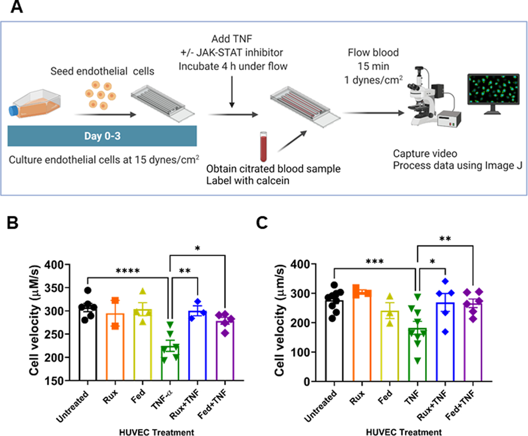
Methods: We assessed endothelial activation in the presence or absence of ruxolitinib or fedratinib by using immunoblots, immunofluorescence, qRT-PCR, and function coagulation assays. Finally, we used endothelialized microfluidics perfused with blood from normal and JAK2V617F+ individuals to evaluate whether ruxolitinib and fedratinib changed cell adhesion.

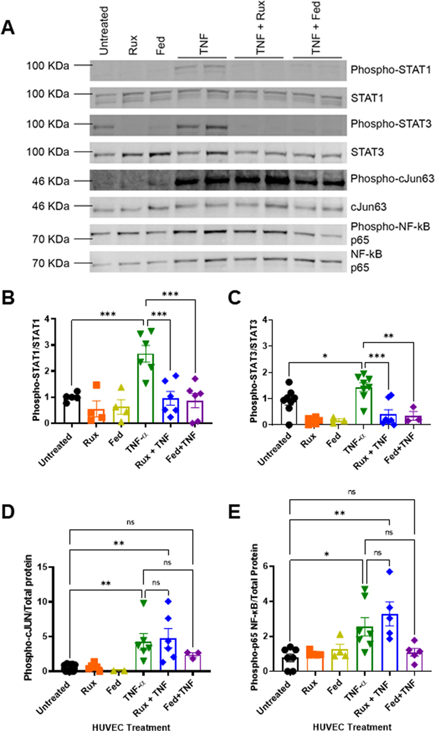
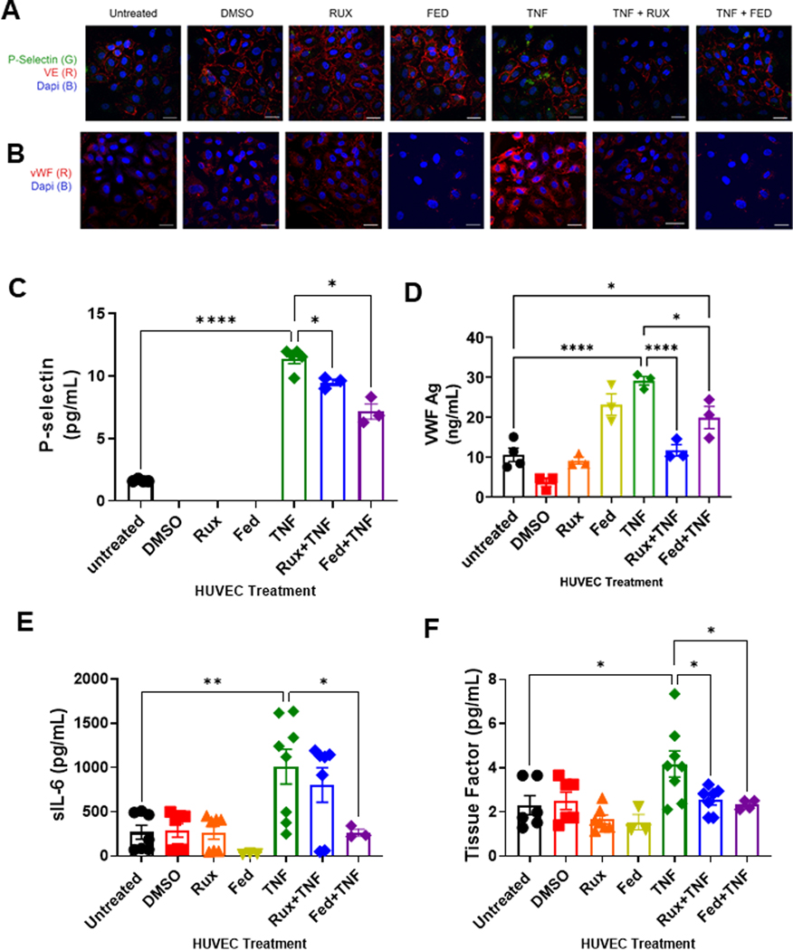
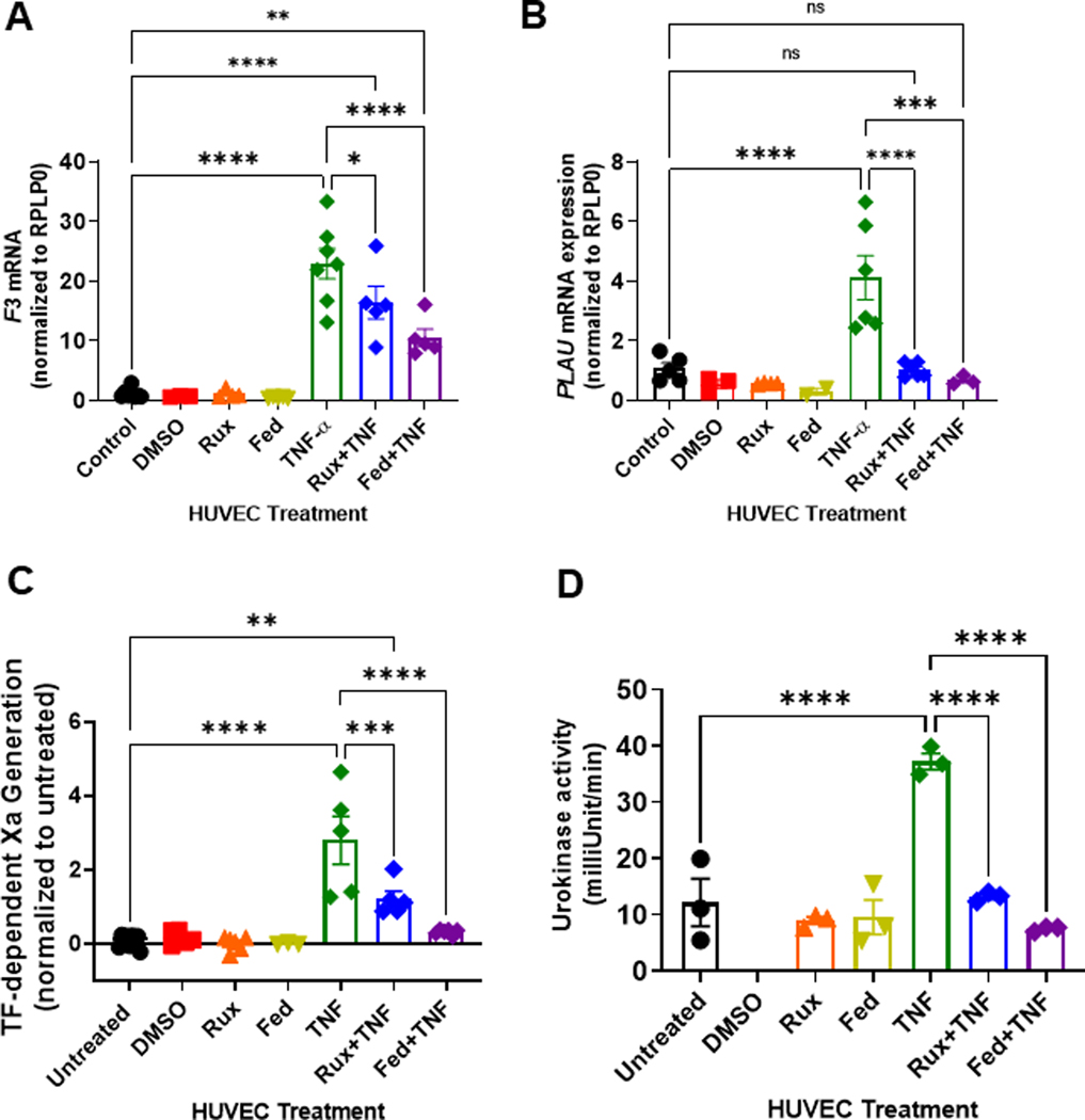
Results: We found that both ruxolitinib and fedratinib reduced endothelial cell phospho-STAT1 and STAT3 signaling and attenuated nuclear phospho-NK-κB and phospho-c-Jun localization. JAK-STAT inhibition also limited secretion of proadhesive and procoagulant P-selectin and von Willebrand factor and proinflammatory IL-6. Likewise, we found that JAK-STAT inhibition reduced endothelial tissue factor and urokinase plasminogen activator expression and activity.

Conclusions: By using endothelialized microfluidics perfused with whole blood samples, we demonstrated that endothelial treatment with JAK-STAT inhibitors prevented rolling of both healthy control and JAK2V617F MPN leukocytes. Together, these findings demonstrate that JAK-STAT inhibitors reduce the upregulation of critical prothrombotic pathways and prevent increased leukocyte-endothelial adhesion.

Keywords: endothelium; janus kinase inhibitors; myeloproliferative neoplasm; thrombosis; tissue factor.


Conflict of interest statement

Declaration of competing interests J.D.B. receives funds from Bayer independent from work herein. G.M.V. receives research funding from CSL Behring and Mitobridge (Astellas). C.B. has received reagents from CTI BioPharma for the conduct of clinical trial NCT 02891603 and a pending patent, WO2017058950A1, for methods of treating transplant rejection. B.C.B. holds patents related to CD4+ T cell pSTAT3 as a marker and therapeutic target of acute GVHD (WO2015120436A2); for the use of JAK inhibitors for rejection and GVHD prevention (WO2017058950A1); and for the use of CD83-targeted chimeric antigen receptor T cells in GVHD prevention, immune tolerance, autoimmunity, and acute myeloid leukemia therapy (WO2019165156). At this time, neither B.C.B. nor the University of Minnesota has received payment related to claims described in the patent. B.C.B. has received honoraria for participating in advisory board discussions for Incyte Corp and CTI BioPharma within the past 5 years. Remaining authors have no conflicts to disclose.

Figures

FIGURE 1
FIGURE 1
Ruxolitinib and fedratinib inhibit TNF-α-mediated STAT1 and STAT3 with attenuation of c-Jun and NF-κB. A. Representative western blots of nuclear lysates from human umbilical vein endothelial cells (HUVEC) treated with 0.4 μM ruxolitinib or 1 μM fedratinib JDB for 30 minutes followed by addition of 10 ng/mL TNF-α for 4 hours. Nuclear extracts collected and separated on 10% Tris-Hcl gel, transferred to PVDF membrane, and followed by total protein stain and immunoprobes against phospho-antibodies. Membranes were stripped and re-probed for unphosphorylated forms. B. Densitometry values from 5 different westerns blots probed for phospho-STAT1 normalized to total STAT1. C. Densitometry values from 5 different western blots for phospho-STAT3 normalized to total STAT3. D. Densitometry values for 5 different westerns blots probed for phospho-C-JUN normalized to total protein values. E. Densitometry values for 5 different western blots probed for phospho-p65 NF-κB to total protein. Error bars represent mean ±SE of means. p values from analysis of variance testing with appropriate post-hoc multiple comparison testing. ns, not significant. * p < .05, ** p < .01, *** p < .001.
FIGURE 2
FIGURE 2
Ruxolitinib and fedratinib decrease TNF-α mediated release of P-selectin and VWF and reduce secretion of IL-6 and tissue factor. HUVEC were treated with 0.4 μM ruxolitinib or 1 μM fedratinib JDB for 30 minutes followed by addition of 10 ng/mL TNF-α for 4 hours. Conditioned media was collected for ELISA. Cells were fixed and stained for the following analyses. (A) Confocal microscopy demonstrating release of P-selectin from endothelial cells. Unpermeablized HUVEC were stained for P-selectin (green) and VE-cadherin (red) and nuclei counterstained with DAPI. (B) Confocal microscopy demonstrating surface expression of VWF. Unpermeablized HUVEC were stained for VWF (red) and nuclei counterstained with DAPI. (C) ELISA assay for soluble P-selectin (sP-selectin). (D) ELISA assay for sVWF antigen. (E) Cytokine array results for sIL-6. (F) Cytokine arrays results for tissue factor. Each dot represents an individual replicate from a total of 3 separate experiments, with n = 2 to 3 replicates per experiment. For sP-selectin, VWF, and sIL-6, several replicates had undetectable levels. Error bars represent mean ±SE of means. p values from analysis of variance testing with appropriate post-hoc multiple comparison testing. ns, not significant. * p < .05, ** p < .01, *** p < .001, **** p < .0001.
FIGURE 3
FIGURE 3
Ruxolitinib and fedratinib decrease TNF-α mediated tissue factor and urokinase mRNA upregulation and decrease tissue factor and urokinase activity. HUVEC were treated with 0.4 μM ruxolitinib or 1 μM fedratinib JDB for 30 minutes followed by addition of 10 ng/mL TNF-α for 4 hours. (A) Tissue factor (Factor 3, F3) mRNA levels were normalized to control. (B) Plasminogen activator urokinase (PLAU) mRNA levels were normalized to control. For activity assays, HUVEC were treated with 0.4 μM ruxolitinib or 1 μM fedratinib for 30 minutes followed by addition of 10 ng/mL TNF-α for 4 hours JDB. (C). Measurement of tissue factor (TF)-dependent Xa generation was performed using Chromagenix Xa solution as described in methods. Results are normalized to untreated cells. (D). For urokinase activity, conditioned media were collected followed by urokinase activity measurement (Abcam). For all graphs, each dot represents an individual replicate from n = 2 to3 separate experiments. Error bars represent mean ±SE of means. p values from analysis of variance testing with appropriate post-hoc multiple comparison testing. ns, not significant. * p < .05, ** p < .01, *** p < .001, **** p < .0001.
FIGURE 4
FIGURE 4
Treatment of endothelium with ruxolitinib or fedratinib prevents TNF-α mediated cell velocity reduction at venous shear rate. A. Experimental schematic for endotheliazed devices and approach to evaluate effects on endothelial activation. HUVEC were cultured under flow for 3 days at 15 dynes/cm2. Endothelialized devices were then treated with ± 10 ng/mL TNF-α and/or ± 0.4 μM ruxolitinib or 1 μM fedratinib for 4 hours. Whole blood from donors was collected in sodium citrated and labeled with calcein. Whole blood was then perfused over endothelial cells for 15 minutes at 1 dynes/cm2. Image created in BioRender.com B. Cell velocity of calcein+ cells obtained from control blood donors (n = 7 donors with n = 3 to 7 replicates per conditions. C. Cell velocity of calcein+ cells obtained from JAK2 V617F+ (n = 9 donors with n = 3 to 9 replicates per condition, red) blood donors. All data ±SE of means. p values from one-way repeated measure ANOVA with Holm–Sidak’s multiple comparisons test. * p < .05, ** p < .01, *** p < .001, **** p < .0001.

Similar articles

Cited by

References

    1. Perner F, Perner C, Ernst T, Heidel FH. Roles of JAK2 in aging, inflammation, hematopoiesis and malignant transformation. Cells. 2019;8. - PMC - PubMed
    1. Cordua S, Kjaer L, Skov V, Pallisgaard N, Hasselbalch HC, Ellervik C. Prevalence and phenotypes of JAK2 V617F and calreticulin mutations in a Danish general population. Blood. 2019;134:469–79. - PubMed
    1. Mehta J, Wang H, Iqbal SU, Mesa R. Epidemiology of myeloproliferative neoplasms in the United States. Leuk Lymphoma. 2014;55:595–600. - PubMed
    1. Hebbel RP, Belcher JD, Vercellotti GM. The multifaceted role of ischemia/reperfusion in sickle cell anemia. J Clin Invest. 2020;130: 1062–72. - PMC - PubMed
    1. Lussana F, Rambaldi A. Inflammation and myeloproliferative neoplasms. J Autoimmun. 2017;85:58–63. - PubMed

Publication types