. 2023 Feb 4;20(1):25.
doi: 10.1186/s12974-023-02712-1.

Endothelial Toll-like receptor 4 is required for microglia activation in the murine retina after systemic lipopolysaccharide exposure

Affiliations

Endothelial Toll-like receptor 4 is required for microglia activation in the murine retina after systemic lipopolysaccharide exposure

Ioanna Tsioti et al. J Neuroinflammation. .

Abstract

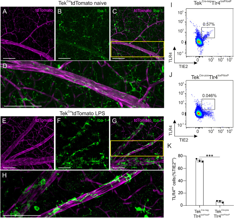
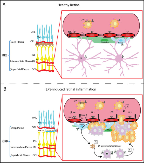
Background: Clustering of microglia around the vasculature has been reported in the retina and the brain after systemic administration of lipopolysaccharides (LPS) in mice. LPS acts via activation of Toll-like receptor 4 (TRL4), which is expressed in several cell types including microglia, monocytes and vascular endothelial cells. The purpose of this study was to investigate the effect of systemic LPS in the pigmented mouse retina and the involvement of endothelial TLR4 in LPS-induced retinal microglia activation.

Methods: C57BL/6J, conditional knockout mice that lack Tlr4 expression selectively on endothelial cells (TekCre-posTlr4loxP/loxP) and TekCre-negTlr4loxP/loxP mice were used. The mice were injected with 1 mg/kg LPS via the tail vein once per day for a total of 4 days. Prior to initiation of LPS injections and approximately 5 h after the last injection, in vivo imaging using fluorescein angiography and spectral-domain optical coherence tomography was performed. Immunohistochemistry, flow cytometry, electroretinography and transmission electron microscopy were utilized to investigate the role of endothelial TLR4 in LPS-induced microglia activation and retinal function.

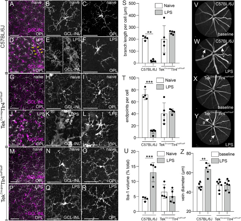
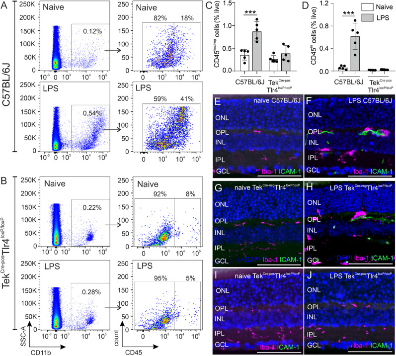
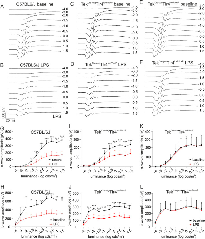
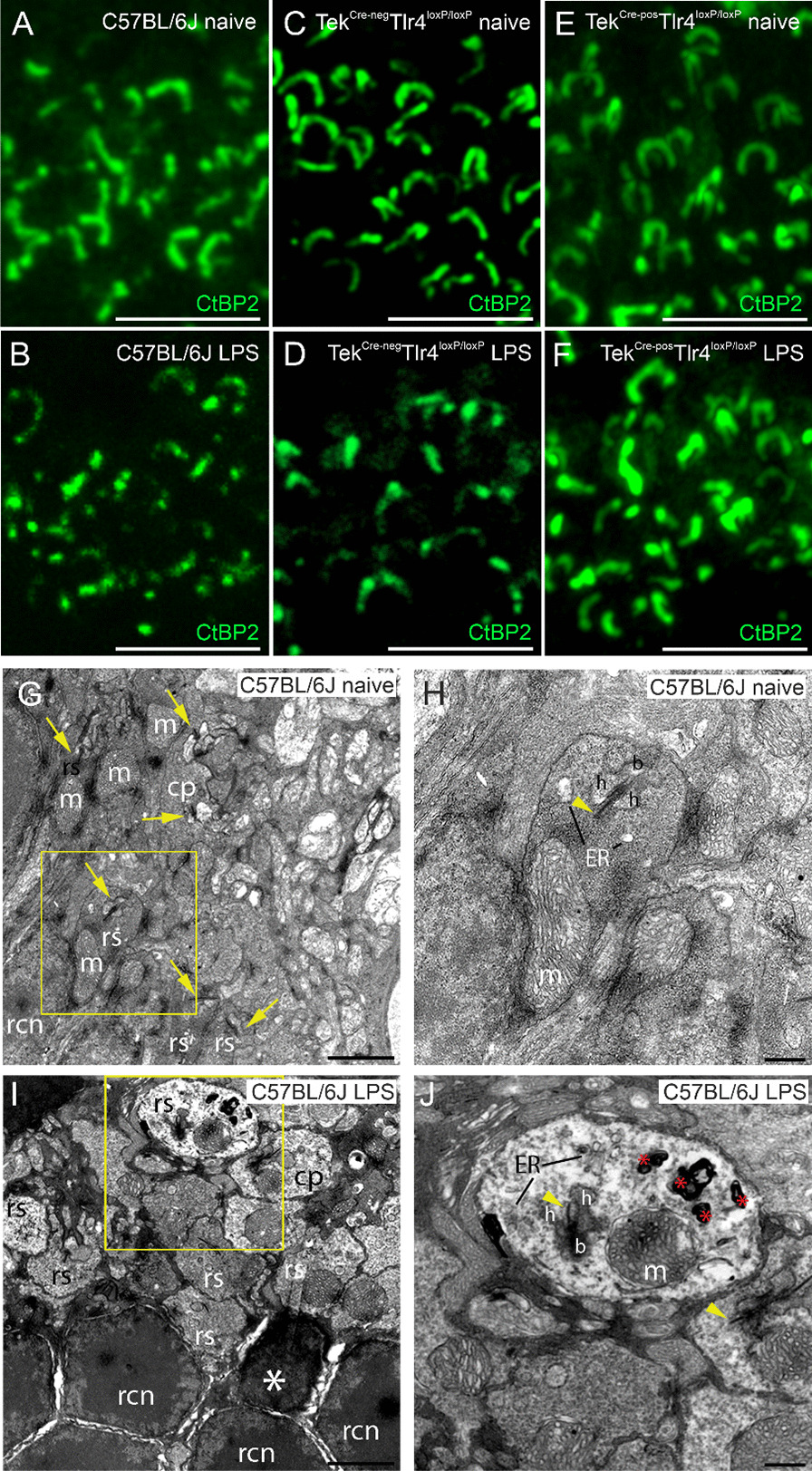
Results: Activation of microglia, infiltration of monocyte-derived macrophages, impaired ribbon synapse organization and retinal dysfunction were observed after the LPS exposure in C57BL/6J and TekCre-negTlr4loxP/loxP mice. None of these effects were observed in the retinas of conditional Tlr4 knockout mice after the LPS challenge.

Conclusions: The findings of the present study suggest that systemic LPS exposure can have detrimental effects in the healthy retina and that TLR4 expressed on endothelial cells is essential for retinal microglia activation and retinal dysfunction upon systemic LPS challenge. This important finding provides new insights into the role of microglia-endothelial cell interaction in inflammatory retinal disease.

Keywords: Endothelial cells; Lipopolysaccharide; Microglia; Monocyte-derived macrophages; Retina; Toll-like receptor 4.


Conflict of interest statement

I.T., Boehringer Ingelheim (F); B.L.S, none; P.E., Novartis Pharma Schweiz (C); M.S.Z., Bayer (F, C), Heidelberg Engineering (S), Novartis (C, I), Boehringer Ingelheim (F); P.M.B., Boehringer Ingelheim (E); D. K., Boehringer Ingelheim (F).

Figures

Fig. 1
Fig. 1
Cre-mediated recombination selectively depletes TLR4 on endothelial cells. Representative retinal whole mounts of TekCretdTomato mice stained with Iba-1 (green) in naïve mice (AC) and LPS-challenged mice (EG). tdTomato (magenta) was selectively expressed in retinal blood vessels and no co-localization with Iba-1 was detected. The yellow squares in C and G are magnified in D and H, respectively. Scale bars: 100 μm. Representative dot plots of single, live, TIE2posTLR4pos cells in the retinas of naïve TekCre−negTlr4loxP/loxP and TekCre−posTlr4loxP/loxP mice (I, J). The majority of TIE2pos cells (72.7%) in the TekCre−negTlr4loxP/loxP retinas express TLR4, while in the retinas of TekCre−posTlr4loxP/loxP mice only 6.4% of TIE2pos cells are TLR4pos (K; n = 3 mice per group). The data were analyzed with 2-tailed unpaired t-test, ***p < 0.001)
Fig. 2
Fig. 2
Systemic LPS fails to trigger microglia activation and retinal vasodilation in endothelial Tlr4-deficient mice. Representative images of Iba-1-stained retinal whole mounts in naïve (A, G, M) and LPS-challenged (D, J, P) C57BL/6J, TekCre−negTlr4loxP/loxP and TekCre−posTlr4loxP/loxP mice, respectively. Iba-1pos cells located in the GCL-INL and the OPL are shown in magenta and white, respectively. Yellow arrows in D indicate the clustering of Iba-1pos cells around a retinal vessel. In naïve C57BL/6J mice, microglia cells have a ramified morphology throughout the retina (B, C). After the LPS challenge, Iba-1pos cells adopt a bushy or amoebic morphology and accumulate around retinal blood vessels in the GCL-INL (E). In the OPL, these cells have an amoebic or rod-like shape (F). The morphology of Iba-1pos cells before or after the LPS challenge in TekCre−negTlr4loxP/loxP mice was comparable to C57BL/6J mice (H, I, K, L). Individual Iba-1pos cells of TekCre−posTlr4loxP/loxP mice had a ramified morphology both in the naïve and the LPS-challenged group (N, O, Q, R). Quantification of average branch length (S; n = 4–5 mice per group), number of endpoints per cell (T; n = 4–5 mice per group) and Iba-1 occupied volume (U; n = 4–5 mice per group) on images obtained from retinal whole mounts. Reduced branch length, less endpoints per cell and increased Iba-1 occupied volume were detected in the C57BL/6J but not the endothelial Tlr4 knockout mice. Representative images of fluorescein angiographs at baseline (V, X) and after the LPS challenge (W, Y) in C57BL/6J and TekCre−posTlr4loxP/loxP mice, respectively and quantification of vein dilation (Z; n = 5–7 mice per group). Vein diameter was significantly increased in LPS-challenged C57BL/6J mice compared to baseline, but not in TekCre−posTlr4loxP/loxP mice. The quantification data are reported as mean ± SD. The data were analyzed separately for each genotype with 2-tailed unpaired t-test (SU) or 2-tailed paired t-test (Z). **p < 0.01, ***p < 0.001. a Artery; v vein; GCL-INL ganglion cell layer-inner nuclear layer; OPL outer plexiform layer. Scale bars: left panel: 100 μm; middle and right panels: 50 μm
Fig. 3
Fig. 3
Effect of LPS on microglia/macrophage numbers in the presence or absence of endothelial Tlr4. Representative dot plots of CD11bpos populations (A, B left panels) gated as CD11bposCD45low/neg microglia and CD11bposCD45hi monocyte-derived macrophages (A, B right panels) in naïve and LPS-challenged C57BL/6J and TekCre−posTlr4loxP/loxP mice, respectively. Quantification of CD45low/neg microglia and CD45hi macrophages expressed as a percentage of the total number of live cells in the samples (C, D) revealed an increase of both populations after the LPS challenge in C57BL/6J mice but not in TekCre−posTlr4loxP/loxP mice (n = 5 mice per genotype and experimental group). The data were analyzed separately for each genotype with 2-tailed unpaired t-test (***p < 0.001). Representative images of retinal sections stained with Iba-1 and ICAM-1 in naïve (E, G, I) and LPS-challenged (F, H, J) C57BL/6J, TekCre−negTlr4loxP/loxP and TekCre−posTlr4loxP/loxP mice, respectively. ICAM-1 immunoreactivity was detected in LPS-challenged C57BL/6J and TekCre−negTlr4loxP/loxP mice but not in TekCre−posTlr4loxP/loxP mice. Scale bars: 200 μm. GCL Ganglion cells layer; INL inner nuclear layer; IPL inner plexiform layer; ONL outer nuclear layer; OPL outer plexiform layer
Fig. 4
Fig. 4
Endothelial Tlr4 depletion rescue the retina from LPS-induced visual dysfunction. Representative waveforms at baseline (A, C, E) and after the LPS challenge (B, D, F) under scotopic conditions in C57BL/6J, TekCre−negTlr4loxP/loxP and TekCre−posTlr4loxP/loxP mice, respectively. Comparison of a- (G, I, K) and b-wave amplitudes (H, J, L) between baseline and LPS in C57BL/6J, TekCre−negTlr4loxP/loxP and TekCre−posTlr4loxP/loxP mice, respectively. Reduced mean a- and b-wave amplitudes were recorded in C57BL/6J and TekCre−negTlr4loxP/loxP mice after the LPS challenge. In TekCre−posTlr4loxP/loxP mice, mean a- and b-wave amplitudes after the LPS challenge were comparable to baseline levels. The data were analyzed with repeated measures 2-way ANOVA followed by Sidak’s post hoc analysis (n = 4–5 mice per genotype, one eye per mouse; *p < 0.05, **p < 0.01, ***p < 0.001)
Fig. 5
Fig. 5
OPL ultrastructure in C57BL/6J mice is affected upon the LPS challenge. Representative z-stack images of retinal sections stained with the ribbon synapse marker CtBP2 in naïve (A, C, E) and LPS-challenged (B, D, F) C57BL/6J, TekCre−negTlr4loxP/loxP and TekCre−posTlr4loxP/loxP mice, respectively. In the LPS-challenged C57BL/6J and TekCre−negTlr4loxP/loxP mice, ribbon synapses lose their characteristic shape and are less abundant in the OPL. Representative transmission electron microscopy images (×8200 magnification) obtained from naïve (G) and LPS-challenged (I) C57BL/6 J mice. Higher magnification (×20,500) of the yellow squares in G, I are shown in H, J, respectively. The LPS exposure led to alterations in the OPL ultrastructure characterized by membrane whorls, electro-lucent cytoplasm and decreased vesicle density in rod spherules. Yellow arrows and arrowheads indicate ribbon synapses and ribbons, respectively. White asterisk indicates an affected rod and red asterisks indicate membrane whorls. b Bipolar cell dendritic process; cp cone pedicle; ER endoplasmic reticulum; h horizontal cell axon tip; m mitochondrion; rcn rod cell nucleus; rs rod spherule. Scale bars: AF 5 μm; G, I 2 μm. H, J 500 nm
Fig. 6
Fig. 6
Proposed mechanism of systemic LPS-induced retinal microglia activation and retinal dysfunction. A. The inner BRB (iBRB) is composed by a deep, an intermediate, and a superficial vascular plexus. In the healthy retina, endothelial cells, that line the blood vessels, are connected by tight junctions forming an effective vascular barrier. Endothelial cells bear TLR4 receptors and they are surrounded by pericytes and perivascular macrophages. Microglia resides in close proximity to the vasculature, while monocytes circulate in the bloodstream. B. Upon the systemic LPS challenge, LPS binds to TLR4 located on circulating monocytes and endothelial cells (1), and induces the expression of the adhesion molecule ICAM-1 from the latter (2). Activated endothelial cells release cytokines and chemokines (3), which may act as chemoattractants causing microglia migration towards the affected vasculature (4). Subsequently, monocyte-derived macrophages are entering the retina through the disrupted BRB and AIF-1 – ICAM-1 interactions (5). Migration of microglia away from retinal neurons may account for disruption of ribbon synapses and impaired retinal function (6). EC endothelial cell; GCL ganglion cell layer; iBRB inner blood retinal barrier; ICAM-1 intercellular molecule 1; INL inner nuclear layer; IPL inner plexiform layer; LFA-1 lymphocyte function-associated antigen 1; LPS lipopolysaccharide; M microglia; MDMs monocyte-derived macrophages; ONL outer nuclear layer; OPL outer plexiform layer; PRs photoreceptors; PVMs perivascular macrophages; RSs ribbon synapses; TJs tight junctions; TLR4 Toll-like receptor 4

Similar articles

Cited by

References

    1. Caso JR, Pradillo JM, Hurtado O, Lorenzo P, Moro MA, Lizasoain I. Toll-like receptor 4 is involved in brain damage and inflammation after experimental stroke. Circulation. 2007;115(12):1599–1608. - PubMed
    1. Hua F, Ma J, Ha T, Xia Y, Kelley J, Williams DL, et al. Activation of Toll-like receptor 4 signaling contributes to hippocampal neuronal death following global cerebral ischemia/reperfusion. J Neuroimmunol. 2007;190(1–2):101–111. - PMC - PubMed
    1. Wang Y, Ge P, Zhu Y. TLR2 and TLR4 in the brain injury caused by cerebral ischemia and reperfusion. Mediat Inflamm. 2013;2013:124614. - PMC - PubMed
    1. Mowry FE, Peaden SC, Stern JE, Biancardi VC. TLR4 and AT1R mediate blood–brain barrier disruption, neuroinflammation, and autonomic dysfunction in spontaneously hypertensive rats. Pharmacol Res. 2021;174:105877. - PMC - PubMed
    1. Ko MK, Saraswathy S, Parikh JG, Rao NA. The role of TLR4 activation in photoreceptor mitochondrial oxidative stress. Investig Ophthalmol Vis Sci. 2011;52(8):5824–5835. - PMC - PubMed