. 2023 Mar:248:109251.
doi: 10.1016/j.clim.2023.109251. Epub 2023 Feb 3.

Ectopic lymphoid structures in the aged lacrimal glands

Affiliations

Ectopic lymphoid structures in the aged lacrimal glands

Jeremias G Galletti et al. Clin Immunol. 2023 Mar.

Abstract

Aging is a complex biological process in which many organs are pathologically affected. We previously reported that aged C57BL/6J had increased lacrimal gland (LG) lymphoid infiltrates that suggest ectopic lymphoid structures. However, these ectopic lymphoid structures have not been fully investigated. Using C57BL/6J mice of different ages, we analyzed the transcriptome of aged murine LGs and characterized the B and T cell populations. Age-related changes in the LG include increased differentially expressed genes associated with B and T cell activation, germinal center formation, and infiltration by marginal zone-like B cells. We also identified an age-related increase in B1+ cells and CD19+B220+ cells. B220+CD19+ cells were GL7+ (germinal center-like) and marginal zone-like and progressively increased with age. There was an upregulation of transcripts related to T follicular helper cells, and the number of these cells also increased as mice aged. Compared to a mouse model of Sjögren syndrome, aged LGs have similar transcriptome responses but also unique ones. And lastly, the ectopic lymphoid structures in aged LGs are not exclusive to a specific mouse background as aged diverse outbred mice also have immune infiltration. Altogether, this study identifies a profound change in the immune landscape of aged LGs where B cells become predominant. Further studies are necessary to investigate the specific function of these B cells during the aged LGs.

Keywords: Aging; B cells; Dry eye; Ectopic lymphoid structures; Lacrimal gland; Tertiary lymphoid organs.


Conflict of interest statement

Declaration of Competing Interest CSDP was a consultant to Spring Discovery from May to August 2022. All other authors declare no conflicts of interest.

Figures

Figure 1.
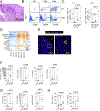
Figure 1.. Increased B cell infiltration in female aged C57BL/6 lacrimal glands.
A. Representative whole histological scans of young and aged female C57BL/6 lacrimal glands stained with H&E. Areas of infiltration are demarcated in dark blue. B. The number of foci (>50 cells) was counted under a 10X microscope lens, and the area of the gland was calculated to generate the focus score on the right. Each dot represents a left lacrimal gland. Kruskal-Wallis with Dunn’s multiple comparison test. P value as shown. Scale bar = 1000 μm. C. Representative merged laser scanning confocal microscopy image of young (3-month-old, 3M) and aged (24-month-old, 24M) lacrimal glands stained with anti-B220 (green), anti-CD3 (white) and propidium iodide nuclear counterstaining (red). Small insets are magnified. Scale bar 50μm (left and middle panel) and 10μm (right panel). D. Representative immunohistochemistry of aged (26 month-old) lacrimal gland stained with either CD19 or CD3 antibodies (brown color). Scale bar 100μm. E. Representative dot plots of CD19 and B220+ stained cells using flow cytometry in lacrimal gland lysates. Cells were gated from CD45+ cells. Numbers in the quadrants represent percent of cells. F. Accumulative data of frequency of CD19+B220, CD19+B220+ and CD19B220+ cells in lacrimal glands. Each dot represents a mouse, n = 6–10. Kruskal-Wallis with Dunn’s multiple comparison test. P value as shown. G. Relative fold of expression of Cd19 and Cd22 in lacrimal gland lysates. Each dot represents a mouse, n = 8. Kruskal-Wallis with Dunn’s multiple comparison test. P value as shown.
Fig. 2
Fig. 2. Increased B cell activation genes in female aged C57BL/6 lacrimal glands.
A. The overall heat map profile for 2220 differentially expressed genes. B. A volcano plot showing the magnitude of change in the lacrimal gland modulated by aging (Log 1.2-fold change) versus the adjusted p-value (−Log10). Some of the most significantly up genes are highlighted. The dotted line indicates a significance of 0.05. C. Detailed heatmap of significantly differentially expressed genes (DEGs) associated with B and T cell activation.
Fig. 3.
Fig. 3.. Germinal center markers in female aged C57BL/6 lacrimal glands.
A. Representative H&E of an aged lacrimal gland (LG) showing germinal-center like infiltration. Scale bar = 50 μm. B. Representative dot plots of the lacrimal gland (LG) and draining lymph nodes (dLN) suspensions stained with GL7 and CD95. Cells were gated as described in the methods. C. Accumulative data of frequency of GL7+CD95+ cells in lacrimal glands (LG) and draining lymph nodes (dLN). Each dot represents a mouse, n = 5. Kruskal-Wallis with Dunn’s multiple comparison test. P value as shown. D. Heatmaps showing DEG related to germinal center formation. E. Representative merged laser scanning confocal microscopy image of aged (24-mont-hold, 24M) lacrimal glands stained with anti-LYVE-1 (green), PNAd (red), and DAPI counterstaining (blue). Scale bar 100μm (left) and 20μm (right panel). F-G. Relative fold of expression of Glycam1, Cxcl13, Ltb, Cxcl12 (F) and Cccl19, Ccr7, Tnfsf13b (G) in lacrimal gland lysates determined by qPCR. Each dot represents a mouse, n = 8. Kruskal-Wallis with Dunn’s multiple comparison test. P value as shown. H. Luminex assay showing protein levels of BAFF and CCL21. Each dot represents a mouse, n = 8. Kruskal-Wallis with Dunn’s multiple comparison test. P value as shown.
Fig. 4.
Fig. 4.. Age-related increased frequency of T follicular helper cells in female C57BL/6 lacrimal glands.
A. Heatmaps showing DEG related to Tfh markers. B. Relative fold expression of Cxcr5, Il21 in lacrimal gland lysates. Each dot represents a mouse, n = 8–10. Kruskal-Wallis with Dunn’s multiple comparison test. P value as shown. C. Representative density plots of CXCR5 and PD-1 were further gated into BCL6 and IL-21 in single-cell suspensions of the lacrimal gland (LG) and draining lymph nodes (dLN). D. Accumulative data of CXCR5+PD-1+ cells and BCL-6+IL-21+ cells in lacrimal gland and dLN. Each dot represents a mouse, n = 8. Kruskal-Wallis with Dunn’s multiple comparison test. P value as shown.
Figure 5.
Figure 5.. Age-related increase in marginal zone-like B cells in female C57BL/6 lacrimal glands.
A. Representative density plots of IgM and CD23 cells in single cell suspensions of lacrimal glands (LG) and draining lymph nodes. Cells were gated sequentially as CD45+, B220+CD93 cells and then IgM and CD23 were plotted. B. Accumulative data of CD93IgM+CD23 (MZB) and CD93IgM+CD23+ (follicular) cells. Each dot represents a mouse, n = 8. Kruskal-Wallis with Dunn’s multiple comparison test. P value as shown. C. Heatmaps showing DEGs related to MZB cell biology.
Fig 6.
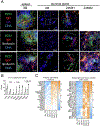
Fig 6.. Increased immunoglobulin production in female aged C57BL/6 lacrimal glands.
A. Merged images of laser confocal microscopy of spleen and lacrimal glands from 3-month (3M) and 24-month-old (24M) stained with B220 (green) and different immunoglobulin isotype antibodies as shown. Square insets in the top row show B220 and IgA single staining. In the second and third rows, there is autofluorescence at the margins of the infiltrates; this has been described as lipofuscin inclusions. Areas of infiltrates in aged lacrimal glands are demarcated by dotted ellipse. Scale bar = 50 μm. B. Normalized raw counts (fragments per million, FKPM) of immunoglobulin genes from bulk RNA Sequencing. p value indicates FDR from ROSALIND® software, calculated as described in the “Methods.” C. Heatmaps showing DEGs related to plasma cells (D) and kappa and variable chain.
Figure 7.
Figure 7.. Aged Diversity Outbred mice display lacrimal gland infiltration and goblet cell loss.
A. Representative scans of whole H&E-stained lacrimal glands of the aged female and male Diversity Outbred mice. Areas of infiltration are demarcated in white. B. Accumulative data of focus score/4mm2 as a measurement of lacrimal gland infiltration (left panel) C. Accumulative data of goblet cell density in Diversity Outbred mice in young (3–4 months) and aged (23–25-months age). Each dot represents one mouse, n = 10/age/group. Kruskal-Wallis with Dunn’s multiple comparison test.
Fig. 8.
Fig. 8.. Increased anti-Ro52/SS-A and anti-la/SS-B antibodies in the sera of aged female C57BL/6 mice.
Serum was collected from female C57BL/6 (B6) mice of different ages. Male sera from NOD.B10.H2b mice (which develop dacryoadenitis) were used as positive controls. Each dot represents one animal. Kruskal-Wallis with Dunn’s multiple comparison test, n = 8–14/group.
Fig. 9.
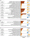
Fig. 9.. Meta-analysis showing shared and unique pathways in Sjögren syndrome and aged lacrimal glands
Meta-analysis comparing RNAseq of Sjögren syndrome (SS) and aging datasets. Twelve different patterns of expression have been obtained and divided into 3 categories: (A) DEGs upregulated or downregulated in both datasets, (B) DEGs altered specifically in SS and (C) DEGs differentially regulated between Aging and SS. −Log10 (P-value) of Top-3 pathways enriched by these genes have been plotted. On the right, a summary of gene expression for that pattern is shown to indicate its trend (upregulated in yellow, downregulated in blue) and its average fold change (Avg. FC) in each dataset.

References

    1. de Paiva CS, St Leger AJ, Caspi RR: Mucosal immunology of the ocular surface. Mucosal immunology 2022:1–15. - PMC - PubMed
    1. Knop E, Knop N: Lacrimal drainage-associated lymphoid tissue (LDALT): a part of the human mucosal immune system. Invest Ophthalmol Vis Sci 2001, 42:566–74. - PubMed
    1. Saitoh-Inagawa W, Hiroi T, Yanagita M, Iijima H, Uchio E, Ohno S, Aoki K, Kiyono H: Unique characteristics of lacrimal glands as a part of mucosal immune network: high frequency of IgA-committed B-1 cells and NK1.1+ alphabeta T cells. Invest OphthalmolVisSci 2000, 41:138–44. - PubMed
    1. Gudmundsson OG, Benediktsson H, Olafsdottir K: T-lymphocyte subsets in the human lacrimal gland. Acta ophthalmologica 1988, 66:19–23. - PubMed
    1. Gudmundsson OG, Bjornsson J, Olafsdottir K, Bloch KJ, Allansmith MR, Sullivan DA: T cell populations in the lacrimal gland during aging. Acta ophthalmologica 1988, 66:490–7. - PubMed

Publication types