A method describing the microdissection of trabecular meshwork tissue from Brown Norway rat eyes
- PMID: 36740159
- PMCID: PMC9991013
- DOI: 10.1016/j.exer.2022.109367
A method describing the microdissection of trabecular meshwork tissue from Brown Norway rat eyes
Abstract
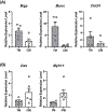
Glaucoma is often associated with elevated intraocular pressure (IOP), generally due to obstruction of aqueous humor outflow within the trabecular meshwork (TM). Despite many decades of research, the molecular cause of this obstruction remains elusive. To study IOP regulation, several in vitro models, such as perfusion of anterior segments or mechanical stretching of TM cells, have identified several IOP-responsive genes and proteins. While these studies have proved informative, they do not fully recapitulate the in vivo environment where IOP is subject to additional factors, such as circadian rhythms. Thus, rodent animal models are now commonly used to study IOP-responsive genes in vivo. Several single-cell RNAseq studies have been performed where angle tissue, containing cornea, iris, ciliary body tissue in addition to TM, is dissected. However, it is advantageous to physically separate TM from other tissues because the ratio of TM cells is relatively low compared to the other cell types. In this report, we describe a new technique for rat TM microdissection. Evaluating tissue post-dissection by histology and immunostaining clearly shows successful removal of the TM. In addition, TaqMan PCR primers targeting biomarkers of trabecular meshwork (Myoc, Mgp, Chi3l1) or ciliary body (Myh11, Des) genes showed little contamination of TM tissue by the ciliary body. Finally, pitfalls encountered during TM microdissection are discussed to enable others to successfully perform this microsurgical technique in the rat eye.
Keywords: Microdissection; RNA isolation; Trabecular meshwork.
Copyright © 2023 Elsevier Ltd. All rights reserved.
Figures


References
-
- Borras T, 2003. Gene expression in the trabecular meshwork and the influence of intraocular pressure. Prog Retin Eye Res 22, 435–463. - PubMed
-
- Erickson-Lamy K, Rohen JW, Grant WM, 1991. Outflow facility studies in the perfused human ocular anterior segment. Exp Eye Res 52, 723–731. - PubMed
-
- Ethier CR, Chan DW, 2001. Cationic ferritin changes outflow facility in human eyes whereas anionic ferritin does not. Invest Ophthalmol Vis Sci 42, 1795–1802. - PubMed
Publication types
MeSH terms
Grants and funding
LinkOut - more resources
Full Text Sources
Medical
Miscellaneous

