Review
. 2023 Feb;151(2):289-300.e4.
doi: 10.1016/j.jaci.2022.12.005.

Updates on the immunopathology and genomics of severe cutaneous adverse drug reactions

Affiliations
Review

Updates on the immunopathology and genomics of severe cutaneous adverse drug reactions

Andrew Gibson et al. J Allergy Clin Immunol. 2023 Feb.

Abstract

Severe cutaneous adverse reactions (SCARs) such as Stevens-Johnson syndrome, toxic epidermal necrolysis (SJS/TEN), and drug reaction with eosinophilia and systemic symptoms (DRESS)/drug-induced hypersensitivity syndrome (DIHS) cause significant morbidity and mortality and impede new drug development. HLA class I associations with SJS/TEN and drug reaction with eosinophilia and systemic symptoms/drug-induced hypersensitivity syndrome have aided preventive efforts and provided insights into immunopathogenesis. In SJS/TEN, HLA class I-restricted oligoclonal CD8+ T-cell responses occur at the tissue level. However, specific HLA risk allele(s) and antigens driving this response have not been identified for most drugs. HLA risk alleles also have incomplete positive and negative predictive values, making truly comprehensive screening currently challenging. Although, there have been key paradigm shifts in knowledge regarding drug hypersensitivity, there are still many open and unanswered questions about SCAR immunopathogenesis, as well as genetic and environmental risk. In addition to understanding the cellular and molecular basis of SCAR at the single-cell level, identification of the MHC-restricted drug-reactive self- or viral peptides driving the hypersensitivity reaction will also be critical to advancing premarketing strategies to predict risk at an individual and drug level. This will also enable identification of biologic markers for earlier diagnosis and accurate prognosis, as well as drug causality and targeted therapeutics.

Keywords: AGEP; DIHS; DRESS; HLA; SCAR; SJS/TEN; T-cell; altered peptide.


Figures

Figure 1.
Figure 1.
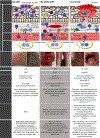
Clinical presentation and immunopathology of SCAR. AGEP, DRESS/DIHS and SJS/TEN are the reaction phenotypes that comprise SCAR. These reactions vary in mortality, immunopathological features, and clinical characteristics. Adapted from Hama et al. 2022. AGEP: CD4+ T-cells secrete IL4, IL5, IL13, IFNγ, TNFα, IL8, IL17, and IL22 (Th17). IL8 drives neutrophil and T-cell recruitment to the epidermis to form sterile pustules. DRESS/DIHS: CD4+ and CD8+ T-cells, plasma DCs, and monocytes are enriched in the dermis. DCs produce CCL17 to recruit CCR4+ Th2 T-cells. Th2 cells and ILC2 produce IL5 to induce activation and migration of eosinophils that drive inflammation. TNFα, IFNγ (Th1), IL-4, IL-5, and IL-13 (Th2) are observed with HHV reactivation and Tregs in lesional skin. SJS/TEN: Cytotoxic CD8+ T-cells accumulate in blisters and release perforin, granzyme B, and granulysin to kill keratinocytes. N, neutrophil; APC, antigen-presenting cell; Th, T helper; DC, dendritic cell; IL, interleukin; TNFα, tumour necrosis factor alpha; IFNγ, interferon gamma; Treg, T regulatory cell; E, eosinophil; TRM, tissue-resident memory T-cell; CCR, chemokine receptor; CCL, C-C motif chemokine ligand; ILC, innate lymphoid cell; NKT, natural killer T-cell; NK, natural killer cell; GM-CSF, granulocyte macrophage colony-stimulating factor; HHV, Human Herpesvirus; CMV, Cytomegalovirus; EBV, Epstein-Barr virus.*5–10% of SJS/TEN-cases with more than 10% skin detachment do not have mucosal erosions. created using Biorender.com
Figure 2.
Figure 2.. Clinical timeline of SCAR.
Timelines showing the time from initiation of a specific drug to typical onset of symptoms and signs of a specific SCAR (latency period). The latency period differs not only between different SCAR reactions but also between SCAR reactions and benign exanthems. Adapted from Hama et al. 2022. DHR, delayed drug hypersensitivity reactions; SCAR, severe cutaneous adverse reaction; AGEP, acute generalized exanthematous pustulosis; DRESS, drug reaction with eosinophilia and systemic symptoms; DIHS, drug-induced hypersensitivity syndrome; SJS/TEN, Stevens-Johnson syndrome/toxic epidermal necrolysis; MDE, morbilliform drug eruption; FDE, fixed drug eruption; SSLR, serum sickness like reactions.
Figure 3.
Figure 3.. Inheritance of HLA alleles and their role in SCAR.
(A) HLA alleles are co-dominantly inherited. (Bi) Antigen processing varies between HLA-Class I (left) and II (right). HLA-class I: Intracellular antigens undergo proteasomal processing, with peptides loaded into the ER for loading on HLA-class I and presentation to CD8+ T-cells. HLA-class II: Extracellular antigens are phagocytosed for antigen processing in endosomes, which fuse with lysosomes from the ER carrying HLA class II for loading and surface presentation to CD4+ T-cells. (Bii) HLA-Class I presentation results in cytotoxicity. Briefly, antigen engages the TCR to enable phosphorylation of CD3 by Lck for binding of ZAP70, Grb2, and PLCγ1. This activates calcium, RAS, MAPK, and PI3K signalling and transcription factors for cell activation and release of cytotoxic granules. HLA, human leukocyte antigen; APC, antigen-presenting cell; Tel, telomeric; Cen, centromeric; ER, endoplasmic reticulum; TAP, tapasin; TCR, T-cell receptor; Lck, lymphocyte-specific protein tyrosine kinase; PI3K, Phosphatidylinositol 3 kinase; P, phosphorylated; ZAP70, Zeta-chain-associated protein kinase 70; LAT, linker for activation of T cells; PLC, phospholipase; Grb, Growth factor receptor-bound protein; SOS, Son of sevenless protein; Ca2+, calcium; RAS, ‘Rat sarcoma virus’ protein; MAPK, mitogen-activated protein kinase. Figure created using Biorender.com
Figure 4.
Figure 4.. HLA-restricted models of drug-induced T-cell activation.
(A) Models of HLA-restricted T-cell activation include the (i) hapten, (ii) pharmacological interaction (PI), and (iii) altered self-peptide repertoire models. Hapten: Drug-antigen binds self-protein before intracellular processing forms drug-modified peptides, which bind irreversibly and covalently to risk HLA. PI: Drug-antigen binds directly, reversibly, and non-covalently to risk HLA or TCR without a need for antigen uptake and processing. Altered self-peptide repertoire: Drug-antigen binds to risk HLA in such way that it alters the repertoire of self-peptides that may bind, which are subsequently seen as immunogenic. (B) Models for the HLA-restricted origins of drug-reactive T-cells in SCAR patient skin include (i) naïve priming and (ii) the heterologous immunity model. Naïve priming: Drug antigen primes the naïve T-cell which generates long-lived TRM, which are re-activated upon subsequent exposure to the same drug. Heterologous immunity: The patient is first infected with a virus, from which peptides are presented by the risk HLA to prime naïve T-cells. In later life, exposure of the same patient to a drug-antigen which shares structural cross-reactivity and HLA-restriction drives re-stimulation of the viral-primed TRM and SCAR. APC, antigen-presenting cell; HLA, human leukocyte antigen; TCR, T-cell receptor; TRM, tissue-resident memory T-cell. Figure created using Biorender.com
Figure 5.
Figure 5.. HLA class I risk is thought to be necessary but not sufficient for predicting many SCAR.
Positive predictive values differ based on drug and specific hypersensitivity phenotype. 100 patients with reaction to each drug are shown, with the percentage of those that will express the defined risk allele coloured purple. The remainder of patients, coloured grey, would be tolerant to the drug despite the presence of the risk allele. For abacavir (left), 55% of those carrying HLA-B*57:01 develop abacavir hypersensitivity. For vancomycin (middle), 20% of those carrying HLA-A*32:01 develop vancomycin-DRESS/DIHS when exposed for greater than 2 weeks. For allopurinol (right), 3% of those carrying HLA-B*58:01 develop allopurinol-DRESS/DIHS or allopurinol-SJS/TEN. HLA, human leukocyte antigen; DHR, delayed drug hypersensitivity reaction; SCAR, severe cutaneous adverse reaction; AGEP, acute generalized exanthematous pustulosis; DRESS, drug reaction with eosinophilia and systemic symptoms; DIHS, drug-induced hypersensitivity syndrome; SJS/TEN, Stevens-Johnson syndrome/toxic epidermal necrolysis.
Figure 6.
Figure 6.. Precision medicine approaches to SCAR.
Approaches target prediction and prevention through risk assessment and genetic screening. Multifaceted approaches will facilitate earlier identification of disease. In vivo, ex vivo, in vitro and genetic studies combined with clinical factors will help to identify the culprit drug and improve future drug safety. Studies looking at disease at the site of tissue damage will inform immunopathogenesis, prognosis, oligoclonal T-cell receptor and identify targeted treatments. Functional studies will be important for defining molecular interactions as well as epitope discovery that along with the oligoclonal TCR (s) identified at the site of tissue damage will help define immunodominance and. HLA specificity of a given drug hypersensitivity reaction without the need for large scale epidemiological studies. HLA, human leukocyte antigen. Figure created using Biorender.com

References

    1. Hama N, Abe R, Gibson A, Phillips EJ. Drug-Induced Hypersensitivity Syndrome (DIHS)/Drug Reaction With Eosinophilia and Systemic Symptoms (DRESS): Clinical Features and Pathogenesis. J Allergy Clin Immunol Pract 2022; 10:1155–67 e5. - PMC - PubMed
    1. Brockow K, Ardern-Jones MR, Mockenhaupt M, Aberer W, Barbaud A, Caubet JC, et al. EAACI position paper on how to classify cutaneous manifestations of drug hypersensitivity. Allergy 2019; 74:14–27. - PubMed
    1. Roujeau JC, Kelly JP, Naldi L, Rzany B, Stern RS, Anderson T, et al. Medication use and the risk of Stevens-Johnson syndrome or toxic epidermal necrolysis. N Engl J Med 1995. Dec 14;333(24):1600–7. - PubMed
    1. Momen SE, Diaz-Cano S, Walsh S, Creamer D. Discriminating minor and major forms of drug reaction with eosinophilia and systemic symptoms: Facial edema aligns to the severe phenotype. J Am Acad Dermatol 2021. Sep;85(3):645–652. - PubMed
    1. Soria A, Bernier C, Veyrac G, Barbaud A, Puymirat E, Milpied B. Drug reaction with eosinophilia and systemic symptoms may occur within 2 weeks of drug exposure: A retrospective study. J Am Acad Dermatol 2020. Mar;82(3):606–611. - PubMed

Publication types