Review
. 2024;22(7):1189-1204.
doi: 10.2174/1570159X21666230203103910.

Emerging Roles of Microglia in Blood-Brain Barrier Integrity in Aging and Neurodegeneration

Affiliations
Review

Emerging Roles of Microglia in Blood-Brain Barrier Integrity in Aging and Neurodegeneration

Simeng Zhang et al. Curr Neuropharmacol. 2024.

Abstract

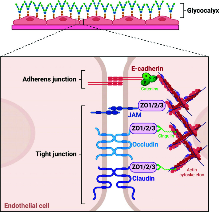
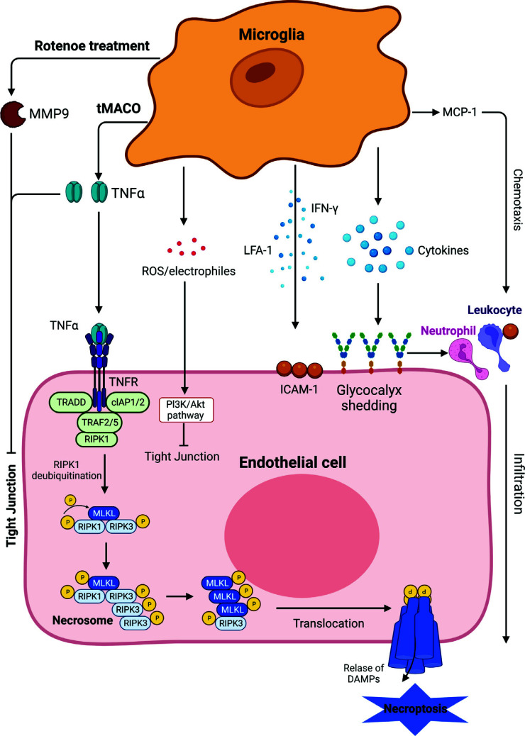
The blood-brain barrier (BBB) is a highly selective interface between the blood and the brain parenchyma. It plays an essential role in maintaining a specialized environment for central nervous system function and homeostasis. The BBB disrupts with age, which contributes to the development of many age-related disorders due to central and peripheral toxic factors or BBB dysfunction. Microglia, the resident innate immune cells of the brain, have recently been explored for their ability to directly and indirectly regulate the integrity of the BBB. This review will focus on the current understanding of the molecular mechanisms utilized by microglia to regulate BBB integrity and how this becomes disrupted in aging and age-associated diseases. We will also discuss the rationale for considering microglia as a therapeutic target to prevent or slow down neurodegeneration.

Keywords: Microglia; aging; blood-brain barrier; neurodegeneration.; neuroinflammation; neurovascular unit.


Conflict of interest statement

The authors declare no conflict of interest, financial or otherwise.

Figures

Fig. (1)
Fig. (1)
Schematic representation of the BBB barriers. The barriers of the BBB include the meningeal barrier, the vascular BBB, the blood-CSF barrier, and the tanycytic barrier. The meningeal barrier is found within the arachnoid matter, and is composed of epithelial cells. The vascular BBB is found at the capillary bed and the adjacent arterioles and venules and is composed of brain endothelial cells. The blood-CSF barrier resides at the choroid plexus and is composed of ependymal cells. The tanycytic barrier separates circumventricular organs from the adjacent area of the brain barrier.
Fig. (2)
Fig. (2)
Schematic structures of the tight junctions (TJs) and adherens junctions (AJs). Major bicellular TJ proteins include three transmembrane proteins: occludins, claudins, and junctional adhesion molecules (JAMs), and cytoplasmatic zonula occludens proteins (ZO1, ZO2, and ZO3). Cadherin adhesion molecules are core AJ components, and all cadherins contain two or more extracellular cadherin domains. The cytoplasmic tail of cadherins binds to other cadherins. The ZOs and catenins interact with membrane proteins to anchor them to the actin cytoskeleton.
Fig. (3)
Fig. (3)
Dual effect of microglia on the BBB integrity. Microglia protect the BBB through phagocytosis and clearance of tau proteins and amyloid fibers, and via secretion of miRNA-126a-5p, which is beneficial for healthy cognitive function. However, microglia can also impair the BBB through NLRP3 inflammasome-mediated neuroinflammation, the production of tau proteins, and amyloid fibers, which contribute to cognitive deficits.
Fig. (4)
Fig. (4)
The effects of microglia on endothelial cells. Microglia may affect endothelial cells in different ways. Rotenone-treated microglia impair the TJs through secretion of MMP-9, and microglia-secreted TNF-α induces endothelial cell necroptosis in transient middle cerebral artery occlusion (tMCAO) mouse model. Stressed microglia-mediated ROS/electrophiles induce TJs impairment through the PI3K/Akt pathway in endothelial cells. Microglia-mediated cytokines may induce peripheral immune cells’ infiltration through upregulation of ICAM-1 and glycocalyx shedding on the cell membrane. Microglia-produced MCP-1 chemotaxis aggravates neutrophil and leukocyte infiltration into the brain. All these effects eventually result in BBB damage.
Fig. (5)
Fig. (5)
Schematic presentation of the effects of microglia on astrocytes. Microglia could communicate with astrocytes via cytokines, which may result in MMP-9 secretion and impair the BBB. Microglia could also secrete IFN-β to inhibit the proliferation of astrocytes or directly phagocytose the astrocyte endfeet, resulting in BBB damage.

References

    1. Banks W.A., Reed M.J., Logsdon A.F., Rhea E.M., Erickson M.A. Healthy aging and the blood–brain barrier. Nature Aging. 2021;1(3):243–254. doi: 10.1038/s43587-021-00043-5. - DOI - PMC - PubMed
    1. Senatorov V.V., Jr, Friedman A.R., Milikovsky D.Z., Ofer J., Saar-Ashkenazy R., Charbash A., Jahan N., Chin G., Mihaly E., Lin J.M., Ramsay H.J., Moghbel A., Preininger M.K., Eddings C.R., Harrison H.V., Patel R., Shen Y., Ghanim H., Sheng H., Veksler R., Sudmant P.H., Becker A., Hart B., Rogawski M.A., Dillin A., Friedman A., Kaufer D. Blood-brain barrier dysfunction in aging induces hyperactivation of TGFβ signaling and chronic yet reversible neural dysfunction. Sci. Transl. Med. 2019;11(521):eaaw8283. doi: 10.1126/scitranslmed.aaw8283. - DOI - PubMed
    1. Bell R.D., Zlokovic B.V. Neurovascular mechanisms and blood–brain barrier disorder in Alzheimer’s disease. Acta Neuropathol. 2009;118(1):103–113. doi: 10.1007/s00401-009-0522-3. - DOI - PMC - PubMed
    1. Sagare A.P., Bell R.D., Zlokovic B.V. Neurovascular dysfunction and faulty amyloid β-peptide clearance in Alzheimer disease. Cold Spring Harb. Perspect. Med. 2012;2(10):a011452. doi: 10.1101/cshperspect.a011452. - DOI - PMC - PubMed
    1. Sweeney M.D., Sagare A.P., Zlokovic B.V. Blood–brain barrier breakdown in Alzheimer disease and other neurodegenerative disorders. Nat. Rev. Neurol. 2018;14(3):133–150. doi: 10.1038/nrneurol.2017.188. - DOI - PMC - PubMed